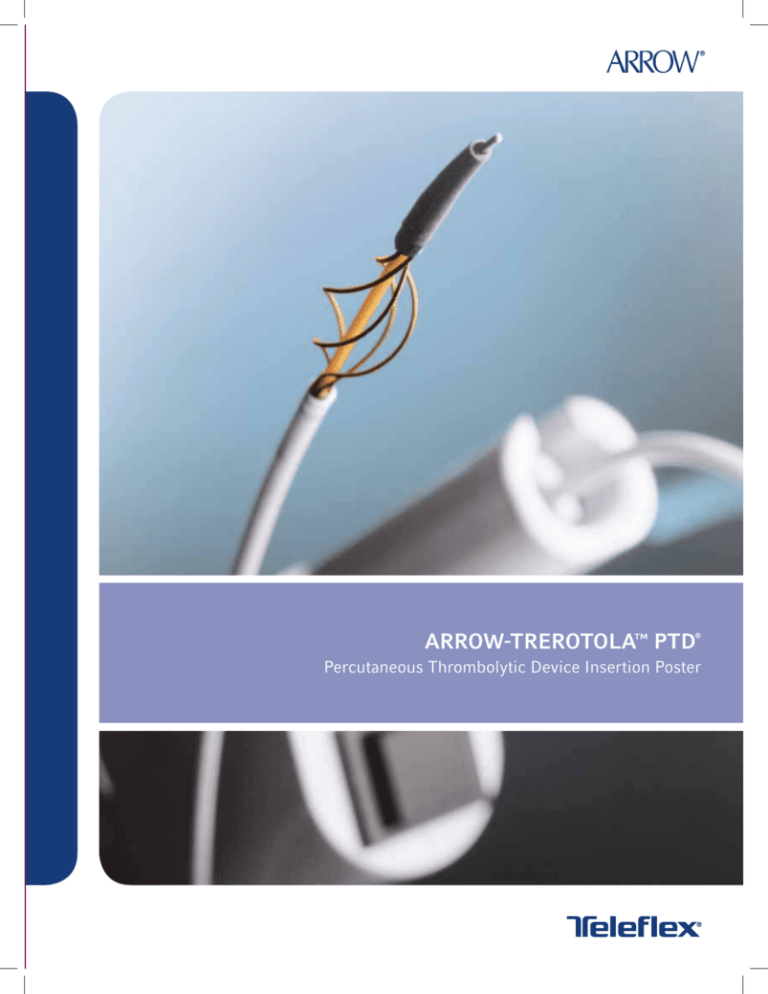
ARROW-TREROTOLA™ PTD
®
Percutaneous Thrombolytic Device Insertion Poster
ARROW–TREROTOLATM PTD®
Percutaneous Thrombolytic Device
ARROW-TREROTOLA
PTD (5 FR.)
ARROW-TREROTOLA PTD
OVER-THE-WIRE (7 FR.)
CATHETER LUMEN SIDEARM
Permits catheter flushing during
preparation and use
SOFT, FLEXIBLE TIP
Designed to easily
maneuver through vessel
ACTIVATED SPINNING BASKET
Macerates the thrombus
UNIQUE EXPANDABLE 9 MM
FRAGMENTATION BASKET
Conforms to variable diameter walls
Shown to easily remove residual
thrombus from dialysis vessel walls6
ARROW-TREROTOLA™ PTD® 5 FR. AND 7 FR. OTW PE
5 FRENCH ARROW-TREROTOLA PTD PRODUCTS
������� #
��������
������
(��)
������������� ������
(��)
������ ��������
(��.)
������� ����� ����
(���)
����/����
��-�����-�
–
–
–
3000
1
��-�����*
65
9
–
–
1
��-�����**
65
9
2/6
–
1
��-�����-���
65
9
2/6 (HF)
3000
1
7 FRENCH ARROW-TREROTOLA OVER-THE-WIRE PTD PRODUCTS
������� #
��������
������
(��)
�������������
������
(��)
���-��-����
���������
������ (��)
������ ��������
(��.)
������� ����� ����
(���)
����/����
��-�����-��
–
–
–
–
3000
1
��-�����-� ***
65
9
0.025
–
–
1
��-�����-��
65
9
0.025
2/7
3000
1
��-�����-����
65
9
0.025
2/7 (HF)
3000
1
��-�����-��
120
9
0.025
2/7
3000
1
PTD ACCESSORY COMPONENTS
����������
�������
������ ����
(��.)
������ ������
(��)
������ �������
������
(��)
��� ���������
�������������
(��)
��-�����
5
2
5
0.038
��-�����
6
2
5
0.038
��-�����-��
6
2
5
0.038
��-�����-��
7
2
5
0.038
���������� ���
������
�����-����
�������
�����-�����
���
����/����
Gray
10
Green
10
Green
5
Orange
5
Does not contain natural rubber latex.
Each product includes:
One Radiopaque Polyurethane Sheath with Integral Side Port/Hemostasis Valve
One Vessel Dilator with SnapLock™ feature
* When ordering this component, the PT-03000-R and CL-08605-HF must also be ordered.
** When ordering this component, the PT-03000-R must also be ordered.
*** When ordering this component, the PT-03009-RW and CL-08705-HF must also be ordered.
Caution: U.S. federal law limits this device to sale by or on order
of a physician. Contents of unopened, undamaged package are
sterile. Disposable. Refer to package insert for current warnings,
indications, contraindications, precautions and Instructions For Use.
W PERCUTANEOUS THROMBOLYTIC DEVICE
X-RAY TESTIMONIALS-AV SYNTHETIC GRAFT
Spot radiograph shows clotted forearm loop graft.
Spot radiograph shows PTD treating arterial limb of graft.
Completed fistulogram after using PTD shows patient graft
with no residual clot.
Completed fistulogram of the venous outflow after using PTD
and performing angioplasty of venous stenosis shows patient
graft and no residual clot.
SUMMARY OF STUDIES TO SUPPORT THE SAFETY OF PTD IN VESSELS
a
b
������
����
����� ����������� �� �����
�������
McLennan G,
Trerotola SO, et al2
Animal
Effects of mechanical thrombolytic device on
canine vein valves
(n=51 valves)
80% exhibited no change in valve reflux function
after PTD use.
77% exhibited insignificant change in reflux time.
McLennan G,
Rhodes CA, et ala
Animal
Effects of mechanical thrombolytic device with
stranded stainless steel basket on venous
endothelium of rabbits (n=30 rabbits)
Stranded stainless steel does not cause
significantly more endothelial loss as compared to
surgical gold-standard Fogarty balloon catheters.
Rocek M,
Peregrin JH, et al3
Human
Effects of mechanical thrombolytic device in
native fistulas (n=10 patients)
90% clinical success,
70% 3 mo. Primary patency,
60% 6 mo. Primary patency.
Rocek M,
Peregrin JH, et alb
(abstract)
Human
Effects of mechanical thrombolytic
device in synthetic grafts and native fistulas
(n=25 patients)
83.3% 12 months patency.
McLennan G, Rhodes CA, et al. Venous endothelial effects of a mechanical thrombolytic device using a stranded stainless steel basket.
(unpublished study)
Rocek M, Peregrin JH, et al. The Arrow-Trerotola percutaneous thrombolytic device and declotting of graft and native fistula
occlusions: (results and one year follow-up. Abstract.)
X-RAY TESTIMONIALS-AV FISTULA
Spot radiograph from arteriogram shows
thrombosed
Spot radiograph shows over-the-wire PTD treating clot in cephalic vein.
Completed fistulogram after using over-the-wire
PTD shows patent fistula with no residual clot.
CLINICAL SUMMARY8
���
�����-�����
Number of grafts treated
64
58
Acute technical success
61/64 (95.3%)
55/58 (94.8%)
64
57
e
Completed procedures
������ ���� (�������)
�� ��������� ����������
�
������
(�����)
�
Barnes Jewish
64
75
(25-209)
Indiana University
15
76
(50-209)
Johns Hopkins
13
58
Methodist
16
University of Pennsylvania
Penn State
������
(�����)
57
85
(50-273)
14
81.5
(60-273)
(30-157)
14
74
(50-172)
80
(25-171)
13
93
(65-131)
10
49
(38-92)
6
65.5
(55-103)
10
84
(67-167)
9
94
(65-220)
1
85
(85-85)
Functional success
Number of grafts requiring adjunctive therapy
for residual thrombus (not including treatment
of the arterial plug)
58/64 (90.6%)
29/64 (45.3%)
51/57 (89.5%)
43/47 (75.4%)
Number of grafts able to dialize at 3 monthsd
25/61 (41.0%)
23/55 (41.8%)
f
c
Excludes one incomplete procedure.
Among acute technical success.
Acute Technical Success: The establishment of patency at the end of the procedure as defined by the restoration of flow.
f Functional Success: The ability to dialize through the graft post procedure.
d
e
WARNINGS:
PRECAUTIONS:
Prior to use, read all package insert warnings, precautions, and instructions.
Failure to do so may result in severe patient injury or death.
During native fistula declotting, an appropriate guidewire should be used with the
OTW device. Excessive vessel tortuosity (i.e. tight stenosis alternate with broader
segments of vessel or sharp angle of anastomosis) may significantly complicate the
thrombectomy procedure. Due to tortuosity, if a guidewire cannot be advanced in
the vessel or a guidewire cannot cross the clot, then an alternative procedure
should be explored.
Sterile, Single use: Do not reuse, reprocess or resterilize. Reuse of device
creates a potential risk of serious injury and/or infection which may lead
to death.
Practitioners must be aware of potential complications associated with
percutaneous dialysis graft thrombolysis including hemorrhage, symptomatic
pulmonary embolism, arterial embolization, allergic reaction to contrast,
and pseudoaneurysm.
The PTD® device is not for use in stents.
Caution should be used when dislodging the plug at the arterial anastomosis
to minimize the risk of arterial embolization. For the OTW device, caution
should be used when declotting synthetic dialysis grafts and AV fistulas.
Arterial and venous sheath tips should not overlap.
Keep the exposed portion of the PTD catheter straight at all times during
the procedure.
Continued unsuccessful aspiration may collapse sheath and graft/fistula. Two
passes are recommended but additional passes may be required to completely
macerate thrombus. The number of passes required could range from 1–8 as
shown in synthetic graft study 8 or 2–7 passes as shown in AV fistula study 3.
Due to lack of excretion associated with hemodialysis patients use of contrast
should be kept to a minimum throughout this procedure.
Do not use unit if the rotator does not activate immediately when the ON/OFF
switch is depressed, and deactivate immediately when the ON/OFF switch
is released.
Potential fatigue failure of PTD torque cable and fragmentation basket may
occur with prolonged activation of PTD device. A withdrawal rate of 1–2 cm/
second is recommended when sharp radii are encountered (i.e. radius of loop
graft, radii < 3 cm).
If assessment reveals venous outflow stenosis greater than 10 cm long, untreatable
central venous stenoses/occlusions, or any large pseudoaneurysm, treatment of
graft should be re-evaluated and alternative treatments should be considered.
Do not advance PTD catheter forward during activation.
Due to the risk of exposure of HIV (Human Immunodeficiency Virus) or other
bloodborne pathogens, health care workers should routinely use universal
blood and body fluid precautions in the care of all patients.
Immediately release activation switch on rotator device if an audible change in
pitch becomes apparent. This will prevent rotator strain and further decrease the
chance of basket breakage.
CONTRAINDICATIONS:
The PTD devices are not recommended in the presence of hemodialysis s access
site/graft infections. They are not recommended for use in venous outflow stenosis
greater than 10 cm long and large pseudo aneurysm.
The non-OTW PTD is also contraindicated for untreatable central venous
stenosis/occlusion.
The OTW PTD is also contraindicated for use in native vessels smaller than 6 mm
in diameter and immature AV fistulas (fistulas that have not been used for at least
one hemodialysis treatment).
REFERENCES:
1. Lajvardi A, Trerotola SO, Strandberg JD, Samphilipo MA, Magee C. Evaluation
of venous injury caused by a percutaneous mechanical thrombolytic device.
Cardiovascular Interventional Radiology. 1995; 18: 172-178.
2. McLennan G, Trerotola SO, Davidson D, Rhodes AC, et al. The Effects of a
Mechanical Thrombolytic Device on Normal Canine Vein Valves. JVIR.
2001;12:89-94.
3. Rocek M, Peregrin, JH, Lastovickova J, Krajickova D, et al. Mechanical
Thrombolysis of Thrombosed Hemodialysis Native Fistulas with use of the
Arrow-Trerotola Percutaneous Thrombolytic Device: Our Preliminary
Experience. JVIR. 2000;11:1153-1158.
6. Trerotola SO, Johnson MS, Shah H, Namyslowski J. Backbleeding technique for
treatment of arterial emboli resulting from dialysis graft thrombolysis. JVIR.
1998;9:141-143.
7. Trerotola SO, Lund GB, Scheel Jr PJ, Savader SJ, Venbrux AC, Osterman Jr FA.
Thrombosed dialysis access grafts: percutaneous mechanical declotting
without urokinase. Radiology. 1994;191:721-726.
8. Trerotola SO, Vesely TM, Lund GB, et al. Treatment of thrombosed
hemodialysis access grafts: Arrow-Trerotola percutaneous thrombolytic device
versus pulse-spray thrombolysis. Radiology. 1998;206:403-414.
9. Winkler TA, Trerotola SO, Davidson DD, Milgrom ML. Study of thrombus from
thrombosed hemodialysis access grafts. Radiology. 1995;197:461-465.
4. Swan TL, Smyth SH, Ruffenach SJ, Berman SS, Pond GD. Pulmonary embolism
following hemodialysis access thrombolysis/thrombectomy. JVIR.
1995;6:683-686.
10. Lazzaro CR, Trerotola SO, Shah H, Namyslowski J, Moresco K, Patel N.
Modified use of the Arrow-Trerotola Percutaneous Thrombolytic Device for
treatment of thrombosed hemodialysis access grafts. JVIR. 1999;10:1025-1031.
5. Trerotola SO, Johnson MS, Schauwecker DS, Davidson DD, Filo, RS, et al.
Pulmonary emboli from pulse-spray and mechanical thrombolysis: evaluation
with an animal dialysis graft model. Radiology. 1996;200:169-176.
11. Trerotola SO, Johnson MS, Shah H, Namylowski J, Filo RS. Incidence and
management of arterial emboli from hemodialysis graft surgical thrombectomy.
JVIR. 1997; 8:557-562.
ARROW-TREROTOLA™ PTD® 5 FR. AND 7 FR. OTW
5 Fr. PTD
HEMOSTASIS HUB
SIDEARM
HEMOSTASIS
HUB
CABLE CABLE
STOP
STOP
ARM
SLOTS
2
1
3
COMBINED 5 FR. & 7 FR.
OTW PTD PROCEDURE
INSTRUCTIONS
1. PATIENT PREPARATION
CATHETER HUB
ASSEMBLY
EXPANDED
FRAGMENTATION BASKET
Pre-medicate, prep, and drape patient per
hospital protocol.
2. INTRODUCER SHEATH PLACEMENT
Figure 1
Refer to Figures A-D below for sheath
placement techniques. Puncture sites should
be at least 10 cm apart. Sheath tips should
not overlap.
Administer local anesthesia and place
venous introducer sheath directed toward
the venous anastomosis. NOTE: In AV fistula,
the venous sheath placement can be optional
depending on the clot burden in vessel. If a
venous sheath is used, it should be placed in
venous limb of fistula and directed toward
central venous outflow. NOTE: If no venous
sheath is used in AV fistula, then go to
ARTERIAL LIMB THROMBOLYSIS listed
below for single sheath procedure to treat
AV fistula.
SIDEARM
SLOT #3
Figure 2
SIDEARM LOCKED
3. PATIENT ASSESSMENT
SLOT 1
SHEATHED, COMPRESSED
FRAGMENTATION BASKET
Figure 3
Under fluoroscopy, assess any existing
central and venous outflow stenosis per
institutional protocol. In an AV fistula, if
there are large thrombosed aneurysms,
a cuff could be considered.
NOTE: If assessment reveals venous outflow
stenosis greater than 10 cm long, untreatable
central venous stenosis/occlusions, or any
large pseudoaneurysm, treatment of graft
should be re-evaluated and alternative
treatments should be considered.
SLOT #2
4. ANTICOAGULATION
UNSHEATHED
FRAGMENTATION BASKET
Figure 4
If graft or fistula is salvageable, administer
heparin (or other appropriate anticoagulant)
intravenously or adhere to hospital protocol.
5. VENOUS LIMB THROMBOSIS
ROTATOR ASSEMBLY
PROCEDURE
Flush catheter lumen through sidearm.
Verify function of rotator drive unit by
pressing ON/OFF switch.
Insert catheter into rotator. (see Figure 1)
Turn arm on cable stop into slot #3 to lock
catheter in place. This arm should remain
locked in this position throughout the
entire procedure.
If using the 7 Fr. OTW device, advance
appropriate spring-wire guide through
venous sheath into venous limb of
graft or fistula.
BRACHIAL ARTERY
RADIAL ARTER
Slide side arm forward to cover and
compress fragmentation basket. Lock into
compressed position by inserting side
arm into slot #1. (see Figure 3)
Unlock and slide side arm from slot #1 to
slot #2. Lock into deployed position by
inserting side arm into slot #2. (see Figure 4)
Depress ON/OFF switch to ensure unit
spins fragmentation basket. Release
switch to stop motor.
IF ANY PART OF THIS SYSTEM FAILS TO WORK,
REPLACE THE MALFUNCTIONING PART AND RETEST.
VEN
ARTERIAL SHEATH
BASILIC VEIN
Figure A – (AV Graft)
TROUBLESHOOTING TIPS
POTENTIAL PROBLEM
Cable breaks:
In proximity of rotator
In proximity of basket
Basket breaks:
POSSIBLE SOLUTIONS
Remove device with attached catheter sheath.
Remove remaining fractured cable section from
introducer sheath.
Remove catheter hub assembly. Use snare
to grasp basket (not tip) and remove through
introducer sheath.
Action depends on site of break. If possible,
remove device through introducer sheath. If
pulling back will cause broken wire to catch on
graft/fistula wall, use snare through opposite
sheath to grasp basket (not tip) of device and
remove.
POTENTIAL PROBL
Introducer sheath p
Catheter basket enta
with second sheath
Sheath exits graft
Repeat clotting in
graft/fistula - long
procedure:
Clot caught in baske
7 Fr. orange cannul
separates from tip:
TW PERCUTANEOUS THROMBOLYTIC DEVICE
r
ould
d
d
ula,
onal
a
d in
d
us
With basket in compressed position, insert
catheter (over guidewire if using the 7 Fr.
OTW device) into venous limb of graft or
fistula. In a graft, advance flexible tip up to,
but not beyond the venous anastomosis. In
a fistula, advance the tip up to the centralmost extent of the clot and expose PTD
basket by sliding the sidearm from slot #1
into slot #2. (see Figures 3 and 4)
PRECAUTION: Continued unsuccessful
aspiration may collapse venous sheath and
graft/fistula. Two passes are recommended,
but additional passes may be required to
completely macerate thrombus. The number
of passes required could range from 1–8 as
shown in synthetic graft study8 or 2–7 passes
as shown in AV fistula study3.
Activate the on/off switch, and slowly
withdraw the deployed rotating basket along
graft or vessel to macerate the adherent clot.
WARNING: Potential fatigue failure of the
PTD torque cable and fragmentation basket
may occur with prolonged activation of the
device. The cumulative activation time of
the PTD catheter should be limited to 30-60
seconds in tight curves. A rapid withdrawal
rate of 1–2 cm/second is recommended when
tight curves are encountered (i.e. radius of
loop graft or vessel < 3 cm).
6. ARTERIAL LIMB THROMBOLYSIS
Administer local anesthesia and place
arterial sheath directed toward arterial
anastomosis.
(If using the 7 Fr. OTW device, position
spring-wire guide through arterial sheath
into graft or fistula.) With catheter basket in
compressed position, insert catheter (over
guidewire if using the 7 Fr. OTW device)
through sheath and into arterial limb of
graft or fistula keeping exposed PTD
catheter straight at all times.
Advance flexible tip of PTD basket up
to, but not beyond arterial anastomosis.
Expose PTD basket by moving sidearm
from slot #1 to slot #2, but do not grip
outer catheter when deploying basket near
arterial anastomosis. If catheter is held, the
fragmentation basket may jump forward and
cause arterial plug to dislodge and embolize.
Allow catheter to slide backward in your
hand. Activate rotator and repeat steps with
two passes.
Remove device in compressed position.
Remove guidewire and aspirate
approximately 5cc of clot through arterial
sheath and note that a closed system may
prevent aspiration. PRECAUTION: Continued
unsuccessful aspiration may collapse arterial
sheath and graft/fistula. Two passes are
recommended, but additional passes may be
required to completely macerate thrombus.
The number of passes required could range
from 1-8 as shown in synthetic graft8 study
or 2-7 passes as shown in AV fistula study3.
Flush catheter lumen and remove any fibrin
from PTD basket. Check device function.
t
w
ble
er
ant)
col.
Release switch when basket reaches tip of
introducer sheath.
Move the PTD basket into the compressed
position and remove from introducer sheath
(leaving the guidewire in place if using the
7 Fr. OTW device). Flush catheter lumen
and manually remove fibrin from the PTD
basket. Re-insert device and repeat passes
as necessary.
Flush catheter with heparinized saline and
remove any fibrin from PTD basket. Inject a
small amount of contrast to ensure adequate
thrombolysis of venous limb. AVOID OVERINJECTION OF CONTRAST TO MINIMIZE
THE RISK OF ARTERIAL EMBOLIZATION.
Remove (guidewire if using the 7 Fr.
OTW device and) catheter with basket in
compressed position. If possible, aspirate
approx. 5cc of clot via the venous sheath
and note that a closed system may
prevent aspiration.
BRACHIAL ARTERY
L ARTERY
RADIAL ARTERY
VENOUS SHEATH
ARTERIAL SHEATH
VENOUS SHEATH
BASILIC VEIN
Option 1
L PROBLEM
Figure B – (AV Graft)
POSSIBLE SOLUTIONS
sheath problems:
sket entangled
Stop rotator. Move device back and forth until free
sheath
from sheath.
s graft
In some cases sheath can be reinserted into graft
over PTD catheter.
ting in
a - long
in basket:
e cannula
rom tip:
Option 2
POTENTIAL PROBLEM
POSSIBLE SO
Basket deployment failure:
Make sure fib
by manually r
deploying bas
Difficulty retracting basket
into catheter outer sheath:
Clean fibrin fr
force to reshe
retracted into
introducer she
continuing pr
Difficulty aspirating
venous clot:
Treat arterial
may be possib
Incomplete maceration
of clot:
Make addition
Venous spasm due to
inadvertent entry into
native vessel:
Administer ni
aliquots IV pu
hospital proto
Make sure heparin level is adequate by
consistently checking activated clotting time
(ACT). Administer additional heparin, if necessary.
Gently pinch basket and pull thrombus toward the
tip of the catheter or rinse expanded basket in saline.
Guidewire should always be used with 7 Fr.
OTW device to ensure stabilization of cannula
within basket.
7a. PLUG REMOVAL PROCEDURE
WITH BALLOON CATHETER
(advance to 7b. for alternate plug
removal procedure using the PTD)
If applicable, use appropriate guidewire
and pass balloon catheter through arterial
sheath and carefully feed past the arterial
anastomosis of graft or fistula.
Re-insert PTD catheter (over guidewire
if using the 7 Fr. OTW device) back into
the arterial limb. Expose PTD basket and
activate rotator to macerate arterial plug
using contrast to guide thrombolysis.
De
w
ar
be
Ac
th
po
tip
Re
cl
Re
(R
O
As
ARTERIAL PLUG
(Remove guidewire if using the 7 Fr. OTW
device) place PTD into compressed position
and remove from sheath.
Aspirate 5-10cc of clot using either sheath
and inject contrast to assess degree of
thrombus removal. Treat residual thrombus
using PTD via both sheaths as needed.
ARTERIAL PLUG
7b. PLUG REMOVAL PROCEDURE
WITH PTD FOR SYNTHETIC
AV GRAFTS10
Inflate balloon and pull arterial plug into
middle of the arterial limb of graft or fistula.
ARTERIAL PLUG
This procedure has been evaluated in 6 mm
forearm loop grafts with brachial artery
anastomosis. PRECAUTION: This technique
may not be applicable to straight forearm
loop grafts with radial artery anastomosis
or tapered grafts.
(Re-insert guidewire if using the 7 Fr.
OTW device).
While in the compressed basket position,
advance the flexible tip through the arterial
anastomosis and into the inflow artery, but
DO NOT ACTIVATE WHILE THE BASKET IS
WITHIN THE INFLOW ARTERY.
COMPRESSED
BASKET
8. A
In
re
vi
Re
us
an
tr
pr
Pe
ve
Ac
ho
Deflate balloon and remove catheter. (remove
wire if not using the 7 Fr. device.)
VENOUS SHEATH
AR
BRACHIAL ARTERY
RADIAL ARTERY
ARTERIAL SHEATH
BASILIC VEIN
Figure C – (AV Graft)
Option 3
Figure D – (AV Fistula)
SIBLE SOLUTIONS
POTENTIAL PROBLEM
POSSIBLE SOLUTIONS
sure fibrin has been removed from basket
anually removing it from wires. While
ying basket, activate device for short bursts.
Intact basket gets stuck:
Remove catheter assembly from rotat
and manually rotate counterclockwise
basket. Manually resheath basket.
n fibrin from basket frequently. Use slight
to resheath device. If device still cannot be
cted into outer sheath, remove device via the
ducer sheath and remove fibrin before
nuing procedure.
Cable stretching:
Remove device from patient and repl
Arterial Embolization (AE)
contributing factors:
nȟȟ(OLDINGȟCATHETERȟȟ
when near arterial plug
Do not grip catheter when deploying
basket near arterial anastomosis to m
RISKȟTHATȟBASKETȟWILLȟJUMPȟFORWARDȟAND
arterial plug to dislodge and emboliz
catheter to slide backward in your ha
nȟȟ/VERINJECTINGȟCONTRASTȟȟ
before clot is macerated
!VOIDȟOVERINJECTINGȟCONTRAST
nȟȟ!RTERIALȟPLUGȟTREATMENTȟȟ
with occlusion balloon
5SEȟOFȟOVERTHEWIREȟOCCLUSIONȟBALLOO
reduce occurrence of AE.
arterial side of graft and venous aspiration
be possible.
additional pass through clot at a slower rate.
nister nitroglycerin, 100 µg/ml. Give in 1 ml
ots IV push to relieve spasm or adhere to
tal protocol.
7 Fr. OTW PTD
Deploy fragmentation basket and slowly
withdraw expanded basket into the
arterial anastomosis until the basket
begins to compress.
HEMOSTASIS HUB
SIDEARM
CABLE
STOP
ARM
1
SLOTS
2 3
EXPANDED
BASKET
EXPANDED
FRAGMENTATION BASKET
Figure 1
SIDEARM
Activate rotator while withdrawing through
the graft/fistula. Move device to compressed
position when you have reached the sheath
tip, (leaving guidewire in place if used).
Remove from sheath, flush catheter, and
clean fibrin from PTD basket.
Repeat passes in above steps as necessary.
TUOHY-BORST
CAP
SLOT #3
Figure 2
SIDEARM LOCKED
ARTERIAL PLUG
SLOT 1
SHEATHED, COMPRESSED
FRAGMENTATION BASKET
Figure 3
(Remove guidewire if using the 7 Fr.
OTW device).
Aspirate 5-10cc of clot using either sheath.
SIDEARM LOCKED
8. ASSESSMENT AND PROCEDURE WRAP-UP
Inject contrast to assess degree of thrombus
removal. Treat residual thrombus using PTD
via both sheaths as needed.
Remove blood pressure cuff or tourniquet if
used. When thrombus removal is complete,
any underlying disease or stenosis should be
treated with balloon angioplasty per hospital
protocol.
Perform final fistulogram. Remove sheaths,
verifying entire length has been withdrawn.
Achieve hemostasis at both sites per
hospital protocol.
SLOT #2
UNSHEATHED, EXPANDED
FRAGMENTATION BASKET
Figure 4
ROTATOR ASSEMBLY
PROCEDURE
Flush catheter lumen through sidearm.
Verify function of rotator device unit by
pressing ON/OFF switch.
Insert catheter assembly into rotator device.
(see Figure 1)
Turn arm on cable stop into slot #3 to lock
catheter into place. This arm should remain
locked in this position throughout the entire
procedure. (see Figure 2)
Compress and cover PTD basket by locking
sidearm into slot #1. Expose PTD basket by
moving sidearm into slot #2.
(see Figure 3 & 4)
Depress ON/OFF switch to ensure unit spins
fragmentation basket. Release switch to
stop motor.
Ensure Tuohy-Borst cap is open and pass
guidewire into rotator unit and through the
flexible tip.
ARTERIAL SHEATH
RADIAL ARTERY
VENOUS SHEATH
IF ANY PART OF THIS SYSTEM FAILS TO WORK,
REPLACE THE MALFUNCTIONING PART AND RETEST.
ula)
TREATMENT OF SYMPTOMATIC ARTERIAL EMBOLIZATION11
rom rotator device
clockwise to free
asket.
Mechanical techniques:
and replace device.
2. Use an over-the-wire balloon (e.g. wedge catheter) to mobilize embolus.
eploying
osis to minimize
WARDȟANDȟCAUSE
embolize. Allow
n your hand.
T
ONȟBALLOONȟMAY
1. Treat with backbleeding - place an embolectomy catheter in artery proximal
to the anastomosis, inflate the balloon and allow backbleeding from the distal
artery to carry embolus back into graft.
3. Thromboaspiration.
If mechanical techniques fail, thrombolysis or surgical thrombectomy may be
alternatives. Asymptomatic emboli may not need treatment.
Teleflex is a global provider of medical products designed to enable
healthcare providers to protect against infections and improve
patient and provider safety. The company specializes in products
and services for vascular access, respiratory, general and regional
anesthesia, cardiac care, urology and surgery. Teleflex also provides
specialty products for device manufacturers.
The Teleflex family of brands includes ����� , ����� ������� ,
�������� , ������ , ������ ��� , ������ , ������� , �����-���� ,
����� , �������� , ��� , ���� , ��� ��� , �������� and ���� ,
all of which are trademarks or registered trademarks of Teleflex
Incorporated or its affiliates. Teleflex Incorporated (����:���) has
annual revenues of approximately $1.5 billion and customers in
more than 130 countries.
®
®
®
®
®
®
®
®
®
®
®
®
For detailed information, see teleflex.com
TELEFLEX MEDICAL
PO Box 12600
Research Triangle Park, NC 27709
Toll Free
Phone
Intl
866.246.6990
919.544.8000
919.433.8088
Teleflex, Arrow, Arrow-Trerotola, PTD and SnapLock are trademarks
or registered trademarks of Teleflex Incorporated or its affiliates.
© 2012 Teleflex Incorporated. All rights reserved. 2012-0731
®
TM
®

