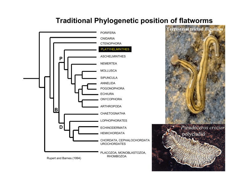
Traditional Phylogenetic position of flatworms
B
Traditional Phylogenetic position of flatworms
Terrestrial triclad Bipalium
PORIFERA
CNIDARIA
CTENOPHORA
PLATYHELMINTHES
P
D
ASCHELMINTHES
NEMERTEA
MOLLUSCA
SIPUNCULA
ANNELIDA
POGONOPHORA
ECHIURA
ONYCOPHORA
ARTHROPODA
CHAETOGNATHA
LOPHOPHORATES
ECHINODERMATA
HEMICHORDATA
CHORDATA, CEPHALOCHORDATA
UROCHORDATES
Pseudoceros crozieri polycladid
PLACOZOA, MONOBLASTOZOA,
RHOMBOZOA
Rupert and Barnes (1994)
The Flatworms – Platyhelminthes- Traditionally with 4 Classes
Trematoda - Parasitic flukes Monogenea - Parasitic flukes (1 gen)
Rugogaster hyrdolagi
Cestoda Taenia pisiformis - Scolex, and anterior suckers of a tapeworm.
Turbellaria
-
A triclad marine flatworm
Phylum Platyhelminthes (Flatworms )
Parasitic or free-living, unsegmented worms
Triploblastic, bilateral symmetrical, spiral cleavage, flattened dorsoventrally
Acoelomates (solid body forms lacking the fluidfilled space, such as coelom, pseudocoel, or hemocoel, between body wall and gut that is seen in more complex metazoans)
Complex but incomplete gut. No gut is some parasitic flatworms (Cestoda)
Cephalization, central nervous system with an anterior cerebral ganglion and sometimes longitudinal nerve cords connected by a ladderlike nervous system
Protonephridia, complex excretory/osmoregulatory structures
Hermaphroditic, complex reproductive structures. Mutually insemination in sexual reproduction . Spermatozoa with two tails
Body plans of triploblastic Metazoans
Acoelomates
Blastocoelomates
Eucoelomates
Class Turbellaria – (Free Living Flatworms)
Bilaterally symmetrical-vary in size from <1 mm to several cm
Cylindrical body in small turbellarians, dorsoventrally flattened in large
Epidermis is ciliated, ciliary locomotion
Secrete solid mucus pellets called rhabdoids
If gut present with one opening (mouth present, no anus), carnivores. Mouth located on ventral surface that opens into the GV cavity.
No circulatory system (diffusion), protonephridia for excretion
Cheliplana sp. (Rhabdocoela)
Connective tissue called parenchyma
Some cephalization
Most likely paraphyletic group
Maiazoon orsaki
Turbellarian vs hydroid epidermis
Rhabdoids (rod-shaped epidermal inclusions) produced by mesenchymal cells
( rhabdites), stored in packets. When released, produce slime (chemical defense)
Gland cell
Gut organization
Digestive system typically a blind sac except where high degree of branching makes return to mouth of wastes difficult
Epithelium consists of gland cells and phagocytic cells
Gut may be simple, unbranched (acoels)- lacks permanent cavity (but with syncitium)
Gut may be highly branched with diverticula – increase surface area for digestion/absorption
Pharyngeal patterns
• Simple pharynx – opening (mouth) directly into digestive syncitium
• Bulbous pharynx – reduction of pharyngeal cavity, separated from parenchyma by muscular septum – can act independently of pressure in body
• Plicate pharynx – muscular pharyngeal tube within a cavity – probably originated as folding of simple
• pharynx
Jawed Proboscis
Cheliplana, Turb.
Dugesia, x-section in the region of the pharynx
Class Turbellaria (Cont’d)
Triclad Dugesia
Most famous example is Dugesia (formerly Planaria ) , a triclad planarian. Most students have worked with them in introductory and invertebrate zoology courses
Oval to elongate in shape
Dorsoventrally flattened, more so with size
Usually quite small (<10 mm) but some very large exceptions ( Rimacephalus arecepta - 60 cm)
Primarily aquatic and most marine, interstitial
• 12 Orders: Acoela, Catenulida, Haplopharyngida,
Lecithoepitheliata, Macrostomida, Nemertodermatida
Polycladida, Proseriata, Tricladida, Rhabdocoela,
Proplicastomata, Proplicastomata. The 2 superorders
Archoophorans and Neoophorans (based on reproductive system) are almost defunct.
Archoophorans- entolecithal (yolk is deposited within the cytoplasm of the ova), development may include planktonic larva, but usually direct
Nucleus
Triclad Bdelloura
Yolk granules
Cell membrane
Class Turbellaria (Cont’d)
Neophorans – ectolecithal (yolk is deposited outside the cytoplasm of the ova), development always direct
Yolk cells
Yolk granules
Most are marine, but some freshwater forms
Yolkless egg
Egg capsule
Larval Forms
4 arms or lobes – Goette’s larva
8 arms or lobes – Mueller’s larva
These eventually resorb arms and develop into small adult worms
Müller's larvae
Müller's larva of
Pseudoceros canadensis
Unidentified Müller's larvae
Archoophoran reproductive plan
Acoels-no separation of gonads by epithelium, ovaries/testes absent
Macrostomida-ovaries/testes present, well developed accessories
Polycladida – multiple branches of ovaries/testes, well developed accessories
Neophorans reproductive plan
Main characteristic – separate vitellarium or yolk producing glands
Brusca and Brusca (2002)
Asexual reproduction;
• Paratomy –formation of 2 or more zooids (buds) along axis of body prior to division
• Architomy – transverse splitting of body into several pieces, each of which regenerates to form a complete individual AFTER fission
Sexual reproduction
Most turbellarians in general are hermaphroditic and have internal fertilization
Dorsoventral muscles may occur and aid in flattening body
Temporary attachment organs allow adhesion to substrate
(duogland organs)
Circular muscles occur below the parenchyma and epithelium - elongation
Longitudinal muscles occur interiorly – contraction in length
Diagonal muscles – twisting
Muscles are usually smooth
Types of Movement
A. Ciliary movement
B. Undulation
C. Muscular creeping
D. Didactically
E. Contraction
G. Peristalsis to mix gut contents
H. Secretion of mucus strings
I. Somersaulting
Dugesia
Detail of the lateral margin of the body of Dugesia showing the adhesive gland on the ventral surface and the differentiation of the cilia between the dorsal and ventral surfaces.
Dorsal
Ventral
Excretion
– Via Protonephridia (major advance over diploblasts) – simple filter cells (flame bulb) with 1 or more internal flagella that draw fluid into cell – wastes empty to the exterior by nephridiopores. Some ammonia excreted but most metabolic waste by diffusion
No protonephridia on Acoela,
Nemertodermatida and some catenulids
Flame cells connected to collecting tubules, protonephridial system in a freshwater turbellarian.
Turbellarian Nervous system
Ladder-like nervous system
Advanced condition
Dugesia
Cephalization of nervous system
Cross linked (segment like)
Anterior end with brain, eyespots and the longitudinal nerve cords
Interstitial turbellarian
Statocyst sensory bristles
Turbellarian nervous systems and organs
Polyclad Planocera
Triclad Crenobia
Inverted pigment cup ocellus (x-sec)
Anterior end of rhabdocoel
Mesostoma (x-sec ) with tactile, chemo- and rheoreceptors
Nervous systems of other Platyhelminthes
Brusca and Brusca (2002)
fluke Probolitrema
Chinese liver fluke
Schistosomiasis
(swimmer’s itch)
Stylochus
Taenia
Tegument organization of flukes and cestodes
Brusca and Brusca (2002)
Class Monogenea (parasitic flukes)
Monogenean flukes do not have a well-developed sucker.
Posterior end with a bulbous structure covered with hooks called an opisthaptor.
Anterior end with buccal sucker (prohaptor)
Most monogeneans are ectoparasites on fish or other aquatic animals, although a few live in the urinary bladders of turtles and frogs.
Their life cycle involves a single host. Eggs hatch into ciliated larvae, which may attach directly to a host or swim freely for a time before attaching.
Hermaphroditic
Adults lack external cilia
Prohaptors and Opisthaptors of Monogeneans
Adhesive organs – adaptations for parasitic life style
Opisthaptor – main organ of attachement
Prohaptor Opisthaptor
Larva of monogeneans
Class Trematoda (digenetic parasitic flukes)
All parasites, most adults parasitize vertebrates, about 9000 species
Body covered with a tegument, a peculiar kind of epidermal arrangement in which the main cell bodies are deep, separated from the cytoplasm that lies next to the exterior by a layer of muscle (but connected to the exterior layer by cellular processes. The tegument lacks cilia in adults.
Exterior layer syncytial (i.e continuous, not broken by cell membranes)
Unlike monogeneans, trematodes have no opisthaptor; with 1 or 2 hookless suckers. Acetabulum. Most are endoparasites.
With a developed alimentary canal (like turbel.), muscular, excretory, and reproductive systems are almost complete.
Complex life cycles, with larval stages parasitizing one or more species that are different from host of adults. Larval stages include miracidium, redia, cercaria, and metacercaria.
Enormous impact on humans (liver flukes, blood flukes causing schistosomiasis).
Human liver fluke
Opisthorchis sinensis
General features of a digenetic fluke
Similar to turbellarian flatworms
• Neophoran
• Diverticulated gut
• Specialized genital organs
Genital pore
Copulating pair of human blood flukeSchistosoma mansoni
Mouth
Oral sucker
Intestine
Ventral Sucker (Acetabulum)
Vitellaria - yolk glands
Testes
Ovary
Miracidium ciliated
Ingested or penetrates snail host
Cercaria
Metacercaria
Redia
Representative Digenean Lifecycles
Sporocyst with redia
Egg - encapsulated, passes in urine, feces
Life cycle of blood fluke
Schistosoma mansoni one of 3 spp, causing schistosomiasis.
Different spp. Affect different organs
(e.g. blood vessels, bladder, liver)
Brusca and
Brusca (2002)
Egg
Cercaria larva
Digenetic Fluke Life stages
Metacercaria
Redia larva
Cercaria larvae
Redia larva
Class Cestoda (tapeworms)
Bodies are long and flat, made up of many segments called proglottids. Each proglottid is a reproductive unit.
Adults lack cilia and their surface is a tegument (as in monogeneans and trematodes), but in cestodes the tegument is covered with tiny projections, microvilli, which increase its surface area and thereby its ability to absorb nutrients from a host.
Digestive tracts are absent completely.
Anterior end with is a specialized segment called a scolex, which is usually covered with hooks or suckers and anchors it to the host.
Proglottids
About 5000 species, all endoparasites.
Require two hosts, with the host of the adult tapeworm a vertebrate.
Intermediate hosts are often invertebrates. Many species inhabit humans.
Proglottids
General features of an intact tape worm
Scolex
Reduced or lost nervous system and gut
Internal parasites
Reproductive machines
Hexacanth larva
Rostellum-on the tip of scolex
Sometimes retractable
Myzorhynchus-Protrucible sucker
3 types of adhesive suckers
Bothria – elongated grooves
Bothridia – 4 leaf-like structures with suckers at anterior ends
Acetabulum – true suckers (4)
Cestode Attachment
Brusca and Brusca (2002)
Phylogeny of Platyhelminthes
Brusca and Brusca
(2002)
2- prohaptor
3-- opisthaptor
4– ventral sucker
5-- tegumental microtriches
6-- scolex
7– loss of digestive tract
8– strobilation of the body
