Chapter 3: The Stimulus and Anatomy of the Visual System
advertisement
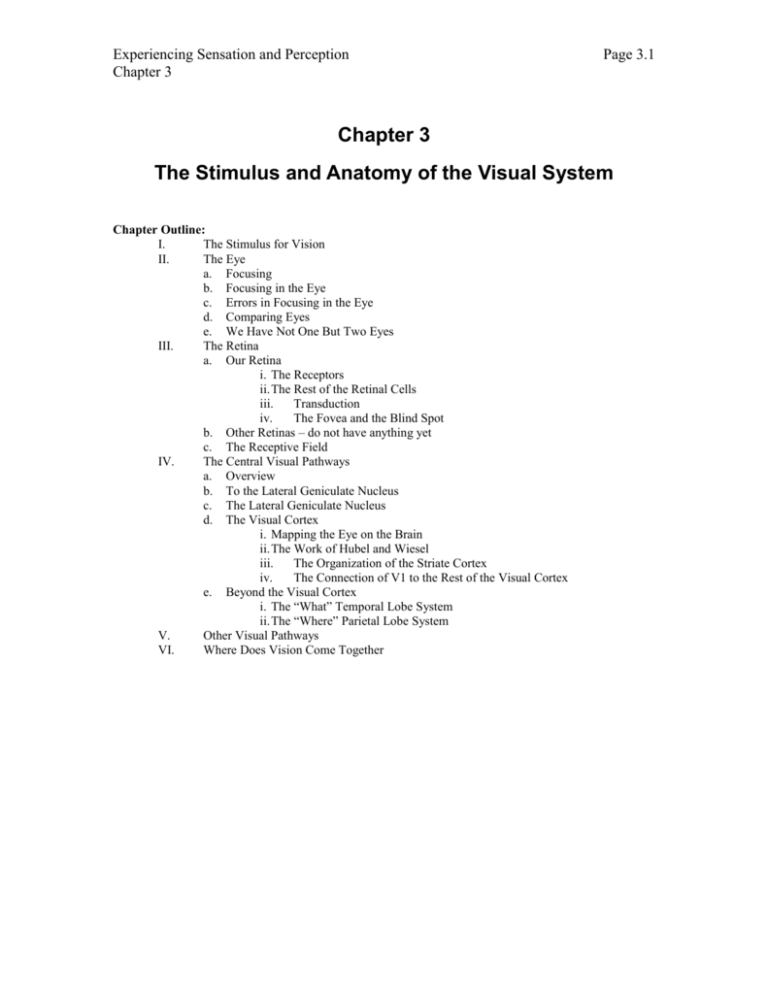
Experiencing Sensation and Perception Chapter 3 Page 3.1 Chapter 3 The Stimulus and Anatomy of the Visual System Chapter Outline: I. The Stimulus for Vision II. The Eye a. Focusing b. Focusing in the Eye c. Errors in Focusing in the Eye d. Comparing Eyes e. We Have Not One But Two Eyes III. The Retina a. Our Retina i. The Receptors ii.The Rest of the Retinal Cells iii. Transduction iv. The Fovea and the Blind Spot b. Other Retinas – do not have anything yet c. The Receptive Field IV. The Central Visual Pathways a. Overview b. To the Lateral Geniculate Nucleus c. The Lateral Geniculate Nucleus d. The Visual Cortex i. Mapping the Eye on the Brain ii.The Work of Hubel and Wiesel iii. The Organization of the Striate Cortex iv. The Connection of V1 to the Rest of the Visual Cortex e. Beyond the Visual Cortex i. The “What” Temporal Lobe System ii.The “Where” Parietal Lobe System V. Other Visual Pathways VI. Where Does Vision Come Together Experiencing Sensation and Perception Chapter 3 Page 3.2 The Stimulus and Anatomy of the Visual System In the first two chapters, we laid the foundation for the study of sensation and perception. Both general theoretical and historical issues were discussed in the first chapter. Some fundamental methods and laws of psychology were discussed in the second chapter. Now it is time to directly encounter our world as experienced through our senses. In doing so, we shall use the organization from the general framework for senses introduced in Chapter 1 (refer back to Figure 1.x). [production: could we repeat that figure here? Would it be hepful? Idea, would it be useful to use icons from that figure to illustrate where in the system each section of this chapter falls on that model?] Visual information abounds that allows us to move about and work in the world with amazing precision. The deceptively simple act of maintaining your balance involves many senses, including your visual system. Try this demonstration: Stand up on one leg. Keep your eyes open and notice how well you balance. Now close your eyes and notice how much more you sway. You might think that this is an artificial demonstration. You might retort that we don’t stand on one leg. But you do, with each step you stand on one leg for a short period of time. In addition to these practical uses of vision, our visual system is also capable of providing experiences that create a sense of wonder in us. Imagine looking across a field and a bright red rose in full bloom against the green foliage of its leaves grabs your attention. The texture, color, and contrast all create an intense experience of breathtaking beauty. It is not inconceivable that both the practical and aesthetic aspects of vision are intertwined. Whether mundane or majestic, most of all visual experiences start with a light source. The most important light source is the sun. Next, in most cases this light reflects off of a surface. Following this step, the light interacts with the anatomical structures of our visual system, starting with our eyes and ending in our brain. There is a close tie between what our visual system detects and the way it is built. So we can begin to learn how the visual system works by examining the physical nature of the stimulus and the anatomical structure of the visual system. The Stimulus for Vision It seems almost trivial to state that visible light is an example of electromagnetic energy (Feynman, 1985, 1995). Visible light is the same basic phenomenon as infrared radiation, ultraviolet radiation, gamma rays, and even the electricity that flows in our walls. Light is made up of particles called photons that also behave in a wavelike manner (see Feynman, 1985, for a more thorough explanation of our current understanding of light in layman’s terms). The wavelike behavior allows us to describe the behavior of light using many concepts that are familiar, so it will be emphasized. What is nice about the wave theory is that all waves behave the same way. So once a wave is understood, say the waves in a pond, all the behaviors known about those waves apply to all others, regardless of what causes each wave. Thus, when sound waves are covered later in the text, these same concepts about waves will apply. Still, the particle description of light will also be important to understand, and it will also be discussed. Consider waves at the beach. The waves are up and down undulations of the water surface. As a result, waves are usually drawn as going up and down (Interactive Illustration 3.x, The Basics of Waves [link to the media]). The waveform is diagramed in this illustration as a sine wave. You might remember sine waves from one of your math classes. This type of gently changing wave is often used to illustrate a simple form of a wave. Going back to the beach, if you watch the waves coming in, you can tell that not all waves are the same. Some are taller than others, and some peaks are closer together, and other peaks are farther apart. These features of waves need to be measured and described so that one wave can be distinguished from other waves. The distance between the peaks of waves is called the wavelength [to glossary] and the height of the wave is called its intensity [to glossary]. The two sliders at the bottom of the screen, Wavelength and Intensity, allow these fundamental measures of a wave to be controlled in the illustration. More formally, wavelength is the distance that represents one complete wave. This one complete wave is called a cycle. You can see one wavelength indicated by pressing the Show Wavelength button below the Wavelength slider. The horizontal red line coming out from the yaxis travels one wavelength. You can still adjust the wavelength of the wave and watch the change in the wavelength. As the cycle gets longer, so does the wavelength. The other fundamental measure of a wave is intensity [to glossary] and intensity refers to the height of the wave on the graph. It also refers to the amount of energy in the wave. As you adjust the wave, the wave gets larger or smaller. In our beach analogy, a low intensity wave would be one caused by Experiencing Sensation and Perception Chapter 3 Page 3.3 dropping a pebble in the water where the bigger waves are the ones you might use to body surf into the shore. As you can see, intensity refers to the strength or energy of the wave. If a wave generated by a pebble hits you, you probably would not feel it. However, if you are hit by a wave you can body surf in you might be knocked around a bit, and a tidal wave can destroy cities. Using the Show Intensity button below the Intensity slider will draw a green line that goes from the middle of the waves to the top of the first peak. The length of this line represents the intensity of the waves. The intensity of the white waves are also varied with the intensity setting of the waves. When the intensity of the slider is at 0, the wave is invisible. When the Intensity slider is at its peak, the wave is the most intense possible on your monitor. If we use the particle description of light, the intensity of light can be thought of as the number of particles that are present in the light. There are several facets related to the intensity of light and the way they are of a concern to understanding the role of light in vision. Open Interactive Illustration 3.x, Light Measure [link to media] for a summary of these measures. When you open the figure, you will see two main panels in the middle, one above the other. Each of these panels is very similar. They both have a light wave coming down from the top at an angle and hitting an object, say a piece of paper, on the right hand portion of the panel. This waveform represents the light from the light source. Most objects that we see in the world do not produce their own light. The light that comes from the wall, a book, or a friend’s face is reflected from some of the few light sources, for example, the sun or a lamp. Thus, this waveform coming down from the top of the panel is the light falling on the surface. The light then going down is now a reflection of that light, and is going away from the surface. With these two panels and the few sliders on the screen which will be explained below, we can describe the different measures of light related to light intensity. The first measure related to light intensity is the illuminance of the light. Illuminance is the intensity of light that falls on a surface such as a piece of paper. On the figure, the illuminance is represented by the light from the source that goes down from the top left of each panel and then falls on the “paper” at the right of the panel. You can control the illuminance by using the slider at the left hand side of the screen. The relative illuminance (going from 0 to 1 for this demonstration) is indicated at the bottom of the slider. At the start, the illuminance is set to a value of 1.00. The second measure of intensity is luminance, which is the light reflecting off of a surface. On the figure, the luminance for each object is the lightwave going down and to the left on the two panels. Now, the luminance is certainly related to the illuminance. Generally, more illuminance means more luminance. To see that, just adjust the Illuminance slider and watch what happens to the luminance values at the right hand side of each panel. However, luminance is not just affected by the illuminance. If the panels are still set up as at the beginning, you will notice that, even though the illuminance is the same on the two panels, they have a different luminance. That is because luminance is also affected by the reflectance of surface reflecting the light. So the luminance of an object is equal to the illuminance times the reflectance. Thus, the greater the reflectance the greater the luminance. You can separately adjust the reflectance, and thus the luminance, of each object using the slider below each panel labeled Reflectance. The value for the reflectance is given at the end of the slider. The reflectance of an object is a very important characteristic of that object. Take the writing on the page of a book. There is the white background that surrounds the black letters. The black regions have a very low reflectance while the white has a very high reflectance. Also, the reflectance of an object does not have to be the same for every wavelength. A red object reflects more long wavelengths than short wavelengths and the opposite is true for a blue objects. The final measure to be derived from the intensity of light is contrast. Contrast refers to the luminance difference between two adjacent regions, for example the luminance difference between the white paper and the black letters. A simple way to measure this contrast it to simply measure the ratio between these two regions. In between the luminance values for each region, the contrast ratio is determined between the two panels. Notice, the ratio is the luminance of the more intense region (Lum(max)) divided by the luminance of the less intense region (Lum(min)). Also, changing illuminance does not alter contrast for these reflected surfaces. The Eye Through evolution, the eye developed into a remarkable structure that serves our needs with exquisite refinement. Open Interactive Figure 3.x, The Eye [link to media NOT DON E]. In this interactive illustration, you can see all of the parts of the human eye. Click on the terms, and you can hear Experiencing Sensation and Perception Chapter 3 Page 3.4 them pronounced. In addition, if you click on the Quiz Me button, you can then see the terms off to the right and you can drag them to the proper locations to test yourself. In the operation of the eye, light travels from the object to the cornea. The cornea is a clear bump on the front of your eye. The light then has to pass through the hole in your eye, the pupil, which is formed by the two muscles of the iris. It is the pigments and the color of the muscles of the iris which determine the color of your eyes. From the pupil, light travels to the back of the eye on the retina where an image is formed on the retina. The image is upside down and backwards. Let us discuss why that is. Focusing Above, it was stated that light will go in all directions from a light source. Open Interactive Illustration 3.x, Basic Focusing [link to media], and we will examine what this means for our ability to see. There are two lights on the left hand side of the screen. One is red and the other green. Currently they are off. In the right side of the screen is a piece of paper viewed from the edge. There are buttons below each light to turn them each on or off. First turn on the red light. Red light goes out in all directions but we are only concerned about the light that will reach the paper, so we will only show that light that will fall on the paper. Notice that since the light spreads out in all directions it covers the entire piece of paper. Now turn on the green light. It also covers the piece of paper, and every portion of the paper has light falling on it from both lights. If this were the retina, then there would be no way to distinguish between the two lights from the stimulation on the retina. There needs to be some process to group or separate the light coming from each light source, so that the dots in the world appear as dots on the paper/retina. This process of gathering and separating the light from a light source is called focusing [to glossary]. There are some buttons at the bottom of the screen that will add some focusing elements to this situation. The first element that we will add is the “pupil”. Press the Add Pupil button. A pupil is just a hole and on the diagram it will be represented by a gap between two light blue or cyan lines. Think of the cyan lines as representing the iris of the eye. The light that reaches the paper has to pass through the gap. The slider below the Add Pupil button controls the width of this pupil. To illustrate how a pupil works, first turn off one of the lights, either one. Now reduce the diameter of the pupil and watch what happens to the spread of the light on the paper. As the pupil narrows, so does the spread of light on the paper. This spread of light is termed the blur circle. Make the pupil as narrow as possible and then turn on the light you just turned off. When the pupil is narrow enough, the two lights will each have their own separate spots on the paper, letting the eye see that there are indeed two different lights in the world. Thus, an eye with a narrow enough pupil could focus light and make an image. This is the way that the pinholes work when you use them to see an eclipse (Figure 3.x). There are also some animals that use only an extremely narrow hole like a pinhole to form their images of the world on their retinas. The chambered nautilus is one example of animal with a pinhole pupil (Dennis, & Wolf, 1995, Falk, Brill, & Stork, 1986; Sinclair, 1985). A diagram of the nautilus eye is shown in Figure 3.x. There is one problem with using a pupil to do focusing – it blocks a large amount of the light that might fall on the retina and thus, pinholes can only be used effectively to focus on an object where there is a lot of light. So another method of focusing would be desirable, one that does not waste so much light. This other method of focusing uses a lens and it works by gathering light. Remove the pupil from the diagram and click on the Add Lens button to add a lens between the lights and the paper, and make sure that only one of the lights is on. Notice that the lens is shaped so that it is wider in the middle than the ends. This lens will slow down the light as the light crosses it, and as a result the lens redirects the light. The light at the ends that hit the lens shows that most clearly. After the light passes through the lens instead of continuing to spread out even more, the light starts coming together. As the light goes further from the lens, eventually the light will come together again into a single point. Use the slider to move the paper back and forth until the light forms this point on the page of paper at the right hand side of the screen. You can then add the other light and see that the two points in the world are now two points on the page. These points of light are in focus. One of the limitations of using a lens to focus an image is that it only provides a clearly focused image at one distance. If you move the lights closer to the lens, using the slider below the Turn Red On/Off and the Turn Green Light On/Off buttons, you will notice that large blur circles appear on the paper. Thus, if you have a pair of binoculars and change from looking at something far away to something close at hand, you will find the image becomes blurry. You need to alter the focusing power of your binoculars. In the example on the screen you can adjust the focusing power by adjusting the thickness Experiencing Sensation and Perception Chapter 3 Page 3.5 of the lens in the lens using the slider below the button you used to add the lens. Adjust the thickness and see if you focus the lights into points on the paper again. When the lens is thicker, it is more powerful and can focus an image at a closer distance. The distance between the object and the pupil much less affects the focusing of a pupil. You can remove the lens and add the pupil and try it out for yourself. This observation begins to explain why it is useful in an eye to have both a pupil and a lens. The lens gathers a large amount of light, but its focusing is very sensitive to the distance of the object. Conversely, focusing by a pupil is much less sensitive to the distance of an object but it wastes a large amount of light. Thus, using both a lens and a pupil allows a compromise between these two focusing techniques thereby optimizing the outcome. This dual focusing system is not only found in the human eye. The more expensive cameras use both a lens and a pupil. The pupil in a camera is called the diaphragm. Focusing in the Eye The human eye takes advantage of both a pupil and lenses to focus images clearly on the retina. The pupil helps focusing, particularly in the daytime, by getting smaller when there is more light around. In addition, there are two lenses in the eye. The cornea [to glossary] is the more powerful lens but it cannot adjust and help to focus on objects that are at different distances. The other lens is called the lens [to glossary] or the crystalline lens and its strength can be adjusted, particularly when young, in the human eye. This process of adjusting the focus for different distances by changing the shape of the lens is called accommodation [to glossary]. This process is very rapid [REF for Speed], although changing accommodation from a near object to a far object is faster than going from a far object to a near object (Kirchhof, 1950). Open Interactive Illustration 3.x, Accommodation [link to media] to see an illustration of how accommodation works. On the right side of the figure is a schematic eye and on the left side is a single point that the eye is to focus. It is focused when it is a dot on the retina. Turn on the light, using the button below the light. As in the Interactive Figure 3.x, Basic Focusing, the light spreads out from the point. Only the light that will actually enter the pupil of the schematic eye is shown. A large pupil is displayed on the schematic eye to maximize the role of the lens in accommodation. In this starting position, the light should be close to focused on the retina. It depends on the resolution of the screen how close to being focused you are. You can adjust the position of the light using the slider below the button that turned on the light. Go ahead and adjust the distance of the light from the eye. Notice that, as in the Basic Focusing figure, the light is only in focus at one distance from the eye. The lens of the eye is just like any other lens, in that at any one time it can focus only one distance clearly. However, you are hopefully saying to yourself, that that is not your experience. You can see both near and far objects clearly. The difference is that the lens can adjust in strength through the process called accommodation mentioned above. Press the Make the Eye Accommodate button below the eye. Now the strength of the lens will track the object and make sure that the object is focused. You can move the object to all of the distances and the lens will adjust. The lens gets fatter and stronger when the object is closer to the eye and slender and weaker when the object is farther from the eye. Compare this to what you did manually with the lens in the Basic Focusing figure (Interactive Image 3.x: Basic Focusing). Errors in Focusing in the Eye You may wear some form of glasses or contact lenses. If you do not wear them, you certainly know someone who does. There are several reasons why it becomes important for a person to wear some form of optical correction. We will focus on only a few of them. First open Interactive Figure 3.x, Visual Corrections [link to media]. This figure will have a schematic eye on the right side of the figure like in the last figure. Across the top of the figure there are three buttons that allow you to determine whether the eye has a need of some form of visual correction or not. On the left side of the screen is the same red light that has been used in the past couple of figures. The goal of this eye is to focus the object on the back of the retina in a point, just as in the accommodation figure (Illustration Figure 3.x: Accommodation). When the illustration is first started, the eye is emmetropic [to glossary], which means that the eye has a normal focus. Turn on the light and use the slider below the red light to adjust the position of the red light so that it is in focus. The emmetropic eye can adjust the lens and keep the image of the figure clear on the retina. In this simulation, the eye does not accommodate. There simply is not enough room to have the eye accommodate on the screen and make the optical errors also possible. Now press the “Myopia” [to glossary] button at the top of the screen. With myopia the eye tends to be too long for the lens. Another way to state this is that the lens is too strong for the length of the eye. Experiencing Sensation and Perception Chapter 3 Page 3.6 The problem can result either because the eye is too long for a standard lens or because the lens is stronger than normal. In this simulation, the eye is too long. When you made the eye myopic, notice that the red light is no longer in focus on the retina. However, if you move the red light closer to the eye, you can find a point where the red light is in focus. Thus, a person with myopia can see objects close to their faces clearly. But, if the object is moved away from the person, the red light will not be in focus. The image for the red light is focused in front of the retina when it is too far away. In an eye capable of accommodation, there is a distance from the lens where the lens cannot get any thinner, and then objects farther away than this point are no longer in focus. This farthest point that a person can see clearly is called the Far Point [to glossary]. In an emmetropic person, this far point is optical infinity or as far as you would want to see. For a myopic person, this far point is much closer. To summarize myopia, the person can see near objects but not far objects clearly. It is for this reason that a myopic person is called “near sighted.” As stated above, myopia results from the fact that the lens is too strong for the eye. To correct the problem, the lens must be weakened. To weaken the lens, a diverging or negative lens is used. A diverging lens is wider at the edges than at the middle; the opposite of our own lens and the lenses that have been used in the figures. As a result, light is spreading out even more after the light has passed through the lens than before. To see these lenses in action, first return the eye to being emmetropic, using the button at the top of the screen. Move the light so that it is in focus on the emmetropic eye. Click on the Myopia lens and see the light focused in front of the retina. Click on the Add Negative Lens button on the bottom of the screen, and a diverging type of lens will be placed in front of the eye. It just so happens that this lens is exactly the right strength to correct the myopia of this eye. Notice that this lens is exactly the opposite shape of the lens in the eye, which is why it is a diverging lens. A diverging lens is a negative lens, and if you have a glasses prescription, it is given a negative number to indicate the strength of the lens, because it takes away from the power of your own eye. The opposite type of optical problem to myopia is “Hypermetropia” [to glossary]. Go back to the figure, remove this lens and click on the Hypermetropia button at the top of the screen. (You might want to make sure that the red light is still in focus on the retina for the emmetropic eye.) In hypermetropia, the eye is too short for the lens. As above, it could be because either the eye is the wrong shape or the lens is the wrong shape. In this simulation, the eye is too short. In this part of the simulation, the light will be allowed to pass behind and out back of the eye. You can see that the light comes to a point behind the eye, well behind the retina. If you move the light farther away from the eye, you will find a point where the red light focuses on the retina and not behind the it (remember focus is when the light is at the smallest point possible). So, this person is the opposite of a myopic individual. This person can see objects in the distance clearly, but not when they are up close. The term “far-sighted” is applied often to people who are hypermetropic. A hypermetropic person had a problem not with their far point but with their Near Point [to glossary]. They cannot focus close enough to themselves to be able to perform many essential functions, such as reading, with ease. Thus, as in all other ways the hypermetopic person is opposite the myopic person, the correction will be opposite as well. This person needs a converging or positive lens, like the lens of our eye and in all of the figures so far. Restore the eye to being emmetropic and put the light at a distance where it is in focus. Then, make the hypermetropic and add the converging lens by clicking on the Add Positive Lens button at the bottom of the screen. This lens will place the red light back in focus on the retina. The next type of optical problem is one that develops as we age, presbyopia [to glossary], literally old eyes. As we age, the lens becomes stiffer, less flexible. The position that the lens stiffens in is relatively thin, suitable for focusing on far but not near objects (Koretz & Cook, 2001). Eventually, objects near the face become blurry and the older adult, perhaps your parent or professor, places the objects farther and farther from their face until the presbyopia exceeds their reach and they need a pair of reading glasses or bifocals. This problem is illustrated in Interactive Illustration 3.x, Presbyopia [link to media]. In this simulation, the eye can accommodate. It is very similar to the Interactive Illustration 3.x, Accommodation, when it first comes up. When you turn the red light on and move it, the eye will track the distance to the object and keep it focused. However, if you press the Make Eye Old button, the lens will only get so fat. For far objects this is not a problem, but as the red light is moved towards the eye, there comes a point whem it is no longer in focus. Presbyopia is different than hypermetropia, because you can’t simply add a single lens and correct the problem. The inability of the lens to fatten is the problem. It is in this case that bifocals may be used. In bifocals, there are two lenses, one for distance vision, through the top half of the lens, and one for close vision through the bottom half of the lens. All eyes eventually become presbyopic. Experiencing Sensation and Perception Chapter 3 Page 3.7 The last type of optical problem is astigmatism [to glossary]. In astigmatism, the lens is not a spherical shape. It is the spherical shape which provides the best focusing. Instead, the lens is shaped more like a cone where it bends the light more strongly in one direction, say in the vertical direction, than it does in the direction that is at a right angle to the first direction, say the horizontal direction. Open up the Animation 3.x, Astigmatism [link to media] to see a simulation of this optical problem. When you open up the animation you see a pattern similar to that used to test for astigmatism. It is a series of lines in many different orientations. Now click on the image. This image will morph to an image that simulates a person with astigmatism tilted at a 45 degree angle to the left. Notice how the lines near this orientation are very blurred while the lines at 90 degrees are not nearly as blurred. Astigmatism makes seeing some orientations blurry, while others are still relatively clear. To correct this problem a complex cone-like lens is used to offset the differences. Sometimes contact lenses are used to directly correct the problem. Comparing Eyes Our experience of the world is a result of our evolutionary history and how our senses have developed. This statement tells us that the way we experience the world is in a way that works for us, but it is not really how the world is. Thus, it is useful to examine how the sensory systems of other animals are structured and work. These comparisions will begin to explore some of the variety of ways the world is experienced and thus get a better sense of how our sensory systems shape our own experience of the world. We will begin this by looking at the structure of eyes of some different animals. Open Interactive Illustration 2.x: Comparing Eye [link to media NOT DONE]. When the figure first comes up you will see a human eye like you saw above. However, there are buttons that will allow you to see the eyes of three other animals: the X, the X, and the X. [to production: the eyes should morph from one to the next and not be abrupt changes, if possible]. In the X, you see an eye that is very differently shaped. It is not circular but the back of the eye is --- I need more here – get stuff from Emily. (How does a slit eye work) We Have Not One But Two Eyes Another way that that eyes differ across species is where they are placed on the head, and where they are pointed. Open Interactive Illustration 3.x: Eye Placement and Optical Axes [link to media NOT DONE YET]. When this figure comes up, you will see a line drawing of a human head with the two eyes indicated. The lines leaving the eyes indicate the optic axis of the eyes. This is the direction that the fovea is pointing for each eye, which indicates the direction that each eye is “looking”. The fovea, which will be discussed below, is the region where we see the most detail. Notice that for our eyes, the optic axes are aligned so that the same object, even the same part of the same object falls on both foveas. The circle in the top part of the figure shows how the visual fields [to glossary] of the two eyes overlap. In the human eye most of the two visual fields overlap, so that most of what we see is considered to be binocular, that is, seen by both eyes. The eye alignment is not the same for all other animals. Click on the Albatross button on the right of the screen. The human head and visual field diagram will morph [to production I would prefer morphing to emphasize the change] into the head of an albatross and their visual field diagram (Martin, XXXX; Martin, 1998). Here the optic axes for the two eyes do not point to the same direction but actually point away from each other. There is only a small binocular field, and that is in the extreme periphery. In addition, the blind area behind the bird is much smaller. A similar pattern is found in some pigeons and owls, though the owls have more binocular overlap (Martin, XXXX; Martin, 1982). The lack of overlap can become even more extreme. Press the Rabbit button on the screen. Now the rabbit eye position and visual fields will appear. Notice that the eyes seem to point to the sides and have some overlap both in the front and behind. These eyes do not have any blind area (REF, XXXX). What can be made of these differences? The rabbit will be discussed first. It is a prey animal, and thus the ability to see in all directions is probably critical for its survival. It has been argued that the albatross, like other birds with similar eye placements, use the narrow binocular field to visually guide its pecking behavior. Notice that this binocular field is right over the bill (Martin, 1998; Martin & Katzir, 1995). In the human, the large degree of binocular overlap is indicative of great need for hand-eye coordination and is also probably related to our hunting ancestry. Many other predatory animals, such as cats and eagles, also have optic axes that point in the same direction and large binocular fields. As predators high on the food chain, a large blind area is not a big risk. Unless, of course you are in a Hollywood movie and then someone is likely sneaking up on you. Of course, the hero will always make some slip-up so that they can be found, and it will be a fair fight at the end. Experiencing Sensation and Perception Chapter 3 Page 3.8 The Retina Our Retina The retina [to glossary], the paper thin structure at the back of the eye, is one of the most remarkable structures to be found anywhere, human-made or natural. The intricacy and precision of the arrangement of the retina are mind-boggling. Though very thin, the retina is composed of several interconnected layers. Bring up Interactive Figure 3.x, The Retina [link to media NOT DONE]. This illustration works similarly to the figure of the eye earlier in this chapter. To examine the retina, let us start with where the light first interacts with the nervous system, the receptor layer. This layer is at the back of the retina. It is there because the receptors use a great deal of energy, and thus needs a great deal of blood supply. Since blood is opaque, it is necessary to bring this large blood supply to the back of the eye in the choroid layer (see Interactive Figure 3.x: The Eye). The receptors are as close to this blood supply as possible. The Receptors. There are two types of receptors, the rods and the cones. These receptors, in the periphery, look pretty much like their names imply (Figure 3.x). In the fovea, there are only cones and they look more like very skinny rods (Figure 3x). There is only one type of rod, while there are three classes of cones (Nathans, Piantanida, Eddy, Shows, & Hogness, 1986; Neitz & Jacobs, 1986, 1990). There are about 120 million rods and only about 7 million cones. It is in the receptors that the very key function of transduction takes place. The rods and cones are not spread evenly across the eye. If you examine Figure 3.x, you will see that the cones are most centrally located in that part of the retina called the fovea. There are no rods in the fovea at all. The rods are mostly in the periphery about 20 degrees off the fovea towards your temples. There is one minor feature that is not shown in Figure 3.x. At the extreme edge of the retina, there is a thin rim of cones (Polyak, 1941). However, to date, there has not been any conclusive determination of the function of these cones (Mollon, Regan, & Bowmaker, 1998). Parenthetically, recent research has apparently discovered a third type of receptor. This receptor seemingly does not play any role directly in our visual experience. Instead, it seems to be important for the regulation or our biological clock necessary to control of our circadian rhythms, like our sleep/wake cycle (REF). The Rest of the Retinal Cells. After the receptors, the cells are arranged in the retina to carry the information from the rods and cones into two different types of pathways: laterally within the eye, and through the retina on a path to the brain. The bipolar cells carry the information from the receptor layer to the ganglion cell layer, and the ganglion cells carry the information from the eye to the brain. The axons of the ganglion cells make up the optic nerve. The horizontal cells and the amacrine cells carry the signals laterally within the eye. You can click on the Show Pathways button in the figure of the retina to see how information is carried from one rod to several ganglion cells. Transduction. Transduction is such a key process in the senses that it will be worth the time to discuss this process in some detail. As you can see from Figure 3.x, the receptor is divided into two sections, the inner and outer segment. It is the outer segment that is of interest here, because it is in the outer segment where transduction takes place. The outer segments of both rods and cones are composed of layers upon layers of horizontally aligned membranes. Embedded on these layers are millions of photopigment molecules. Actually, these photopigments are composed of two molecules bound together, a protein called an opsin [to glossary] and an altered form of vitamin A called retinal [to glossary] (Bowmaker, 1998). The name of the photopigment in the rods is rhodopsin. The photopigments in the cones are similar to rhodopsin, and there are three classes of these cone photopigments (Bowmaker, 1998). Thus, you can see that it is the nature of the photopigment that is central to determining how the receptor behaves with regard to light. These different receptors tend to absorb different wavelengths of light, as we will see, and it is the differences in the opsins that are key to these differences. The retinal is the same in all of the photopigments known. As an aside, the role of retinal in the rhodopsin explains one piece of folk knowledge that you may have heard about. You may have been told that eating lots of carrots will help your night vision. Well, carrots are very rich in vitamin A, which is the source of retinal. If you have a healthy diet, you will probably not improve your vision by eating more carrots, but diets that are deficient in vitamin A can result in night blindness (Wald, 1968). In the photopigment that is ready to respond to light, retinal is in a bent form, called 11-cis, that fits into the opsin. When a photon of light is absorbed by the photopigment, the rhodopsin straightens out Experiencing Sensation and Perception Chapter 3 Page 3.9 into a different form, called 11-trans. This new form of the retinal cannot stay bound to the opsin so the photopigment breaks apart (Wang, Schoenlein, Peteanu, Marthies, & Shank, 1994). This straightening of the bond takes place in an unbelievably short period of time (Schoenlein, Peteanu, Mathies, & Shank, 1991). Oddly enough, the effect of this absorption of the photon by the photopigment makes the receptor have a more negative voltage inside relative to outside the receptor (Tomita, 1970). This is called a hyperpolarization [to glossary]. What makes this hyperpolarization so interesting, is that in the rest of the nervous system, stimulating a neuron is associated with decreasing the negative voltage, not increasing it. Even more of a puzzle, this hyperpolarization inhibits the activity of the receptor, just as if it were part of the rest of the nervous system. This hyperpolarization actually causes the receptor to release less of the neurotransmitter, which the receptor uses to communicate with the rest of the retina. Thus, oddly, light is inhibitory. So the question naturally arise, how does light act to stimulate the visual system so we can see? It turns out that the neurotransmitter of the rods, probably glutamate, is inhibitory to the other cells of the retina, that is, it stops the responses of these bipolar and horizontal cells (Schiells, & Falk, 1987). Thus, light inhibits the release of an inhibitory neurotransmitter, and ultimately, excites the visual system. In birds and some reptiles, the story is a little different. These animals have an additional feature not found in the discussion of the receptor and transduction above. These animals have brightly colored oil droplets that restrict the wavelengths of light that reach the photopigment. It is the oil droplets in these animals’ cones that determine the range of wavelengths that are absorbed by their photopigments. In addition, they may have 4 or 5 types of cones. In several birds, there seems to be one cone that can absorb light very strongly in the region of light we call ultraviolet (Bowmaker, Heath, Wilkie, & Hunt, 1997). The process of transduction at the level of the photoreceptor proceeds as described above. The Fovea and the Blind Spot. Now, the retina is not the same over all of the back of the eye. There are subtle differences throughout the eye, but there are two areas of special note (see Interactive Illustration 3.x: The Retina). These are the blind spot and the fovea. The existence of the blind spot or Optic Disk makes more sense, since we have considered the energy needs of the receptors. It arises because the ganglion cell axons, on the surface of the retina, have to cross through the retina to exit the eye. The retina is pushed aside where the neurons exit the eye, and there are not any receptors, so we don’t see in that region. Open up Interactive Illustration 3.x, The Blind Spot [link to media] to demonstrate the blind spot. This is a very simple figure. It consists of a plus sign on the left hand side of the screen. There is a line going to the right, and an X that breaks the line along the way. The slider at the bottom of the screen moves the X around. View the fixation mark from about 2 feet away and move the slider until the X disappears. Notice what else happens. The line is seen as continuous. Consider this. What does this observation imply about why we don’t see the blind spot all of the time? The fovea is the other special region of the retina. We have already seen one way that is it special: there are only cones in that region. This region is also on the optic axis [to glossary]. The optic axis is the central point in the eye. When you tell someone to look at you, you want your image to fall on their fovea. Note again, this means that you want them to be using their cones when they look at you. There are other differences between the fovea and the rest of the retina as well. The other layers of the retina are pushed to the side (Figure 3.x) so only the retinal layers are hit by the light. This observation seems to imply that while having the other retinal cells, such as the bipolar or amacrine cells, are not a major problem for vision, their existence must impact vision in some way, these cells would not be pushed to the side in the retina. Now that we have discussed the layout of the other cells, it is useful to discuss how these other cells respond. From basic psychology courses and the appendix at the back of the book, it should be clear that neurons send signals by what is called an action potential. However, only the ganglion cells in the eye actually generate action potentials. The rest of the cells only generate some form of graded potentials (Barlow, 1982). We saw earlier that the receptors respond to light with a hyperpolarization. However, the cells of the retina after the receptors respond with a hypopolarization or a reduction in the negative voltage inside the cell. Thus, the inhibitory effect of light at the level of the receptor is turned to an excitatory response in the bipolar and horizontal cells. Interactive Illustration 3.x, Responses of Retinal Cells [link to the media NOT DONE (there is a static form given for now in the figures at the end)], shows a summary of these responses. When you open the figure, you are presented with a very schematic representation of the retinal cells. The receptors are at the bottom of the screen, which is the equivalent of the back of the eye. Next to each cell is a small graph that is plotting the voltage of the inside of the cell, indicated by the dashed line linking each graph to a cell. Using the button at the top of the screen, you can Experiencing Sensation and Perception Chapter 3 Page 3.10 stimulate the receptors with a brief light pulse. As the signal propagates through, the retinal cells are highlighted. Also notices how the voltages change. In the receptors, the voltage will become more negative while the bipolar and horizontal cells will become more positive. The amacrine cells also become more positive, but there is a spike-like feature on the response. Finally, the ganglion cell will generate an action potential. Receptive Fields As was noted above, each receptor is connected to several different ganglion cells. Another observation that needs to be added to this is that there are only about 1.1 million ganglion cells, far fewer than the number of receptors, or even just cones. Just imagine how big your optic nerve would be if it had to contain 100 times as many axons. Therefore, each ganglion cell must receive inputs from many different receptors. Try the Show Inputs button on the receptor figure to see the number of receptors that might input into one single ganglion cell. This leads to a general feature of sensory systems: cells (at all levels) usually respond to inputs from a number of receptors. In addition, these inputs are usually organized in some fashion. In this case, all of the inputs are from receptors that are next to each other on the retina, which form a grouping of adjacent receptors. This grouping is called a receptive field [to glossary]. In the retina, the first receptive fields studied were the receptive fields of the ganglion cells. Kuffler (1953) was the first researcher to study these remarkable cells. He used what was a very innovative technique in his day, single cell recording. In single cell recording for a cell in the visual system, the animal is anaesthetized and put into a stereotaxic instrument to hold the animal’s head precisely in place (Figure 3.x). With the eyes open, a very fine electrode, so fine it can pierce a single axon or, more, often cell body of a neuron without doing damage to the cell, is inserted into a ganglion cell. Then the action potentials can be recorded from the neuron. Now, neurons fire action potentials even when there is not a stimulus. This is the background firing rate and is of great assistance to investigators. They move the electrode in. The output of the electrode is sent through an amplifier and sent to a speaker that the investigator listens to. When the investigator hears click in the loudspeaker, then the research knows that the electrode has penetrated a neuron. Open Experiment 3.x, Ganglion Cell Receptive Fields [link to the media]. In this interactive experiment you will get to simulate what Kuffler did in studying the receptive fields of the ganglion cells. On the left half of your monitor, you will see a screen where the stimuli will be presented to our imaginary cat in the experiment. In addition, there is a bar graph which will show the neuron's activity. The stimulus is a dot of light on the dark screen and it is first positioned in the upper left hand corner of the screen. This stimulus is outside of the receptive field, but note that there is still some firing by the neuron as shown on the bar graph plotting the firing rate of the cell. Move the stimulus around, and observe what happens to the firing rate. You move the stimulus by using the sliders or by clicking and/or dragging your mouse around on the stimulus screen. If you observe a clear increase in the firing rate, click on the Excite Resp (+) button found below the stimulus screen. If the firing rate clearly decreases, then click on the Inhib Resp (-) button which will put a minus on the screen. You can also use the plus and minus keys on your keyboard to make these marks. If you press an incorrect button, you can remove the last mark that you made by pressing the Remove Last (del) button, or the delete key. As you make more observations, see if you can observe a pattern to the pluses and minuses that you are placing the graph. After you check this receptive field, click on the New Cell button and try it again. Is the pattern of responses identical for both cells? What you have found is that in the first cell all of the plusses are gathered around in a central circle. The minuses will be around on the outside. On the second cell, the situation was reversed. These two outcomes represent the two basic types of cells discovered by Kuffler. Figure 3.x shows the idealized patterns of these cells, when circles are drawn around the two regions of a receptive field. These receptive fields are called center-surround receptive fields. You can see how these idealized circles match up to your marks on the Experiment 3.x, Ganglion Cell Receptive Fields [link to the media]. After you have added 20 marks on your screen, watch the counter on the screen, you can press the Show Cell button above to the New Cell button. Later it became clear that not all ganglion cells are the same. Open up Experiment 3.x, Ganglion Cell Types [link to media NOT DONE YET]. This is a very similar set up to last interactivity. However, Experiencing Sensation and Perception Chapter 3 Page 3.11 the stimuli will be very different. You will be stimulating the entire receptive field with a light circle that will fill the central region. When you stimulate an on-center cell, the firing rate will increase and when you stimulate an off-center cell the firing rate will go down. Also notice that the firing rate of this cell will stay high as long as the cell is stimulated. Now click on the Transient Cell button. When you stimulate this cell, the stimulation occurs when the cell first comes on, but then the cell firing rate returns to the background firing rate. Then when you turn the stimulus off, the firing rate again increases. These differences in the response of cells to these stimuli have lead researchers to postulate that there are at least two different types of cells. These ideas date back to the ground breaking research of Enroth-Cugell and Robson (1966; 1984) who called their two types of cells X and Y cells. The X cells give the sustained response seen above and the Y cells give the on-off or transient response seen in the figure. Enroth-Cugell and Robson studied their cells in cats and those terms for those cells are still used when referring to cats. More recent terms applied, and more often in the study of primates, are the terms parvo [to glossary] and magno [to glossary], respectively (Rodieck & Breening, 1983). (parvo and magno are Latin for small and large, respectively). The names refer to the cell sizes, and the x/parvo cells have smaller cell bodies, smaller extent of dendrites, and axons with a smaller diameter than the y/magno cells. The Central Visual Pathways It seems that one principle of the sensory systems is that there are several different directions that sensory information travels when it enters the central nervous system. This diversity of pathways is a product of several factors, not the least of which is our evolutionary heritage. Let us take a simple example. Amphibians, birds and reptiles do not have cortexes as part of their brains (REF, 19XX). Yet, some of these animals have highly developed visual systems. For example, the eyes of the eagle are very acute in terms of seeing small objects in the distance. Yet, eagles, like all birds, do not have a visual cortex. As mammals evolved with progressively more extensive cerebral cortexes, new areas of the brain that were involved in visual processing also evolved. Instead of simply removing the old pathways, new structures were added to the brain to perform new visual functions. This point also leads to a second reason for the multiple brain targets for sensory signals. Sensory information performs many different functions, and as a result, there are many different brain regions involved. However, there are some targers in the central nervous system that attract researchers’ attention more than others. One target is primary sensory cortex and it performs the most complex processing and more importantly the processing in these brain locations seems vital to our being aware of the visual information. When these structures are destroyed, the person will claim to be blind or deaf or unable to smell, even though other brain regions may still be processing inputs from vision or audition or olfaction. In this section, we will review the central visual pathway. In the following section, one of the other pathways will be examined as a contrast. Overview Look at Figure 3.x, a dissection of the human brain that shows the central visual pathway to the primary visual cortex. [to production – could this image be converted into an interactive image like that of the eye and retina?] The axons of the retinal ganglion cells leave the eye and form the optic nerve. The stub of the optic nerve is visible in the dissection. The two optic nerves travel towards each other and meet at the optic chiasm. This name seems really odd until you realize that the capital form of the Greek letter chi is shaped like a capital X. So, the chiasm is named to indicate its shape and is not such a strange name after all. From the optic chiasm, the same axons that originated in the retinal ganglion cells continue but are now inside the brain. To indicate this change in the location of the axons from the eye, the pathway is now called the optic tract. These axons then travel to and synapse in a part of the thalamus called the lateral geniculate nucleus (abbreviated LGN). Again, the LGN is named for its appearance. If you look closely at the figure, you will see that the LGN is bent, kind of like the bend in your knee. Genu is derived from the Latin for knee, as in genuflect, and this resemblance gives the LGN its name (Oxford English DictionaryOnline, 2001). The cells in the LGN send their axons around the Optic Radiations to the visual cortex. The cerebrum, or the large region on the top of the brain, is covered by a thin layer of cells about ¼ of an inch thick, called the cortex. It is in the cortex where the most complex sets of connections and neural processing take place. Now, let us examine the details of these pathways. Experiencing Sensation and Perception Chapter 3 Page 3.12 To the Lateral Geniculate Nucleus The optic chiasm plays an interesting role in the path from the eye to brain. Open Interactive Illustration 3.x, From Eye to LGN [link to media] to assist you in following this discussion. This figure is a very schematic representation of the way that we see the world, and how the signals travel back to the brain. On the far left side of the screen are the words Right World on top and Left World on the bottom. There is a cross in the middle. The cross represents the fixation point or the position where the two foveas of the eyes are looking. The cross is red on top (like the words Right World) and green on the bottom (like the words Left World). Recall that the images of the world are upside down and backwards on the retina. Therefore, the right half of the world (the red half) is going to fall on the left side of the retina, and the opposite will be true for the left half of the world (the green half). Next, there are lines from the two sides of the world to their corresponding parts of the retinas. These are very schematic representations of the retinas, and they are broken in half right where the fovea would be to make clear how the two halves of the world fall on the different halves of the retina. The portion of the optic nerve that carries information from one of the indicated portions of the retina is the curved line that leaves the retina to the right and goes to the LGN shown on the right side of the figure. Thus, there are two lines that represent each optic nerve. The optic nerves then travel to the optic chiasm. At the optic chiasm, the visual system undergoes a partial decussation [to glossary]. When the retina is split in half at the fovea, the side of the retina that is towards the nose is called the nasal half of the retina, as indicated on the figure. The outer halves of the two retinas are nearer to the temples and are called the temporal halves of the retina. At the optic chiasm, the part of the optic nerve that come from the nasal sides of the two retinas cross while the part of the optic nerve that comes form the temporal sides of the retina do not cross. Press the Nasal Retina button at the bottom of the screen and see the halves of the optic nerves turn the color of their corresponding half of the world. This operation will make it clear how these fibers cross. Pressing the Temporal Retina button will show that the temporal halves of the retinas do not decussate. This partial decussation has an interesting effect on the signals from each eye. As a result of the partial decussation at the optic chiasm, the images from this left half of the world for both eyes end up on the right side of the brain. Press the Left World button and check this out. The opposite is true for the images on the left half of the world which can be seen by pressing the Right World button. You can see the same outcome by pressing the Right LGN and Left LGN button, respectively. Notice that the Right LGN button and the Left World button lead to the same effect on the image. The same is true for the Left LGN and Right World buttons. Try the other buttons until you are clear about this pattern of connections. The Lateral Geniculate Nucleus The primate lateral geniculate nucleus is composed of six layers as shown in Figure 3.x. There are two types of layers that get their names from the sizes of their somas, or cell bodies. The two eyes project to alternating layers of the LGN. The layers of the LGN labeled 1, 4, and 6 in the figure receive their inputs from the eye on the opposite side of the body. This is called the contralateral [to glossary] eye where contra is derived from the Latin word for opposite. Layers 2, 3, and 5 receive their inputs from the eye on the same side of the body, which is called the ipsilateral [to glossary] eye. If you go back to the Interactive Figure 3.x, From Eye to LGN figure, you can check this fact out. Notice that the LGN as drawn on the figure shows all 6 layers. Layer 1 is the bottom layer as shown in Figure 3.x. Press the Nasal Retina button. Remember it is the nasal retina that crosses so that each LGN received input from the contralateral nasal retina. When you pressed the Nasal Retina button, layers 1, 4, and 6 are drawn with the same color as the nasal retina pathway that connects to that LGN. Now press the Temporal Retina button and the other layers are colored, again in the same color as the pathway. The top four layers are made up of cells with relatively small cell bodies which are called the parvocellular layers. The bottom two layers are made up of cells with relatively large somas and are called the Magnocellular layers. There are a few cells between the these main layers called the Koniocellular layers. Their functions has not been clearly determined so they will not be further discussed. It is a truism in the study of anatomy and physiology of the brain, that if an anatomical difference is observed, then a difference in the actions of those cells will also be observed. Both magnocellular and parvocellular cells show the standard center-surround response that were observed for the ganglion cells. Experiencing Sensation and Perception Chapter 3 Page 3.13 In the ganglion cells, there were the x and y cells that also had the terms parvo and magno applied to them. The use of similar terms is not really designed to confuse students. The parvocellular cells respond very similarly in the LGN as they do in the retina. The same relationship between the magno cells of the retina and LGN is also observed. One additional characteristic is often attributed to the LGN cells. In those animals with color vision, the parvocellular cells show particularly strong color responses. The Visual Cortex The first stop in the cortex is known as both the Striate Cortex and V1. The cortex of the human brain is a remarkable structure that is only gradually being understood. While these discoveries have led to great advances, the mystery of the cortex is still great. What can be said is that the entire cortex made up of six horizontal layers. Each of these layers can be identified on the basis of their anatomy, and it is the differences in the thickness of these layers that allow the different regions of the cortex. The fourth layer from the top receives sensory input. In some regions of the cortex, this layer is practically absent, such as in the regions of the cortex that control motor movements. However, in sensory areas of the cortex, this region is very important and very large. This visual information is the most complex of all of the senses, and the greatest number of fibers enter the visual cortex of any of the sensory areas. Thus, this region is the largest in the visual cortex. This size can be seen when the cortex of the brain is stained. Looking at Figure 3.x, which is a cross section of the cortex at the striate cortex. The dark stripe that goes parallel to the surface of the brain is the fourth layer where the majority of the axons from the LGN synapse. Mapping the Eye on the Brain. One of the important characteristics of the visual cortex is that it is organized. The organization of the cortex has already been discussed to some extent by describing the layers, and how they operate differently. On the other hand, other features of the organization of the cortex are only now being discovered. Still, one of the first aspects of the organization to be discovered is one of the most important. Each striate cortex receives input from one LGN. As we saw above that means that each cortex will receive the input from the opposite half of the visual world. The left striate cortex will receive input from the left half of each retina, which corresponds to the right half of the world. Now each point of this left half of each retina is projected to a distinct point on the cortex. More than that, adjacent locations on the retina project to adjacent points on the striate cortex. Take both the retina and the striate cortex and lay them out flat. Think of the retina as a terrain and the striate cortex as a map of the terrain. There is a point by point relationship between the retina and the striate cortex as is seen on a topographic map. As a result, the cortex is said to have a retinotopic map of the retina. Just as maps of the world are not exactly the same as the original terrain, neither is the striate cortex a precisely exact map of the retina. Some regions of the retina get to take up a much greater proportion of the cortex than others. In particular, the fovea takes up a huge proportion of the cortex. Examine Figure 3.x. Figure 3.x(a) shows a stimulus pattern. Now through some advanced techniques, it is possible to determine which cells of the visual cortex have been active. This method usually involves administering a radioactive form of glucose to an animal. When neurons fire a lot they use a lot of glucose, and the radioactive molecules will build up in those cells that fire the most. These cells can then be imaged by techniques that measure radioactivity. Figure 3.x (b) is a picture of the right striate cortex that sees the left half of the figure in 3.x (a). The fovea is on the left hand edge of the brain slice shown. Notice the small circle, which was seen by the fovea, is now nearly a straight line as the fovea takes up nearly half of the striate cortex. Also notice, that taking the enlargement of the fovea into account, the original stimulus figure is visible on the brain. This reading of the image on the brain is much like learning to read a topographic map. This mapping of sensory information on the cortex will be a feature of all of the senses that we will discuss in this text. It is reasonable to ask why the fovea would take up so much of the cortex. The fovea is less than 1% of the retina takes up over 50% of the cortex (Mason & Kandel, 1991). That is a lot of the brain’s real estate on the cortex dedicated to a small portion of the retina. It would seem reasonable to conclude that this region of the retina is very important. We shall see that the fovea is, indeed, a very special region of the retina. The Work of Hubel and Wiesel. When scientists started trying to determine how the cells in the striate cortex behaved, there were a lot of surprises in store. They initially approached the cortex as they had the ganglion cells of the retina and the LGN. This approach proved not to be a good way to understand these cells as was discovered by David Hubel and Torsten Wiesel (1963, 1965, 1977, 1979). They used the technique of single cell recording and found some stark differences between the responses of the LGN and ganglion cells to those in the cortex. Experiencing Sensation and Perception Chapter 3 Page 3.14 Open up Experiment 3.x, Simple and Complex Cells [link to media], and you will be able to explore the nature of these cells as they are discussed. When you open up this figure, you will have a selection of stimuli that you can choose in a drop down menu on left hand side of the screen. To make this task easier, let us assume that you, acting the researcher in this experiment, already know where the receptive field of the cell you are recording from is located. That region of the retina will be stimulated by any stimulus displayed in the central area of the screen. Now your task will be to try to understand this cell. You want to know what types of stimuli make it fire quickly and why type of stimuli keep it from firing and even what type of stimuli have not impact at all on how fast the cell fires. To summarize all this verbiage, you wish to characterize the behavior of this cell. To perform this characterization you will have at your beck and call a set of stimuli in some ways like what Hubel and Wiesel used in their first studies (Hubel and Wiesel, 1959, 1962, 1965). The firing rate of the cell is indicated by the bar graph on the right hand side of the figure. Now, the goal of the experiment is to determine what features of a stimulus makes the cell fire either more or fire less strongly. In particular, is there a stimulus that will make the cell fire much more strongly than any other stimulus? If such a stimulus can be identified, the next step is to try to figure out the important characteristics of the stimulus that leads to the firing rate. These characteristics can be said to be what the cell is responding to or selecting. In other words, this stimulus can that causes the cell to fire faster than any other stimulus can be considered, in a way, the stimulus that the cell is looking for in the environment and then informing the rest of the brain when it is present. Notice, that even when the figure comes up and is being stimulated, there is a background firing rate. The bar is not at zero. So, as before, some stimuli will increase the firing rate and others will decrease the firing rate. The experiment comes up with your recording from a simple cell as indicated on top of the stimulus area. This type of cell is the first type of cell that Hubel and Wiesel (1959, 1962) described. First, pick the white dot stimulus by selecting it on the pull down menu on the left. Using the sliders around the window that represents the screen being presented to the animal, you can move this dot around. The slider on the bottom of this window moves the dot left to right, and the slider on the right moves the dot up and down. It is also possible to move the dot by clicking in the stimulus window area and dragging the dot. Move the dot around. You may find some regions that increase the cells firing rate and some others that might decrease the firing rate. But there is not a big change in the firing rate. The dot stimulus caused a much bigger response in the ganglion cells and the cells of the LGN. The dot stimulus is not an effective way to stimulate the cells of the cortex. Hubel and Wiesel found that elongated stimuli that looked like bars seemed to be particularly effective stimuli for these cells. One of the features that these cells selected for was the orientation of the stimulus. Click the Center Stimulus button centered below where the stimuli appear and select the white bar stimulus from the stimulus pull down menu. For now, keep the stimulus in the center of the screen, and if you accidentally move the bar out of the center, just recenter the stimulus using the Center Stimulus button. To simplify the simulated experiment, we will have all of the receptive fields centered on our stimulus window. Next, rotate the bar around, while the bar is at the center of the stimulus field. You will either find an angle where the firing rate will go very high, or an angle where the firing rate will tend to go toward zero. If the firing rate tends to go towards zero and you can’t find an angle that leads to a very high firing rate, try the black bar stimulus from the stimulus list. Hubel and Wiesel (1959, 1962) found some cells that wanted a dark bar on a light background, and others that responded to a white bar on a dark background. Thus, both stimuli are provided. One or the other bar will cause the firing rate to go very high at one orientation, but not others. This selective firing rate to the orientation shows the selectivity of the cell to orientation. The next feature that the cell selects for is the position of the bar in the receptive field. Like the center-surround receptive fields, where the stimulus is located in the receptive field is important. Now that you have the bar aligned for the cell, move it around and see that as you move it away from the center of the receptive field, the firing rate drops. Examine Table 1 for a summary of the characteristics that these simple cells are selecting. The center-surround receptive fields seen in the retinal ganglion cells and the lateral geniculate nucleus are also shown. Notice that in addition to what has been simulated in the experiment, the width of the bar, but not the length of the bar, is important. If you press the New Cell button, you can get a new simple cell to try to stimulate. You can imagine that you are now recording from a different cell which may or may not select a different orientation, or maybe a white bar instead of a black bar this time. Experiencing Sensation and Perception Chapter 3 Page 3.15 Now pick a complex cell by clicking on the button labeled Try Complex Cell. Try the same bar stimulus. You will get a weak response to the bar as long as you are in the correct orientation, but it does not seem to matter where the bar is placed in the receptive field. This receptive field does not seem to be selecting for the location of the bar. As long as the bar is in the receptive field, it gets the same response. Now try a moving stimulus, by selecting the moving bar from the stimulus list. The orientation slider now controls the direction of motion as well as the orientation of the bar. To simplify the simulation, the motion of the bar is always perpendicular to the orientation of the bar. Rotate the direction of motion, and you will find that there is one direction of motion that leads to a very strong response. Thus, these cells can be said to be selecting for the orientation of motion. Now, moving stimuli will stimulate centersurround and simple cells, but they are not sensitive to the direction of motion. You can try it if you wish. Complex cells, on the other hand, are very sensitive to the direction of the stimuli’s movement. Now examine Table 1 to see the different features of the world that each of these cells are selecting. Each type of cell responds to a different feature about the world. It is very suggestive to try to associate these different types of receptive fields with different features of the world. For example, simple cells with their precise sensitivity to position, orientation, and width might be thought to be critical to the determination of an object’s form. Complex cells, on the other hand, might be thought to be responding to an object’s motion. As shall be clear in the next two sections, these hypotheses are only approximate. Perhaps you noticed something peculiar about the receptive fields in the LGN versus the receptive fields in the striate cortex. If all of the receptive fields in the LGN are of the center surround type, where did the simple cell’s elongated receptive fields come from? Open Interactive Illustration 3.x, From Center Surround to Simple Cells [link to media NOT DONE]. In this figure, you have on the right hand portion a schematic diagram of several LGN cells feeding into one simple cell. On the left hand side of the figure you have a portion of the retina and a diagram of several center-surround receptive fields. It is your task to arrange those receptive fields into a pattern that would yield a very strong response to a bar stimulus in the cortex. You move them by clicking on one, and then dragging it to where you want that receptive field to be. You can then test your arrangement by pressing the Stimulate button. Use the sliders around the retina just as you did in Experiment 3.x, Simple and Complex Cells, to move the bar around the retina and see where you get the maximal response. The response is indicated on the graph below the diagram of the connections on the retina. When you reach the Maximal Response level on the graph, you will have an optimal arrangement of the receptive fields. You can also take a short-cut and press the Do it for me button and it will arrange the receptive fields into one possible working arrangement. Then you can check that arrangement by putting the bar stimulus in the optimal location and see the response. Give it a try. As you try, think of this hint: the best stimulus is a line. How can you make a line out of dots using the fewest dots possible? The Organization of the Striate Cortex. Perhaps the greatest increase in understanding the striate cortex has come in understanding the organization of the striate cortex. The retinotopic organization of the cortex has long been known, but a much richer picture of the organization of the retina has been found. Again, the story begins with the work of Hubel and Wiesel (1962, 1965). When Hubel and Wiesel were recording from single cells, they recorded from several cells in a single penetration of the electrode into the brain. They would record from one cell, and then move the electrode slowly further into the brain until they found another cell. Then they would record from that cell and determine its receptive field, and repeat the procedure again. If their electrode entered the brain perfectly perpendicular to the brain surface, they found a very interesting pattern. All of the cells responded to a bar in the same location on the retina, as if they had the same receptive field. Well, that finding was not too surprising, since we know about the retinotopic map. But Hubel and Wiesel also found that the cells all selected for bars of the same orientation as well as location. See Figure 3.x for a diagram illustrating these types of results. This vertical arrangement of cells that all responded to cells in the same orientation in the same retinal location, Hubel and Wiesel called a column. Next, they noticed that adjacent columns responded to lines that were only tilted slightly different from each other. In fact, the columns formed an organized pattern according to their orientation (see Figure 3.x). It was later discovered that orientational selectivity goes in one direction on the cortex, while the inputs from the two eyes goes in the other direction along the cortex. Some cells prefer to respond to inputs from one eye, and other cells prefer to respond to the other eye. This is called the ocular dominance [to glossary] of the cell. So this gives of a picture of orientational selectivity varying in one direction along Experiencing Sensation and Perception Chapter 3 Page 3.16 the surface of the cortex while ocular dominance varies at a right angle to the orientational selectivity. Figure 3.x shows an image of an actual cortex that shows this organization of the cortex (REF XXXX). Possible Interactivity: Columns – move recording and determine orientation – move in other way and see input eye change. The view of the cortex remained relatively unchanged until Wong-Riley (1979) used a new stain to examine the cortex. Figure 3.x show what the cortex looks like under this stain. Notice the irregular dark patches seen on the surface of the cortex. These patches have been given the creative name of blobs (Horton, 1984; Livingstone & Hubel, 1984). The light areas between the blobs are cleverly called interblobs. These regions stain differently, and thus indicate that there are differences between how these cells functions. There is one further distinction to be seen. The layer 4 where all of the cells synapse when they come in from the LGN is a complex structure of its own. This layer can be subdivided and of these sublayers, one behaves very differently from the others, and it is called Layer 4B. Recall that in the LGN there were two types of cells, the magnocellular and the parvocellular. These two layers divide into three types of cells in the cortex; the blobs, the interblobs, and the cells in Layer 4B. The connections from the LGN to the cortex are shown diagrammatically in Figure 3.x. The interblob cells respond as the simple cells that we have described above. The blobs show color responses, and the layer 4b respond well to moving stimuli and stimuli of very low contrast. Now, we need to integrate the blobs, interblobs, and layer 4b into the organization of the striate cortex that has been discussed. This organization is seen in the hypercolumns, one of which is shown in Figure 3.x. The orientational selectivity goes in one direction, and the ocular dominance goes at right angles. In each set of orientation columns for each eye, there are two blobs. Layer 4B runs throughout all of the cortex and cuts across all of the columns. This hypercolumn is one functional unit, processing all of the information from one region of the cortex. Adjacent hypercolumns process information from adjacent regions of the cortex and it is these hypercolumns that make up the topographical map that was discussed above. What portions of the hypercolumns that are active at any point in time will indicate the features of what is stimulating that region of the retina. Interactive Illustration 3.x, Simple Hypercolumns [link to media NOT DONE] attempts to illustrate this concept. In this figure you will see on the right, a small collection of simplified hypercolumns showing a small area of the retina. You are looking down on top of them. There are five different orientations in this simplified illustration, and inputs from both eyes. But the blobs are left out of this simulation. On the left you can chose from a set of stimuli with which you can stimulate this region of the cortex. When you select a stimulus, it will be shown below the menu list that had that item. Then the on the hypercomulum display, each of the columns that are active will have its background turn red. The purity of the red will indicate how strongly that column is active. First try the square. The hypercolumns along the outside all have columns that become active and in these hypercolumns only one of the columns becomes at all active, either the vertical or horizontal. The plus will give a similar result for the square but except that it is the hypercolumns that go though the middle that are active. Now try the circle. There are not circular elements in the receptive fields, so its results are more complicated. First, more than one column in the active hypercolumns become active because more that one orientation can respond to a curve. However, one column will become the most active. That is the orientation that forms the tangent with that portion of the curve. From these simple examples, the idea of how these hypercolumns can convey information about the visual scene is explained. But recall that the striate cortex is also called V1. If there is a V1 there will be at least a V2. In fact, there are at least 5 major regions of the visual cortex in the primate brain. The Connection of V1 to the Rest of the Visual Cortex. Figure 3.x shows the visual areas that have been identified, and where they fall on the human brain. Each of these five regions has their own retinotopic map. Each of the maps has features that are related to their needs. V1 has the most precise map, while all of the other visual regions have less precise maps. However, these other maps will extract other relevant information from the visual stimulation. V1, or striate cortex, is where the vast majority of the cells from the LGN send their inputs. This is where the visual information first hits the cortex. The visual signals leave V1 going many different directions. First, from V1 the information will travel to V2. It is worthwhile to take a closer look at V2. V2 is shown stained in Figure 3.x. Notice that there are three distinct types of staining regions, the thick and thin stripes that both stain dark, and there are the lighter interstripes. The three regions in V2 match directly up with the three different types of cells in V1. The blobs connect to the thin stripes. Layer 4b connects to the thick stripes, and the interblobs connect to the interstripes (Zeki, 1993). It is possible to Experiencing Sensation and Perception Chapter 3 Page 3.17 deduce something of how these regions of V2 respond, based on their inputs from V1. Thin stripes will have color responses, thick stripes will be sensitive to motion, and the interstripes will be sensitive to shape and position. V2 is the last region in the cortex to receive all of the visual inputs in this central visual pathway. V3, V4, and V5 only receive inputs from some of the cell types in V1 and V2. V1 and V2 send inputs in parallel to these regions. Figure 3.x will summarize these connections in a simplified fashion. It is worthwhile to speculate on the functions of these regions, but a fuller discussion of these regions will be saved for when the perceptual function associated with that region of the visual cortex is being discussed. For example, V4 will be discussed in Chapter 6 on color vision. Possible Interactivity: click on regions – trace connections – predict functions? Beyond the Visual Cortex The visual cortex is not the end of the story for visual information in the brain. To be able to use what we see to think and act, the results of the processing by the visual cortex needs to be made available to other regions of the brain. Two pathways for visual signals traveling to other regions of the brain have been identified to date. There is one pathway that travels to the temporal lobe of the brain, and another pathway that travels to the parietal lobe of the brain (see Figure 3.x) These systems have been described as the “What” and “Where” pathways respectively (DeYoe & Van Essen, 1988; Mishken, Ungerleider, & Macko 1983). Let us take a closer look at each of these systems. The “What” Temporal Lobe System. Pathways from V3 and V5 (with a smaller contribution from V4) of the visual cortex send fibers to the temporal lobe in what is known as the ventral pathway (Zeki & Shipp, 1988). In the temporal lobe, it seems that the cells that respond to visual information, are responsive to complex features from these signals. While some of the cells in the temporal lobe are sensitive to basic visual features such as size, shape, color, orientation, and direction (Desimone, Albright, Gross, & Bruce, 1980; Desimone, Schein, Moran, & UngerLeider, 1985), the studies that are attracting the most find responses to such complex features hands, or paws, or faces in cells along the superior temporal gyrus (Bruce, Desimone, & Gross, 1981; Gross, Rochas-Miranda, & Bender, 1972). What was interesting about these cells, is that the object that the cell responded to did not need to be in any particular orientation or location (e.g. Gross et al., 1972 and macaque monkey’s and their paws). More recently, some researchers have come to find that many of these cells respond to particularly social aspects of visual stimuli. For example, Fried and colleagues tested this idea during brain surgery when stimulating regions of the temporal lobe both with research and surgical purposes in mind. On the research side of the findings, they discovered that when the right temporal lobe is stimulated, the patients could not identify facial expressions, although other visual functions remain unaffected (Fried, Mateer, Ojemann, Wohns, & Fedio, 1982). These findings have been followed up with brain imaging studies that support them (Puce, Allison, Bentin, Gore, & McCarthy, 1998). These findings have been extended by Brothers and Ring (1993), who dicovered that some cells in the temporal lobe respond selectively to the social meaning of mouth movements (yawn). Moreover, many of these cells are connected to the amygdale, which is thought to be involved with social and emotional responding (Brothers & Ring, 1990). The “Where” Parietal Lobe System. V4 and V5 send information to the parietal cortex in what is known as the dorsal system (Zeki & Shipp, 1988). The responses to cells in this region tend to be related to spatial information (Pohl, 1973). The parietal lobe is associated with a number of spatial processes. For example, lesions here often cause people to loose the ability to localize objects in space, or to determine the relationships between objects (REF, XXXX). For example, lesions in this region of the right parietal cortex may cause people to act as if nothing exists on the left side of the world. In a task to copy a simple drawing like a clock, the patient may only draw the right half of the clock. One of the interesting findings is that some receptive fields can only be mapped for some cells when the animal is gazing in a particular direction (Anderson & Mountcastle, 1983). That is, the cell’s receptive fields are “gaze locked” to when the animal is looking in a particular direction. This is a very spatial behavior. Other cells have receptive fields that respond to features in actual physical space, as opposed to being mapped on to the retina like cells in the visual cortex (areas V1-V5) (Battaglini, Fattori, Galetti, & Zeki, 1990). These examples give a sense of the very spatial or “where” nature of the responses of cells in the parietal cortex. Experiencing Sensation and Perception Chapter 3 Page 3.18 Other Visual Pathways The pathway via the LGN to the visual cortex is only one of several visual pathways in the brain. Vision is used for several functions, so it is not surprising that there are other pathways in the brain. In addition, evolution has some role to play in the multiple pathways. As was stated above, birds, amphibians, and reptiles do not have cortexes. They lack any cortex, yet still have visual areas in the brain. As our ancestors evolved and developed more complex brains, evolution developed new visual regions, especially the cortical pathway we have just been discussing. However, the evolutionarily older structures remain and still are used as visual centers. We will discuss one such pathway, the tecto-pulvinar pathway, which goes from the retina to the superior colliculus and then to the pulvinar nucleus of the thalamus (REF, XXXX). The retina sends some of its axons to the superior colliculus at the back of the midbrain. It turns out that only y or magno cells send their axons back to the superior colliculus. This distinction between what types of cells send their axons to the LGN and the superior colliculus shows that in mammals two visual regions will function very differently. Some of the differences between the central pathway and tecto-pulvinar pathway have been uncovered in the studies of what is called blindsight [to glossary]. This phenomenon was discovered by Lawrence Weiskrantz, Elizabeth Warrington and colleagues in patients who had lost one of their visual cortexes for one reason or another (Weiskrantz, Warrington, Sanders, & Marshall, 1974). Recall from Interactive Illustration 3.x, From Eye to LGN [link to media] that if one of the cortices is gone, the person will only see half of the world with their other cortex. For example, if the left striate cortex is gone, which receives all of the output from the left LGN, then only the right cortex will be working, and it only sees the left half of the world. These patients have what is called a hemianopia [to glossary]. These people will report that they are blind in that proper half of the visual world, say the right half if they have lost the left visual cortex. However, they still have some surprising visual abilities remaining. For example, they can point very accurately to objects present in their blind region (Weiskrantz et al., 1974). It has been proposed that the superior colliculus plays an important role in our ability to orient ourselves relative to the world. This topic will be revisited later when we return to orientation later in the text. Where does Vision Come Together? As a result of our discussions of brain regions and visual pathways, you might be wondering where all of vision comes together. Where in the brain do we see? If you are asking such very natural questions, you are probably falling afoul of what Daniel Dennett referred to as the error of the “Cartesian Theater” (Dennett, 1991). The idea of a Cartesian Theater comes from, according to Dennett, Descartes’ idea of a rational mind or soul distinct from the brain. This mind does our perceptions and cognitions for us. Our feeling of some part of us distinctly observing ourselves certainly comes the way that we experience consciousness. Our experience is, for the most part, of a unitary self. However, in the brain a very different view of how it operates is emerging as a result of many different lines of investigation, and certainly the research in vision is one of the most important. Yes, once upon a time, as visual processing proceeded through the brain, each level of processing got increasingly more specific. This belief led to the expectation of cells that might respond to different objects, sometimes referred to as the “grandmother cell” that would fire when you see your grandmother. The research by Gross et al. (1972) was taken at the time as demonstrating a move towards finding that elusive grandmother cell. Well, as can be seen from the discussion, the grandmother cell was never found. In fact, nowhere in the brain has it been found where all of the visual information converges to a single location (Dennett, 1991; Zeki, 1993). Remember, V1 and V2 are the last areas to have all of the visual information, and that is only for the central visual pathway (Zeki, 1993). It now appears that vision happens with the simultaneous or approximately simultaneous activation across all of the visual areas. This distributed activation of the brain is seen in the visual areas partly from the fact other that visual areas project back to V1 and V2 as well as V1 and V2 projecting up to the other visual areas (Zeki, 1993). The visual areas of the cortex project back even to the thalamus (REF, XXXX), and the visual portions of the temporal and parietal lobes have strong projections to each other (Zeki, 1993). These forward and backward connections are thought to play a role in synchronizing a response in the brain, so that our perceptual experiences are of whole objects and not fragmented. Thus, our experience of vision is distributed at least across the areas of the cortex we have discussed so far. This position is supported by the finding that when we imagine seeing something, using mental imagery, the same regions of the cortex are active as when we actually see a real Experiencing Sensation and Perception Chapter 3 Page 3.19 object (Kosslyn et al. 1993). Of course, that raises the problem of how do we know a real object from an imagined object. But that is for another text. So in the brain, it appears that each region performs part of the task of visual perception. We shall see the same pattern in the other senses. One way to think of these findings is that examining each region is like a facet of a diamond. Each facet may be beautiful in itself, but it is incomplete. Moreover, the complete diamond is more beautiful than the individual facets. It takes all of the facets to actually constitute the entire diamond and to be meaningful. In the subsequent chapters on vision, we shall examine many of the facets individually, building up to as complete a diamond as is possible in this course. Summary This chapter has covered many topics, starting with the nature of the visual stimulus that begins the process of vision. From there, the nature of the human eye was discussed and compared with eyes of other animals. Our eye is sensitively tuned to our needs. Then the discussion proceeded to the retina where transduction takes place. The retina also has two very important features – the fovea where we have our keenest vision and the optic disk where the axons of the retinal ganglion cells leave the eye. In the retina, are found also the first of the very important receptive fields, the center-surround receptive fields. In the retinal ganglion cells, the fields are organized in a center-surround fashion. From there the visual information proceeds to the lateral geniculate nucleus. This structure is part of the thalamus and is composed of six layers. From the thalamus, the information goes to the visual cortex. The visual information can be seen to be segregated into different streams of information. First, in the retina, there are two different types of retinal ganglion cells, called either X and Y cells or parvo and magno cells. This same distinction is seen in the LGN. The parvo cells respond to fine features in a sustained fashion. The magno cells respond best to moving stimuli, responding in a transient fashion. In the striate cortex these streams split into three processing units, the blobs, interblobs, and Layer 4B. The blobs respond to color, the interblobs seem best to respond to form, and the Layer 4B cells respond best to motion. From the striate cortex, the visual information divides and goes to other visual areas. From there, the visual signals travel to visual regions in the temporal and parietal cortex. This pathway however, is only one of several. The tecto-pulvinar pathway was also discussed. This pathway is made up of magno cells from the retina that travel to the superior colliculus and then to the pulvinar nucleus of the thalamus. This pathway seems well suited to determining orientation and position. Now that the physiological underpinning of the visual system has been discussed, we are in a good position to start trying to see what is accomplished by all of this complex wiring. Key Terms Experiencing Sensation and Perception Chapter 3 Page 3.20 Table 1. Selective Characteristics for Different Types of Receptive Fields Center Surround Simple Cells Complex Cells Yes Yes No as long as in receptive field Orientation No Yes Yes Width Yes Yes Yes Length Weakly No No Direction of Motion No No Yes Characteristic Position Experiencing Sensation and Perception Chapter 3 Figure 3.x, The eye of the chambered nautilus. From Falk,, Brill, & Stork, (1986). Page 3.21 Experiencing Sensation and Perception Chapter 3 Page 3.22 Figure 3.x. The relative density of the rods and cones throughout the eye. Data from Osterberg (1935). Will need to get permission. Experiencing Sensation and Perception Chapter 3 Page 3.23 Figure 3x. Images of the December 25, 1999 solar eclipse through pinholes. See the bite out of each of the light circles in the shadow, representing the extent to which the moon covers the sun. Experiencing Sensation and Perception Chapter 3 Page 3.24 Figure 3.x. An image of rods and cones in the retina (from Lewis, Zeevi, & Werblin, 1969) (Permission needs to be obtained). Experiencing Sensation and Perception Chapter 3 Figure 3.x: Types of receptors (From Cornsweet 1971, should be redrawn). Page 3.25 Experiencing Sensation and Perception Chapter 3 Page 3.26 Figure 3.x. The structure of a receptor. There are two basic parts of the receptor, the outer and the inner segment. In the inner segment is the nucleus and many of the life sustaining functions of the cell. The outer segment, composed of the many horizontal layers of membrane, is where transduction takes place. Adapted from Dowling, J. E. (1967). The organization of vertebrate visual receptors. In J. M. Allen (Ed.), Molecular Organization and Biological Function. New York, NY: Harper and Row. Experiencing Sensation and Perception Chapter 3 Page 3.27 Figure 3.x. Responses of retinal cells. Notice how the cells do not show the typical spikes seen in neurons (other than the ganglion cells). These are graded potentials, and they will change their response size to the stimulus. While the amacrine cells show spikes, these spikes do not have all the requirements necessary to call action potentials. From Barlow (1982). No permission obtained at this point. Experiencing Sensation and Perception Chapter 3 Figure 3.x A stereotaxic instrument used for rodents. (photo by author) Page 3.28 Experiencing Sensation and Perception Chapter 3 _ + _ + _ Page 3.29 + (a) (b) Figure 3.x. Ganglion Cell center-surround receptive fields as found by Kuffler (1953). a) An on-center cell. b) an off-center cell. Experiencing Sensation and Perception Chapter 3 Page 3.30 Optic Nerve Optic Chiasm Lateral Geniculate Nucleus Optic Tract Optic Radiations Striate Cortex Figure 3.x A photograph of a dissection showing the elements of the visual system (from Gluhbegovic, N. & Williams, T. H. (1980). The Human Brain: A Photographic Atlas, Hagerstown, MD: Harper and Row). (Permission needs to be obtained) Experiencing Sensation and Perception Chapter 3 Layers Parvocelluar Page 3.31 6 5 4 3 Magnocelluar Layers 2 1 Figure 3.x. The Lateral Geniculate Nucleus. (adapted from Zeki, S. (1993). A vision of the brain. Oxford: Blackwell). (Permission needs to be obtained). Experiencing Sensation and Perception Chapter 3 Page 3.32 The Layer 4 that gives the striate cortex its name Figure 3.x. The Striate Cortex. (adapted from Zeki, S. (1993). A vision of the brain. Oxford: Blackwell). (Permission needs to be obtained). Experiencing Sensation and Perception Chapter 3 Page 3.33 (a) (b) Figure 3.x, An example of retinotopic mapping. A macaque monkey was presented the image shown in (a). The part surrounded by the black rectangle was visible to the brain slice shown in (b). The pattern is very clear, but the area around the fovea takes up a much greater proportion of the brain section (nearly half). Tootell, R. B., Silverman, M. S., Switkes, E., de Valois, R. L. (1982). Deoxyglucose analysis of retinotopic organization in primate striate cortex. Science, 218, 902-904. (Permission is needed to be obtained). Experiencing Sensation and Perception Chapter 3 Page 3.34 Figure 3.x, An illustration of a single column. The electrode track is shown penetrating several cells. On the right hand portion of the figure, the orientation of the bar that gave the strongest response in the cell is shown. See how all the bars are in the same orientation. Figure 3.x. A representation of the orientational selectivity of adjacent columns. Figure 3.x. The pattern seen on the striate cortex as seen by the cytochrome oxidase stain used by Wong and Riley (). Image from Zeki, S. (1993). A vision of the brain. Oxford: Blackwell. (Permission still needs to be obtained). Experiencing Sensation and Perception Chapter 3 Page 3.35 Figure 3.x. Connections from the LGN to the striate cortex. (Temporarily from Woods and Krantz (2001). Used by permission) Figure 3x. Hypercolumns in V1. [NOTE TO ME: Adapt like this which is from Zeki (1993)]. Experiencing Sensation and Perception Chapter 3 Page 3.36 Figure 3.x. The visual areas of the cortex. (We might want to simplify so that the sub areas do not show). From Zeki, S. (1993). A vision of the brain. Oxford: Blackwell. (Permission still required) Figure 3.x V2 stained with cytochrome oxidase. It shows its characteristic features, two types of dark staining lines, the thick (K) and thin stripes (N), and the light interstripes (I). From Zeki, S. (1993). A vision of the brain. Oxford: Blackwell. (permission needs to be obtained.). Figure 3.x. The connections among the different regions of the visual cortex. Summarize from Zeki (1993). Figure 3.x. The flow of visual signals from the visual cortex to both the temporal lobe and parietal lobes of the brain.
