Open Journal of Dentistry and Oral Medicine 3(4): 91-93, 2015
DOI: 10.13189/ojdom.2015.030401
http://www.hrpub.org
Peripheral Giant Cell Granuloma Reaching an Impressive
Overgrowth: A Case Report
Bacem AE Ottoman
Department of Maxillofacial Surgery and Diagnosis, Cairo University, Egypt
Copyright © 2015 by authors, all rights reserved. Authors agree that this article remains permanently open access under the
terms of the Creative Commons Attribution License 4.0 International License
Abstract Among the commonest reactive lesions of the
oral cavity is the peripheral giant cell granuloma. PGCGs
rarely exceed 3 cm in their greater dimension. Although they
are encountered at any age, the fourth to sixth decades are
more frequent. The rarity of this reported case is the
impressive size of PGCG and its unusual epidemiological
find.
depositions were also evident (Figure 3).
Keywords
Peripheral Giant Cell Granuloma,
Mandibular Overgrowth, Oral Vascular Lesions
1. Introduction
Peripheral giant cell granuloma (PGCG) is a relatively
uncommon and unusual hyperplastic connective tissue
response to an injury of gingival tissues; representing an
exuberant reparative process. The feature that sets this lesion
apart from the others is the appearance of multinucleated
giant cells, but the reason for their presence remains
unknown. Surgical excision is the classical treatment of
choice [1].
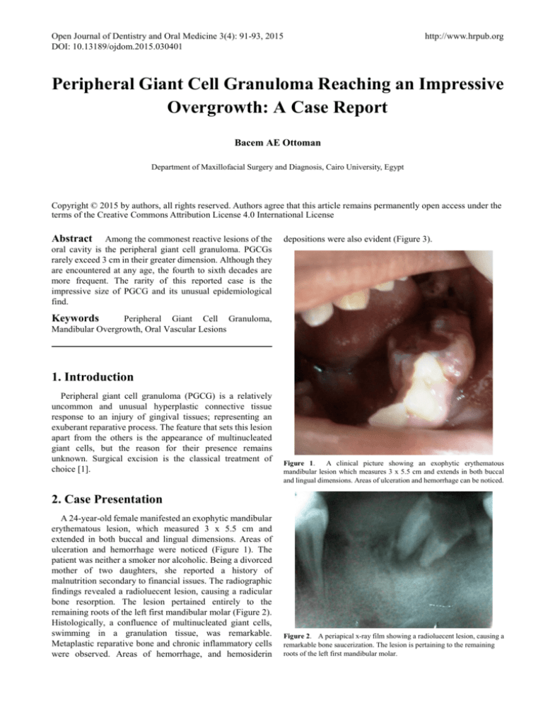
Figure 1.
A clinical picture showing an exophytic erythematous
mandibular lesion which measures 3 x 5.5 cm and extends in both buccal
and lingual dimensions. Areas of ulceration and hemorrhage can be noticed.
2. Case Presentation
A 24-year-old female manifested an exophytic mandibular
erythematous lesion, which measured 3 x 5.5 cm and
extended in both buccal and lingual dimensions. Areas of
ulceration and hemorrhage were noticed (Figure 1). The
patient was neither a smoker nor alcoholic. Being a divorced
mother of two daughters, she reported a history of
malnutrition secondary to financial issues. The radiographic
findings revealed a radioluecent lesion, causing a radicular
bone resorption. The lesion pertained entirely to the
remaining roots of the left first mandibular molar (Figure 2).
Histologically, a confluence of multinucleated giant cells,
swimming in a granulation tissue, was remarkable.
Metaplastic reparative bone and chronic inflammatory cells
were observed. Areas of hemorrhage, and hemosiderin
Figure 2. A periapical x-ray film showing a radioluecent lesion, causing a
remarkable bone saucerization. The lesion is pertaining to the remaining
roots of the left first mandibular molar.
92
Peripheral Giant Cell Granuloma Reaching an Impressive Overgrowth: A Case Report
Figure 3.
(a) Photomicrograph showing the classical histological
appearance of PGCG; characterizing a myriad giant cells with artifactual
spacing, fibroblasts, and inflammatory infiltrates dashing a granulation
background where RBCs and reparative islets are observed. (H&E stained,
Original magnifications 40X)
The lesion was treated by surgical excision. There evinced
a pulsatile gushing hemorrhage, at the surgical scene:
unusual find. The patient had lost approximately 300-400ml
of her blood before stitching up the wound site. Fluid therapy
was commenced immediately before the discharge of the
patient. In 10 month, no evidence of recurrence is observed.
3. Discussions
Peripheral giant cell granulomas (PGCGs) typically
manifests as a firm, soft, bright nodule, either sessile or
pedunculated, with occasional surface ulceration. The color,
ranges from dark red to purple or blue. PGCGs are
encountered at the interdental papilla, edentulous alveolar
margin or marginal gingival level. Secondary ulceration
caused by trauma may result in the formation of a fibrin clot
over the ulcer. Most of these lesions do not exceed 2 cm in
diameter, occur at any age, and tend to show a female
predilection (1.5-2:1) [2-5]. Few PGCGs reached a size
greater than 5 cm, where factors such as deficient oral
hygiene, malnutrition, long-standing irritand or xerostomia
coexisted. Accordingly, there appears a close relationhip
between such predisposing factors and the concomitant
imperative role in the lesional growth [6].PGCGs are usually
observed in the mandible more than the maxilla where molar
and premolar areas. On removal, PGCGs are highly vascular
[7]. PGCGs presumably arise from periodontal ligament or
periosteum, and they sporadically induce alveolar bone
resorption [8]. The newly secreted collagen acts as a scaffold
for infiltration of cells to the site of injury. Moreover, some
degradation occurs providing some space for new
vasculature to start an angiogenesis [9]. The physiognomic
presence of such giant cells, which induce no conspicuous
damage to the surrounding structures, accounts for the high
clinical vascularity of such lesion since the newly secreted
collagen acts as a scaffold for infiltration of cells with some
degradation which should provide some space for new
vasculature to start a remarkable angiogenesis [10].
The characteristic histopathological features oinclude a
non-encapsulated highly cellular mass with abundant giant
cells, inflammation, interstitial hemorrhage, hemosiderin
deposits, mature bone or osteoid. Incipient lesions may bleed
and induce minor changes in gingival contour but large ones
adversely affect normal oral function. Pain is not a common
finding, unless they interfere with occlusion, where they may
ulcerate and become infected [11].
In contrast to the classical negative appearance of PGCGs,
some cases revealed radiographically erosive underlying
bone, with a cup-shaped radiolucency [1-3].
In an immunohistochemical and ultrastructural study,
Carvalho et al concluded that PGCGs of the jaws are
composed mainly of cells of the mononuclear phagocyte
system and that Langerhans cells are present in two thirds of
the lesions. Vimentin, alpha I-antichymotrypsin and CD68
were expressed in both the mononuclear and multinucleated
giant cells. Dendritic mononuclear cells, positive for S-100
protein, were noted in 67.5% of the lesions, whereas
lysozyme and leucocyte common antigen were detected in
occasional mononuclear cells. Ultrastructural examination
showed mononuclear cells with signs of phagocytosis and
sometimes interdigitations with similar cells. Others
presented non-specific characteristics and the third type
exhibited cytoplasmic processes and occasional Birbeck
granules. Some multinucleated giant cells showed oval
nuclei, abundant mitochondria and granular endoplasmic
reticulum whereas others presented with irregular nuclei and
a great number of cytoplasmic vacuoles [12].
In the most recent study by Bo Liu et al [13] in situ
hybridization was carried out to detect the mRNA expression
of the newly identified receptor activator of nuclear
factor-kappaB ligand (RANKL) that is shown to be essential
in the osteoclastogenesis, its receptor, receptor activator of
NF-kappaB
(RANK), and its
decoy receptor,
osteoprotegerin . They concluded that RANKL, OPG and
RANK expressed in these lesions may play important roles
in the formation of multinucleated giant cells.
A study by Willing et al [14] revealed that the stromal
cells secrete a variety of cytokines and differentiation factors,
including monocyte chemoattractant protein-1, osteoclast
differentiation factor, and macrophage-colony stimulating
factor. These molecules were monocyte chemoattractants
and are essential for osteoclast differentiation, suggesting
that the stromal cell stimulates blood monocyte immigration
into tumor tissue and enhances their fusion into
osteoclast-like, multinucleated giant cells. Furthermore, the
recently identified membrane-bound protein family, a
disintegrin and metalloprotease, is considered to play a role
in
the
multinucleation
of
osteoclasts
and
macrophage-derived giant cells from mononuclear precursor
cells.
Surgical resection, with extensive clearing of the base of
Open Journal of Dentistry and Oral Medicine 3(4): 91-93, 2015
Peripheral giant cell granuloma. A report of five cases and
review of the literature. Med Oral Patol Oral Cir Bucal.
2005;10(1):53–7.
the lesion to avoid relapse, is the mainstay treatment
modality. However, intralesional injection of corticosteroid
was reported to be also successful [1,2].
[7]
Katsikeris N, Kakarantza-Angelopoulou E, Angelopoulos
AP. Peripheral giant cell granuloma. Clinicopathologic study
of 224 new cases and review of 956 reported cases. Int J
Oral
Maxillofac
Surg.
1988;17(2):94–9
DOI:
10.1016/S0901-5027(88)80158-9.
[8]
Ross M, Pawlina W: Histology: A Text and Atlas: With
Correlated Cell and Molecular Biology. Philadelphia:
Wolters Kluwer/Lippincott Williams & Wilkins Health,
201:158-78.
[9]
Abe E, Mocharla H, Yamate T, Taguchi Y. Monolages
SC.Meltrinalpha, a fusion protein involved in multinucleated
giant cell and osteoclast formation. Calcif Tissue.
1999;64:508–15.
4. Conclusions
The peripheral giant cell granuloma can reach a massive
size in younger population. The lesion has, quite commonly,
rich blood supply, and tends to ulcerate on interfering with
occlusion.
REFERENCES
[1]
Regezi J, Sciubba J, Jordan R: Oral Pathology. Clinical
Pathologic Correlations, Saunders, Philadelphia, Pa, USA,
5th edition, 2007.
[2]
Neville B, Damm D, Allen C, Bouquot J. Oral and
maxillofacial pathology. St. Louis: Saunders/Elsevier; 3rd
edition 2009.
[3]
Kaffe I, Ardekian L, Taicher S, Littner MM, Buchner A
(1996). Radiologic features of central giant cell granuloma
of the jaws. Oral Surg Oral Med Oral Pathol Oral Radiol
Endod. 81: 720-6.
[4]
[5]
[6]
Whitaker SB, Waldron CA (1993). Central giant cell lesions
of the jaws. A clinical, radiologic, and histopathologic study.
Oral Surg Oral Med Oral Pathol. 75: 199-208.
Stavropoulos F, Katz J (2002). Central giant cell granulomas:
a systematic review of the radiographic characteristics with
the addition of 20 new cases. Dentomaxillofac Radiol. 31:
213-7.
Chaparro-Avendano AV, Berini-Aytes L, Gay-Escoda C.
93
[10] Ottoman B: Giant Cells in Giant Cell Reparative Granuloma:
Physiognomic or Pathognomic Relevance? World J Pathol.
2015, 4:6.
[11] Motamedi MH, Eshghyar N, Jafari SM, Lassemi E, Navi F,
Abbas FM, et al. Peripheral and central giant cell
granulomas of the jaws: a demographic study. Oral Surg
Oral Med Oral Pathol Oral Radiol Endod. 2007;103(3):e39–
43.
[12] Carvalho Y, Loyola A, Gomez R, Araújo V: Peripheral giant
cell granuloma. An immunohistochemical and ultrastructural
study. Oral Dis. 1995;1(1):20-5.
[13] Bo Liu, Shi-Feng Yu, Tie-Jun Li. Multinucleated giant cells
in various forms of giant cell containing lesions of the jaws
express features of osteoclasts. J Oral Pathol Med.
2003;32:367.
[14] Willing M, Engels C, Jesse N, Werner M, Delling G, Kaiser
E. The nature of giant cell tumor of bone. J Cancer Res Clin
Oncol. 2001;127:467–74

