Ratiometric Fluorescent Probes for Detection of Intracellular Singlet
advertisement
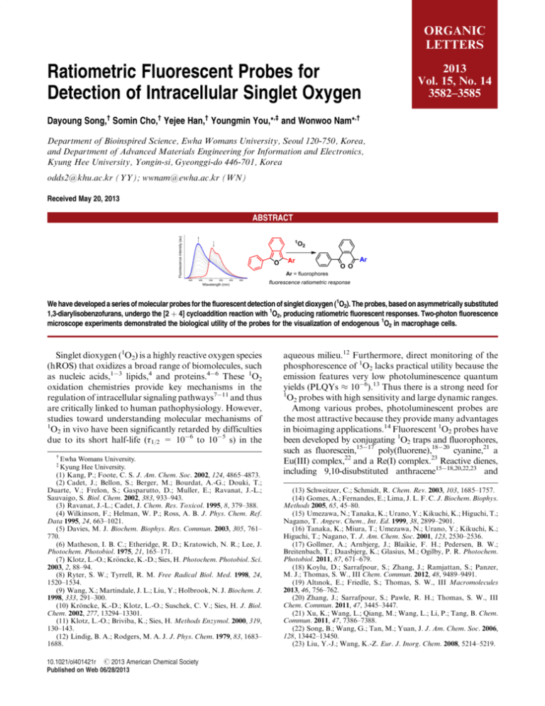
ORGANIC LETTERS Ratiometric Fluorescent Probes for Detection of Intracellular Singlet Oxygen 2013 Vol. 15, No. 14 3582–3585 Dayoung Song,† Somin Cho,† Yejee Han,† Youngmin You,*,‡ and Wonwoo Nam*,† Department of Bioinspired Science, Ewha Womans University, Seoul 120-750, Korea, and Department of Advanced Materials Engineering for Information and Electronics, Kyung Hee University, Yongin-si, Gyeonggi-do 446-701, Korea odds2@khu.ac.kr (YY); wwnam@ewha.ac.kr (WN) Received May 20, 2013 ABSTRACT We have developed a series of molecular probes for the fluorescent detection of singlet dioxygen (1O2). The probes, based on asymmetrically substituted 1,3-diarylisobenzofurans, undergo the [2 þ 4] cycloaddition reaction with 1O2, producing ratiometric fluorescent responses. Two-photon fluorescence microscope experiments demonstrated the biological utility of the probes for the visualization of endogenous 1O2 in macrophage cells. Singlet dioxygen (1O2) is a highly reactive oxygen species (hROS) that oxidizes a broad range of biomolecules, such as nucleic acids,13 lipids,4 and proteins.46 These 1O2 oxidation chemistries provide key mechanisms in the regulation of intracellular signaling pathways711 and thus are critically linked to human pathophysiology. However, studies toward understanding molecular mechanisms of 1 O2 in vivo have been significantly retarded by difficulties due to its short half-life (τ1/2 = 106 to 105 s) in the Ewha Womans University. Kyung Hee University. (1) Kang, P.; Foote, C. S. J. Am. Chem. Soc. 2002, 124, 4865–4873. (2) Cadet, J.; Bellon, S.; Berger, M.; Bourdat, A.-G.; Douki, T.; Duarte, V.; Frelon, S.; Gasparutto, D.; Muller, E.; Ravanat, J.-L.; Sauvaigo, S. Biol. Chem. 2002, 383, 933–943. (3) Ravanat, J.-L.; Cadet, J. Chem. Res. Toxicol. 1995, 8, 379–388. (4) Wilkinson, F.; Helman, W. P.; Ross, A. B. J. Phys. Chem. Ref. Data 1995, 24, 663–1021. (5) Davies, M. J. Biochem. Biophys. Res. Commun. 2003, 305, 761– 770. (6) Matheson, I. B. C.; Etheridge, R. D.; Kratowich, N. R.; Lee, J. Photochem. Photobiol. 1975, 21, 165–171. (7) Klotz, L.-O.; Kr€ oncke, K.-D.; Sies, H. Photochem. Photobiol. Sci. 2003, 2, 88–94. (8) Ryter, S. W.; Tyrrell, R. M. Free Radical Biol. Med. 1998, 24, 1520–1534. (9) Wang, X.; Martindale, J. L.; Liu, Y.; Holbrook, N. J. Biochem. J. 1998, 333, 291–300. (10) Kr€ oncke, K.-D.; Klotz, L.-O.; Suschek, C. V.; Sies, H. J. Biol. Chem. 2002, 277, 13294–13301. (11) Klotz, L.-O.; Briviba, K.; Sies, H. Methods Enzymol. 2000, 319, 130–143. (12) Lindig, B. A.; Rodgers, M. A. J. J. Phys. Chem. 1979, 83, 1683– 1688. † ‡ 10.1021/ol401421r r 2013 American Chemical Society Published on Web 06/28/2013 aqueous milieu.12 Furthermore, direct monitoring of the phosphorescence of 1O2 lacks practical utility because the emission features very low photoluminescence quantum yields (PLQYs ≈ 106).13 Thus there is a strong need for 1 O2 probes with high sensitivity and large dynamic ranges. Among various probes, photoluminescent probes are the most attractive because they provide many advantages in bioimaging applications.14 Fluorescent 1O2 probes have been developed by conjugating 1O2 traps and fluorophores, such as fluorescein,1517 poly(fluorene),1820 cyanine,21 a Eu(III) complex,22 and a Re(I) complex.23 Reactive dienes, including 9,10-disubstituted anthracene1518,20,22,23 and (13) Schweitzer, C.; Schmidt, R. Chem. Rev. 2003, 103, 1685–1757. (14) Gomes, A.; Fernandes, E.; Lima, J. L. F. C. J. Biochem. Biophys. Methods 2005, 65, 45–80. (15) Umezawa, N.; Tanaka, K.; Urano, Y.; Kikuchi, K.; Higuchi, T.; Nagano, T. Angew. Chem., Int. Ed. 1999, 38, 2899–2901. (16) Tanaka, K.; Miura, T.; Umezawa, N.; Urano, Y.; Kikuchi, K.; Higuchi, T.; Nagano, T. J. Am. Chem. Soc. 2001, 123, 2530–2536. (17) Gollmer, A.; Arnbjerg, J.; Blaikie, F. H.; Pedersen, B. W.; Breitenbach, T.; Daasbjerg, K.; Glasius, M.; Ogilby, P. R. Photochem. Photobiol. 2011, 87, 671–679. (18) Koylu, D.; Sarrafpour, S.; Zhang, J.; Ramjattan, S.; Panzer, M. J.; Thomas, S. W., III Chem. Commun. 2012, 48, 9489–9491. (19) Altınok, E.; Friedle, S.; Thomas, S. W., III Macromolecules 2013, 46, 756–762. (20) Zhang, J.; Sarrafpour, S.; Pawle, R. H.; Thomas, S. W., III Chem. Commun. 2011, 47, 3445–3447. (21) Xu, K.; Wang, L.; Qiang, M.; Wang, L.; Li, P.; Tang, B. Chem. Commun. 2011, 47, 7386–7388. (22) Song, B.; Wang, G.; Tan, M.; Yuan, J. J. Am. Chem. Soc. 2006, 128, 13442–13450. (23) Liu, Y.-J.; Wang, K.-Z. Eur. J. Inorg. Chem. 2008, 5214–5219. histidine,21 served as the 1O2 traps, and fluorescence responses were obtained after the [2 þ 4] cycloaddition reactions between 1O2 and the traps. The utility of these probes has been limited because their monotonic fluorescence turnon or turn-off responses suffer from artifacts associated with sensor localization and instrumental drifts.1517,19,2123 In addition, some fluorophores, such as chlorinated fluorescein, convert ground-state dioxygen (3O2) into 1O2 under photoexcitation,17,24 which would severely alter the cellular 1 O2 levels. Furthermore, probes examined for their utility for the detection of biological 1O2 are extremely rare.21,22 These drawbacks present a strong rationale for development of new probes capable of ratiometric fluorescent detection and minimized 1O2 photosensitization. 1,3-Diphenylisobenzofuran (DPBF) is an attractive platform for the construction of 1O2 probes because it rapidly reacts with 1O2 at a rate constant of 9.6 108 M1 s1 in water25,26 and because it does not photosensitize 1O2 due to effective suppression of the triplet state population.27 However, the 1O2 products, an endoperoxide or 1,2-dibenzoylbenzene, are nonfluorescent. Thus DPBF itself offers little practical utility for 1O2 detection. We envisioned that the Scheme 1. Ratiometric Fluorescent Probes for Detection of 1O2 Scheme 2. Synthesis of StPBF, a Ratiometric Fluorescent Probe for 1O2a a The synthetic routes to other probes are shown in the Supporting Information. Synthetic routes to StPBF are depicted in Scheme 2. The syntheses of PPBF and PyPBF are described in the Supporting Information (Scheme S1). The fluorophore, a 4-(diphenylamino)stilbene unit, was obtained through the two-step synthesis, comprising a HornerWadsworth Emmons olefination followed by a Pd(II)-catalyzed Heck reaction. The Re(I) dimer catalysis protocol established by the group of Kuninobu and Takai was employed for the construction of the isobenzofuran unit.28 The 1O2 probes and their precursors were characterized by standard spectroscopic methods, and the data fully agreed with the proposed structures (Supporting Information, Figures S1S12). Table 1. Photophysical Data for the 1O2 Probes displacement of the phenyl moiety of DPBF with fluorescent chromophores will produce ratiometric fluorescent 1O2 responses: the π-conjugation over 1-phenylisobenzofuran and the chromophore would be shortened by the 1O2 reaction to give hypsochromically shifted fluorescence emission instead of fluorescence turn-off (see Scheme 1). To test this idea, we designed a series of 1O2 probes comprising phenanthrene (PPBF), pyrene (PyPBF), and 4-(diphenylamino)stilbene (StPBF) as fluorophores. Herein, we report the design, syntheses, sensing properties, and biological applications of the novel ratiometric fluorescent 1O2 probes. Fluorescent responses of the probes were compared to correlate 1O2 sensitivity and the extent of π-conjugation. (24) Gandin, E.; Lion, Y.; Vorst, A. V. d. Photochem. Photobiol. 1983, 37, 271–278. (25) Onitsuka, S.; Nishino, H.; Kurosawa, K. Tetrahedron 2001, 57, 6003–6009. (26) Aubry, J.-M.; Pierlot, C.; Rigaudy, J.; Schmidt, R. Acc. Chem. Res. 2003, 36, 668–675. (27) Zhang, X.-F.; Li, X. J. Lumin. 2011, 131, 2263–2266. Org. Lett., Vol. 15, No. 14, 2013 DPBF þ 1O2a PPBF þ 1O2a PyPBF þ 1O2a StPBF þ 1O2a λabs (nm, log ε) λems (nm)b PLQY (%)c τobs (ns)d FI ratioe 415 (4.26) 298 (3.78) 411 (4.35) 324 (3.96) 438 (4.40) 358 (4.22) 450 (4.50) 382 (4.16) 455 ndf 476 370 512 398 505 435 72 ( 7.4 ndf 6.4 ( 1.0 0.78 ( 0.12 28 ( 2.7 6.1 ( 0.5 39 ( 6.0 41 ( 8.2 1.46 ndf 1.38 0.425 1.48 1.17 2.29 1.58 turn-off 80 352 14 a O2-saturated THF solutions containing 10 μM probe and 100 nM photosensitizer were photoirradiated under 365 nm for 100 s. b λex = 415 nm (DPBF), 331 nm (PPBF), 358 nm (PyPBF), and 350 nm (StPBF). c Photoluminescence quantum yield relative to 9,10-diphenylanthracene (PLQY = 0.90, cyclohexane) as a standard. d Fluorescence lifetime determined through picosecond (λex = 377 nm) time-correlated single photon counting techniques. e Fluorescence intensity (FI) ratio: PPBF, FI370 nm/FI476 nm; PyPBF, FI398 nm/FI512 nm; StPBF, FI435 nm/FI505 nm. f Not detected. As expected, the 1O2 probes exhibited significant red shifts in their fluorescence peak wavelengths relative to DPBF (10 μM in THF, Δν = νems(DPBF) νems(1O2 probe): PPBF, 970 cm1; StPBF, 2180 cm1; PyPBF, 2450 cm1) 3583 due to the effective conjugation to the fluorophores. The probes are highly fluorescent with photoluminescence quantum yields (PLQYs) of 6.439%. To evaluate 1O2 sensing properties, we employed a biscyclometalated Ir(III) complex, IrF (Scheme 1), as a 1O2 photosensitizer because it has Figure 1. (a) Fluorescence response of 10 μM StPBF toward 1O2. The arrows indicate the direction of the spectral changes. The inset photos show fluorescence emission of the StPBF solution before (top) and after (bottom) the 1O2 generation. (b) Plots of observed reaction rates (kobs) as a function of the concentration of the 1O2 probes. been proved to be highly efficient for 1O2 generation.29 As shown in Figure 1, 100 s photoirradiation (λex = 365 nm) of an O2-bubbled (30 min) THF solution containing 10 μM StPBF and 100 nM IrF led to fluorescence color changes from green to blue. The fluorescence peak wavelength shifted from 505 to 435 nm, and the corresponding fluorescence intensity (FI) ratio at 435 nm versus 505 nm (i.e., FI435 nm/ FI505 nm) increased by a factor of 14. The clear isosbestic point at 490 nm indicates the absence of any fluorescent byproduct. By contrast, the ratiometric change was not observed without IrF or photoirradiation (Supporting Information, Figure S13a). In addition, the addition of an 1O2 scavenger, 1 mM NaN3,30 abrogated the fluorescent 1O2 response (Supporting Information, Figure S13b). These results unambiguously indicate that the fluorescence change was attributed to 1O2. PPBF and PyPBF displayed similar fluorescence ratiometric responses to 1O2 with changes in the fluorescence intensity ratios of factors of 80 (PPBF, FI370 nm/FI476 nm) and 352 (PyPBF, FI398 nm/FI512 nm), respectively (Table 1 and Supporting Information, Figure S14). The UVvis absorption spectra of 10 μM probes displayed hypsochromic shifts (Δν = νabs(before reaction) νabs(after reaction): StPBF, 3960 cm1; PyPBF, 5100 cm1; PPBF, 6530 cm1) upon the reaction with 1O2, indicating products with wide band gap energies (Supporting Information, Figure S15). Positive mode ESIMS spectra for the 1O2 reaction product of PPBF revealed peaks at m/z = 409 and (28) Kuninobu, Y.; Nishina, Y.; Nakagawa, C.; Takai, K. J. Am. Chem. Soc. 2006, 128, 12376–12377. (29) You, Y.; Lee, S.; Kim, T.; Ohkubo, K.; Chae, W.-S.; Fukuzumi, S.; Jhon, G.-J.; Nam, W.; Lippard, S. J. J. Am. Chem. Soc. 2011, 133, 18328–18342. (30) Harbour, J. R.; Issler, S. L. J. Am. Chem. Soc. 1982, 104, 903– 905. 3584 425 mu (Supporting Information, Figure S16). These values correspond to [1,2-diketone þ Na]þ and [endoperoxide þ Na]þ, respectively (see Scheme 1). It appeared that the dominant 1O2 reaction product was 1,2-diketone because 13 C NMR spectra (100 MHz) for the product displayed two carbonyl peaks at δ 197.3 and 197.4 ppm (acetone-d6; Supporting Information, Figure S17). HPLC chromatograms acquired during the 1O2 reaction displayed the complete conversion of PPBF to a single product (Supporting Information, Figure S18). Indeed, the experimental UVvis absorption spectrum of the product is in good agreement with a simulated spectrum of the 1,2-diketone of PPBF (TDDFT, B3LYP/6-311þG(d,p)//uB3LYP/6-311þG(d,p):CPCM (THF); Supporting Information, Figure S19 and Table S1). Taken together, the studies provide strong evidence that the probes react with 1O2 to yield the 1,2-diketone having hypsochromically shifted fluorescence emission. The observed rates (kobs) for the reaction between 1O2 and the probes were determined by monitoring the temporal changes in the fluorescence intensity ratios (StPBF, PyPBF, and PPBF) or the total fluorescence intensity (DPBF) during the continuous photoirradiation. The probe concentration was varied in the range of 220 μM to keep the pseudo first-order kinetics of the probe. As shown in Figure 1b, the plot of kobs as a function of the probe concentration follows a linear line. The largest slope is observed for StPBF (6.25 M1 s1), and the slope decreases in the order of PyPBF (3.71 M1 s1) > PPBF (0.557 M1 s1) > DPBF (0.252 M1 s1). Although the slope cannot be equated with the reaction rate constant under the experimental conditions, the results reveal that the 1O2 reactivity increases in proportion with the extent of π-conjugation. By contrast, the oxidation potentials of the probes displayed a weak correlation with the 1O2 reactivity (Supporting Information, Figure S20). Figure 2. Fluorescence 1O2 selectivity of StPBF over other reactive oxygen species (O2•, 1 mM KO2; ClO, 1 mM NaOCl; 1 mM t-BuOOH; 1 mM H2O2; t-BuO•, 1 mM FeSO4 þ 100 μM tBuOOH; •OH, 1 mM FeSO4 þ 100 μM H2O2) and chemical oxidants (CAN, 1 mM (NH4)2CeIV(NO3)6; DDQ, 1 mM 2,3dichloro-5,6-dicyano-benzoquinone). The 1O2 probes exhibited great selectivity toward 1O2 over other reactive oxygen species, including O2•, ClO, Org. Lett., Vol. 15, No. 14, 2013 Figure 3. Fluorescent detection of intracellular 1O2 in RAW 264.7 macrophage cells. Cells treated with 10 μM PyPBF for 30 min were visualized through dual channels: channel 1, λex = 750 nm (two photon) and λems = 377481 nm; channel 2, λex = 750 nm (two photon) and λems = 490568 nm; ratio image, channel 1/channel 2. The cells were subsequently incubated with 2 μM phorbol myristate acetate (PMA, 30 min) to stimulate endogenous 1 O2 generation (middle panels). Control experiments were performed for the cells preincubated with 1O2 scavenger, 2 mM histidine (30 min; bottom panels). t-BuO•, •OH, and H2O2, and chemical oxidants, such as (NH4)2CeIV(NO3)6 and DDQ (see Figure 2 for StPBF and Supporting Information, Figure S21 for other probes). Although the 1O2 probes formed nano aggregates in aqueous solutions (pH 7.4, 50 mM HEPES þ 0.1 M KCl þ 10 vol % THF or CH3CN) as observed in the FESEM image (Supporting Information, Figure S22), their fluorescent 1O2 responses were retained (Supporting Information, Figure S23). Having established the 1O2 sensing capabilities of the probes, we performed two-photon fluorescence microscopic experiments to visualize endogenous 1O2 in RAW 264.7 macrophage cells. We chose PyPBF because it exhibited the best cell permeability. PyPBF was nontoxic to the cells as judged from the bright-field images (Figure 3). RAW 264.7 cells treated with 10 μM PyPBF (30 min) produced intense fluorescence emission at λems = 490568 nm (channel 2) under two-photon excitation (λex = 750 nm), whereas the fluorescence emission due to the 1,2-diketone of PyPBF at λems = 377481 nm (channel 1) was barely detected (Figure 3). Subsequent stimulation of the cells by 2 μM phorbol myristate acetate (PMA)31 provoked a (31) Kanofsky, J. R.; Hoogland, H.; Wever, R.; Weiss, S. J. J. Biol. Chem. 1988, 263, 9692–9696. (32) Tomita, M.; Irie, M.; Ukita, T. Biochemistry 1969, 8, 5149–5160. Org. Lett., Vol. 15, No. 14, 2013 fluorescence increase in channel 1, indicating endogenous production of 1O2. The corresponding increases in the fluorescence intensity ratios of the channel 1 images over the channel 2 images were greater than the standard deviations (Supporting Information, Figure S24). Additional micrographs are included in Supporting Information, Figure S25. As expected for the 1O2 probe, pretreatment with 2 mM histidine, an intracellular 1O2 scavenger,32 suppressed the 1,2-diketone fluorescence. To the best of our knowledge, this is the first visualization of intracellular 1 O2 by ratiometric fluorescent signaling. To summarize, we developed a series of ratiometric fluorescent probes for the detection of 1O2. The probes underwent an irreversible reaction with 1O2, producing hypsochromically shifted fluorescence. The ratiometric responses were not affected by the probe concentrations (Supporting Information, Figure S26), demonstrating the benefit of the ratiometric fluorescent signaling. The probes were capable of detecting 1O2 among various reactive oxygen species. We further established that greater 1O2 sensitivity could be accomplished using the isobenzofuran platforms with elongated π-conjugation. Finally, biological utility was demonstrated by dual channel two-photon fluorescent visualization of endogenous 1O2 in macrophage cells. Acknowledgment. This work was supported by the CRI (W.N.), GRL (2010-00353) (W.N.), and WCU (R31-2008000-10010-0) (W.N.) programs from the National Research Foundation (NRF) of Korea. Supporting Information Available. Experimental details; scheme depicting synthetic routes to PPBF and PyPBF; figures displaying copies of 1H and 13C NMR spectra of the 1O2 probes and their precursor, fluorescence spectra obtained without IrF nor photoirradiation, or in the presence of NaN3, fluorescence 1O2 responses of PPBF and PyPBF, UVvis absorption changes of the 1O2 probes, the ESIMS spectra, 13C NMR spectrum of the reaction product of PPBF, HPLC chromatograms, TDDFT calculation results, cyclic voltammograms, 1O2 selectivity of PPBF and PyPBF, the FESEM image of the aggregates of PyPBF, fluorescent 1O2 responses and selectivity of PyPBF in buffer solution, calculated fluorescence intensity ratios for the fluorescence micrographs, more cell images, and the fluorescent 1O2 responses of StPBF at different probe concentrations; tables listing the summary of TDDFT calculation results and the Cartesian coordinates of the optimized geometries. This material is available free of charge via the Internet at http://pubs.acs.org. The authors declare no competing financial interest. 3585
