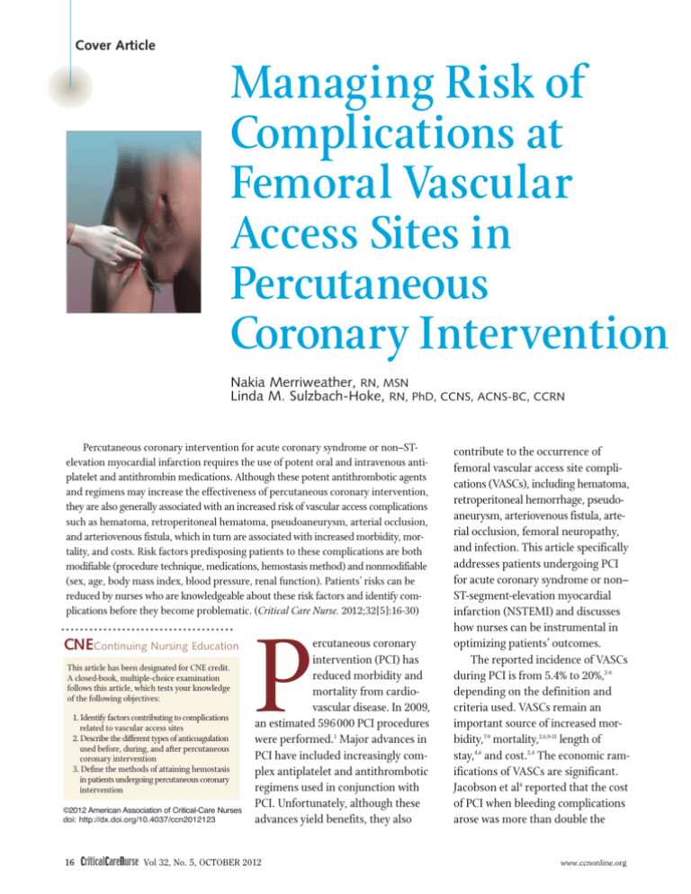
Cover Article
Managing Risk of
Complications at
Femoral Vascular
Access Sites in
Percutaneous
Coronary Intervention
Nakia Merriweather, RN, MSN
Linda M. Sulzbach-Hoke, RN, PhD, CCNS, ACNS-BC, CCRN
Percutaneous coronary intervention for acute coronary syndrome or non–STelevation myocardial infarction requires the use of potent oral and intravenous antiplatelet and antithrombin medications. Although these potent antithrombotic agents
and regimens may increase the effectiveness of percutaneous coronary intervention,
they are also generally associated with an increased risk of vascular access complications
such as hematoma, retroperitoneal hematoma, pseudoaneurysm, arterial occlusion,
and arteriovenous fistula, which in turn are associated with increased morbidity, mortality, and costs. Risk factors predisposing patients to these complications are both
modifiable (procedure technique, medications, hemostasis method) and nonmodifiable
(sex, age, body mass index, blood pressure, renal function). Patients’ risks can be
reduced by nurses who are knowledgeable about these risk factors and identify complications before they become problematic. (Critical Care Nurse. 2012;32[5]:16-30)
CNEContinuing Nursing Education
This article has been designated for CNE credit.
A closed-book, multiple-choice examination
follows this article, which tests your knowledge
of the following objectives:
1. Identify factors contributing to complications
related to vascular access sites
2. Describe the different types of anticoagulation
used before, during, and after percutaneous
coronary intervention
3. Define the methods of attaining hemostasis
in patients undergoing percutaneous coronary
intervention
©2012 American Association of Critical-Care Nurses
doi: http://dx.doi.org/10.4037/ccn2012123
P
ercutaneous coronary
intervention (PCI) has
reduced morbidity and
mortality from cardiovascular disease. In 2009,
an estimated 596000 PCI procedures
were performed.1 Major advances in
PCI have included increasingly complex antiplatelet and antithrombotic
regimens used in conjunction with
PCI. Unfortunately, although these
advances yield benefits, they also
16 CriticalCareNurse Vol 32, No. 5, OCTOBER 2012
contribute to the occurrence of
femoral vascular access site complications (VASCs), including hematoma,
retroperitoneal hemorrhage, pseudoaneurysm, arteriovenous fistula, arterial occlusion, femoral neuropathy,
and infection. This article specifically
addresses patients undergoing PCI
for acute coronary syndrome or non–
ST-segment-elevation myocardial
infarction (NSTEMI) and discusses
how nurses can be instrumental in
optimizing patients’ outcomes.
The reported incidence of VASCs
during PCI is from 5.4% to 20%,2-6
depending on the definition and
criteria used. VASCs remain an
important source of increased morbidity,7-9 mortality,2,6,9-11 length of
stay,4,6 and cost.2,4 The economic ramifications of VASCs are significant.
Jacobson et al4 reported that the cost
of PCI when bleeding complications
arose was more than double the
www.ccnonline.org
costs of uncomplicated PCI ($25371
vs $12279).4 Interventions aimed at
reducing the risk of adverse events
are likely to improve both financial
and clinical outcomes.
Removing femoral sheaths and
managing related complications after
PCI are predominantly the responsibilities of nurses in many acute and
critical care settings.3,5,12,13 Therefore,
it is essential for nurses to understand
the causes of and predisposing risk
factors for VASCs. These risk factors
can be categorized as modifiable
and nonmodifiable.
Table 1
Femoral puncture location and associated complicationsa
Femoral puncture location: definition
Complications
Low stick: puncture below the femoral bifurcation
Pseudoaneurysm
Hematoma
Arteriovenous fistula
High stick: puncturing the inferior epigastric artery
Retroperitoneal hemorrhage
Posterior wall puncture: puncture through the back
wall of the artery
Retroperitoneal hemorrhage
a
Based on data from Turi,7 Ragosta,8 Baim and Simon,15 Kamineni and Butman,18 and Rashid and Bailey.19
Exterior
iliac artery
Modifiable Risk Factors
The primary modifiable risk
factors for VASCs are femoral access;
medications administered before,
during, and after the procedure; and
hemostasis method. Although the
interventional cardiologist controls
the femoral access and medications
ordered, the hemostasis method is
controlled by the nurse unless a
vascular closure device is deployed.
Common
femoral artery
Inguinal
ligament
Profunda
femoris
Femoral
vein
Superficial
femoral artery
Femoral Access
Percutaneous entry through
the femoral artery and vein
approach for PCI is preferred
because of the large diameter of
those vessels,14 which improves the
speed and simplicity of the procedure.15 VASCs at the femoral site
Inferior
epigastric
artery
Figure 1 Anatomical landmarks in relation to the femoral vessels.
are often associated with the location of the femoral puncture,7,15,16
the number of attempts,7,16 and
Authors
Nakia Merriweather is a cardiology nurse in the echocardiography laboratory at the Hospital
of the University of Pennsylvania, Philadelphia.
Linda M. Sulzbach-Hoke is a clinical nurse specialist on a 48-bed progressive care unit at the
Hospital of the University of Pennsylvania, providing nursing care to adult cardiac patients.
Her research and several of her publications support evidence-based nursing practice, specifically in patients undergoing percutaneous coronary intervention.
Corresponding author: Linda Hoke, Cardiac Intermediate Care Unit, Founders 10, Hospital of the University of
Pennsylvania, 3400 Spruce Street, Philadelphia PA 19104 (e-mail: linda.hoke@uphs.upenn.edu).
To purchase electronic or print reprints, contact The InnoVision Group, 101 Columbia, Aliso Viejo, CA 92656.
Phone, (800) 899-1712 or (949) 362-2050 (ext 532); fax, (949) 362-2049; e-mail, reprints@aacn.org.
www.ccnonline.org
catheter size17 (Table 1). To facilitate
vessel entry and effective compression, the puncture should be above
the femoral bifurcation but 1 or 2 cm
below the inguinal ligament,7,8,15,19
which extends from the anterior
superior iliac spine to the pubic
tubercle (Figure 1). Many major
and potentially lethal VASCs are
related to punctures either too high
or too low below the inguinal ligament.8 Table 2 describes the clinical findings of VASCs and the
associated management.
CriticalCareNurse Vol 32, No. 5, OCTOBER 2012 17
Table 2
Vascular access site complications and management
Complication
Hematoma
Incidence:
5% to 23%20
Retroperitoneal
hemorrhage
Incidence:
0.15% to 0.44%20
Pseudoaneurysm
Incidence:
0.5% to 9%20
Arteriovenous fistula
Incidence:
0.2% to 2.1%20
Description
Clinical findings
Management
The most common vascular
access site complication
A collection of blood located in
the soft tissue21
Occurs because of blood loss at
the arterial and/or venous access
site or perforation of an artery
or vein22
May occur if the arterial puncture
is below the femoral bifurcation
so the femoral head is not
available to assist with compression7,8,15,18
Swelling surrounding the puncture
site (visible)21
Area of hardening under the skin
surrounding the puncture site
(palpable)21
Varies in size20
Often associated with pain in the
groin area that can occur at rest or
with leg movement22
Can result in decrease in hemoglobin
and blood pressure and increase in
heart rate, depending on severity22
Apply pressure to site21
Mark the area to evaluate for any
change in size21
Provide hydration21
Monitor serial complete blood cell
counts21
Maintain/prolong bed rest21
Interrupt anticoagulant and
antiplatelet medications if
necessary21
Blood transfusion, if indicated21
If severe, may require surgical
evacuation20
Many hematomas resolve within a
few weeks as the blood dissipates
and is absorbed into the tissue21
Bleeding that occurs behind the
serous membrane lining the
walls of the abdomen/pelvis21
May occur if the arterial wall
puncture is made above the
inguinal ligament, resulting in
perforation of a suprainguinal
artery21 or penetration of the
posterior wall7,8,15
Can be fatal if not recognized
early21
Moderate to severe back pain22
Ipsilateral flank pain20
Vague abdominal or back pain22
Ecchymosis and decrease in
hemoglobin and hematocrit are
late signs21
Abdominal distention21
Often not associated with obvious
swelling21
Hypotension and tachycardia20
Diagnosed by computed tomography20
Provide hydration21
Perform serial blood cell counts21
Maintain/prolong bed rest21
Interrupt anticoagulant and antiplatelet medications if necessary20
Blood transfusion, if indicated14
If severe, may require surgical
evacuation20
A communicating tract between
the tissue and, usually, one of
the weaker walls of the femoral
artery, causing blood to escape
from the artery into the surrounding tissue14
Possible causes include difficulty
with arterial cannulation, inadequate compression after sheath
removal, and impaired hemostasis21
May occur if the arterial puncture
is below the femoral bifurcation
so the femoral head is not
available to assist with compression7,8,15,18
Swelling at insertion site22
Large, painful hematoma21
Ecchymosis22
Pulsatile mass22
Bruit and/or thrill in the groin22
Pseudoaneurysms can rupture, causing abrupt swelling and severe pain21
Suspect nerve compression when
pain is out of proportion to size of
hematoma21
Nerve compression can result in
limb weakness that takes weeks
or months to resolve21
Diagnosed by ultrasound20
Maintain/prolong bed rest21
Small femoral pseudoaneurysms
should be monitored; they commonly close spontaneously after
cessation of anticoagulant therapy20
Large femoral pseudoaneurysms
can be treated by ultrasoundguided compression, surgical
intervention, or ultrasound-guided
thrombin injection21
A direct communication between
an artery and a vein that occurs
when the artery and vein are
punctured14
The communication occurs once
the sheath is removed20
Risk factors:
Multiple access attempts7
Punctures above or below
proper site level7
Impaired clotting21
Can be asymptomatic14
Some arteriovenous fistulas
Bruit and/or thrill at access site20
resolve spontaneously without
Swollen, tender extremity22
intervention21
Distal arterial insufficiency and/or
Some arteriovenous fistulas
deep venous thrombosis can result
require ultrasound-guided
in limb ischemia21
compression or surgical repair20
Congestive heart failure20
Confirmed by ultrasound21
Continued
18 CriticalCareNurse Vol 32, No. 5, OCTOBER 2012
www.ccnonline.org
Table 2
Continued
Complication
Arterial occlusion
Incidence: <0.8%20
Femoral neuropathy
Incidence: 0.21%
23
Infection
Incidence: <0.1%20
Description
Clinical findings
Management
Occlusion of an artery by a
thromboembolism20
Most common sources: mural
thrombus originating in cardiac
chambers, vascular aneurysms,
or vascular atherosclerotic
plaques20
Thromboemboli can develop at
sheath site or catheter tip;
embolization occurs during
sheath removal20
Prevention or at least reduction
can be obtained by anticoagulation, vasodilators, and nursing
vigilance20
Classic symptoms include the 5 Ps:
Pain22
Paralysis20
Parasthesias22
Pulselessness22
Pallor22
Doppler studies help localize the area20
Angiogram is required to identify
exact location of occlusion site20
Treatment depends on size/type of
embolus, location, and patient’s
ability to tolerate ischemia in
affected area20
Small thromboemboli in wellperfused arterial areas may
undergo spontaneous lysis20
Larger thromboemboli may require
thromboembolectomy, surgery,
and/or thrombolytic agents20
Distal embolic protection devices (ie,
filters) may be placed if necessary21
Nerve damage caused by injury
of the femoral nerve(s) during
access and/or compression of
nerves by a hematoma23
Pain and/or tingling at femoral access
site22
Numbness at access site or further
down the leg22
Leg weakness22
Difficulty moving affected leg23
Decreased patellar tendon reflex22
Identification and treatment of the
source23
Treatment of symptoms23
Physical therapy23
Colonization by a pathogen
Causes:
Compromised technique14
Poor hygiene14
Prolonged indwelling sheath
time14
Femoral access closure device
(closure devices have been
linked with increased occurrence of infection)14
Pain, erythema, swelling at access
site14
Purulent drainage at access site20
Fever14
Increased white blood cell count21
Treatment of symptoms (eg, pain)21
Antibiotics20
High sticks are significantly linked
with retroperitoneal hemorrhage
resulting from the likelihood of
puncturing the inferior epigastric
artery.7,8,15,18,19 However, punctures
below the proper access points do
not eliminate the risk of retroperitoneal hemorrhage; penetration of
the posterior wall of the artery during femoral puncture can also cause
retroperitoneal bleeding (Table 1).7,8,15
Low sticks can predispose patients
to pseudoaneurysm, hematoma, and
arteriovenous fistula.7,8,15,18,19 When
the groin is accessed at or below the
level of the femoral bifurcation, the
femoral sheath is put into vessels that
are smaller than the common femoral
www.ccnonline.org
artery. Depending on the size of the
sheath used, these vessels may not be
large enough to accommodate the
sheath. As a result, access below the
femoral bifurcation is more likely to
lead to a VASC.7,8,15,18 When the groin
site is accessed under optimal conditions, the femoral head can be used
after sheath removal to achieve effective compression of the site and prevent bleeding complications. With
low sticks, the femoral head is not
available to assist with compression.
Instead, pressure is placed against
soft tissue, making effective hemostasis less probable. This can predispose
patients to hematoma and pseudoaneurysm.7,8,15,18 Finally, low sticks are
near the bifurcation vessels to other
blood vessels. Various vein branches
that run along or anterior to the
bifurcation may be accessed during
arterial puncture, resulting in an
arteriovenous fistula.7,8,15,18
Repeat or multiple punctures of
the artery increase the likelihood
that another artery or vein will be
punctured, causing the development
of VASCs.7 Increased sheath size
increases the risk for vascular trauma
and VASCs. Grossman and colleagues17 found that PCIs performed
with 7F and 8F guides compared
with 6F guides were associated with
more use of contrast medium, renal
complications, bleeding, VASCs,
CriticalCareNurse Vol 32, No. 5, OCTOBER 2012 19
transfusions, major adverse cardiac
events, and deaths.
Medications
As techniques for performing
PCI have progressed through the
years, so has the approach to anticoagulation before, during, and
after the procedure. Combinations
of oral and intravenous antiplatelet
and antithrombin therapy are universally used for patients with
acute coronary syndrome, including unstable angina and NSTEMI,
who are undergoing PCI. These
agents (shown in Table 3) are critical in reducing rates of mortality,
adverse ischemic events (such as
recurrent myocardial infarction),
short- and long-term complications
of PCI, and other major adverse
cardiac events.35-37
Antithrombins inhibit the coagulation factors that act in a complex cascade to form fibrin strands
as part of the process of hemostasis. The antithrombins consist of
unfractionated heparin (UFH), low
molecular weight heparin (LMWH),
and direct thrombin inhibitors
(DTIs; eg, bivalirudin, argatroban).
Antiplatelet agents can prevent the
formation of blood clots by inhibiting the activation of platelets. In
so doing, these agents prevent
blood clotting, usually in high-flow
areas of the circulation such as the
arterial circulation. They have little effect in inhibiting thrombosis
in the venous circulation. The antiplatelet agents include glycoprotein
IIb/IIIa inhibitors, adenosine
diphosphate inhibitors (eg, clopidogrel and prasugrel), and aspirin.
It is the nurse’s responsibility to
know the mechanism of action of
each medication, verify and double
check the type and dosage of medication(s) prescribed, and monitor
the patients’ reactions to the medication(s) to reduce VASCs.
Unfractionated Heparin. The anticoagulant effect of UFH is mediated
directly by inhibiting thrombinactivated conversion of fibrinogen
to fibrin. UFH is used to prevent
thrombosis before and during PCI.
Heparin binds to thrombin, preventing the conversion of fibrinogen
to fibrin.38,39 During the PCI, heparin
activity is monitored by measuring
activated clotting time, with a goal
of maintaining times greater than
200 s.24 Activated clotting time indicates the effectiveness of high-dose
heparin therapy by measuring the
intrinsic clotting activity of the whole
blood. Activated clotting time lacks
correlation with results of other
coagulation tests and is used to
demonstrate the inability to coagulate rather than to quantify the
ability to clot.
Low Molecular Weight Heparin.
LMWH, like UFH, exerts its anticoagulant activity by activating antithrombin, which plays a role in
restricting thrombus formation.
Although it consists only of fragments of UFH, it is just as effective
and has a half-life 2 to 4 times longer
than that of standard heparin.39
Because LMWH has little effect on
measurements of activated clotting
time, it should not be used as a guide
to anticoagulation therapy.27 Sheath
removal when followed by manual
or mechanical compression may be
performed 4 hours after the last intravenous dose or 6 to 8 hours after the
last subcutaneous dose.28 LMWH is
a safe and effective alternative to
unfractionated heparin.27,28,40,41
Although some studies have shown
20 CriticalCareNurse Vol 32, No. 5, OCTOBER 2012
a modest excess of bleeding, the
advantages of convenience should
be balanced.42,43
Direct Thrombin Inhibitors. DTIs
may be used as an alternative to
UFH and LMWH. DTIs exert their
effect by interacting directly with
the thrombin molecule without the
need for a cofactor. Unlike heparin,
DTIs do not rely on antithrombin to
provide anticoagulation but function by inhibiting thrombin that is
bound to fibrin or fibrin degradation products.44 In addition, unlike
heparin, DTIs have an antiplatelet
effect.45,46
Their pharmacokinetic profile
precludes the need to measure activated clotting times during the procedure or before sheath removal.
Additionally, it has been reported
that the most commonly used DTI,
bivalirudin, is associated with lower
frequency of bleeding at the access
site (4%) than are UFH or LMWH
(plus glycoprotein IIb/IIIa
inhibitors; 7%) in patients undergoing PCI (P<.001).47 DTIs offer many
advantages over heparin, including
the inhibition of both circulating
and clot-bound thrombin; a more
predictable anticoagulant response;
inhibiting thrombin-induced platelet
aggregation; and absence of induction of immune-mediated thrombocytopenia.44,48
Glycoprotein IIb/IIIa Inhibitors.
These inhibitors prevent the final
pathway of platelet aggregation by
attaching to fibrinogen and other
proteins, blocking platelet aggregation and preventing thrombosis.39
Three parenteral agents—abciximab,
eptifibatide, and tirofiban—are currently approved for clinical use by
the Food and Drug Administration.15
They are often used in conjunction
www.ccnonline.org
Table 3
Dosing of anticoagulant and antiplatelet agents used in percutaneous coronary intervention (PCI)
Drug
Dosing
Comments
Anticoagulant agents
Bivalirudin (Angiomax)
Loading dose: 0.75 mg/kg intravenous bolus (wait 30
minutes if patient received unfractionated heparin)
Maintenance dose: 1.75 mg/kg per hour infusion during
PCI24
With addition of 600 mg clopidogrel, can be
administered with or without unfractionated
heparin for antithrombotic treatment in PCI
and acute coronary syndrome24
Enoxaparin
Loading dose: 30 mg intravenous bolus
Maintenance dose: 1 mg/kg subcutaneous every 12 hours
If patient received initial anticoagulant therapy <8 hours
before PCI: no additional therapy
If last subcutaneous dose >8 hours ago: 0.3 mg/kg intravenous bolus25,26
Creatinine clearance <30 mL/min: 1 mg/kg every
24 hours26
No anticoagulation monitoring available27
Risk for heparin-induced thrombocytopenia26
Only partial reversal with administration of
protamine sulfate26
Unfractionated heparin
Loading dose: 70-100 IU/kg intravenous bolus; if target
values for activated clotting time are not achieved,
administer additional heparin boluses 2000 to 5000 IU28
Loading dose on glycoprotein IIb/IIIa inhibitor: 60 U/kg
intravenous bolus28
Administer for target activated clotting time of 200-250
seconds (target activated clotting time may vary by
method of measurement)24
Dosing must take into account whether patient received
initial medical therapy
Monitor by using activated partial thromboplastin time or activated clotting time at bedside26,29
Reverse with protamine sulfate26
Risk for heparin-induced thrombocytopenia29
Intravenous antiplatelet agents
Abciximab (ReoPro)
Administer with aspirin and heparin30
Loading dose: 0.25 mg/kg intravenous bolus
Platelet aggregation returns to normal 24-48
Maintenance dose: 0.125 µg/kg per minute (maximum,
hours after discontinuation26
10 µg/min) infusion during percutaneous coronary inter30
Monitor activated clotting time, activated partial
vention and for 12 hours after PCI
thromboplastin time, hemoglobin, platelet
count when given with heparin30
Eptifibatide (Integrilin)
Loading dose: 180 µg/kg intravenous bolus
Maintenance dose: 2 µg/kg per minute during PCI; continue
18-24 hours after PCI
Creatinine clearance <50 mL/min: Reduce both loading
and maintenance dose infusion rate by 50%24
Administer with aspirin and heparin31
Platelet aggregation returns to normal 4-8
hours after discontinuation26
Monitor activated clotting time, activated partial
thromboplastin time, hemoglobin, platelet
count when given with heparin31
Tirofiban (Aggrastat)
Loading dose: 0.4 µg/kg per minute intravenous for 30
minutes
Maintenance dose: 0.1 µg/kg per minute infusion during
percutaneous coronary intervention and continued for
18-24 hours after PCI
Creatinine clearance <30 mL/min: Reduce both loading
and maintenance dose infusion rate by 50%24
Administer with aspirin and heparin32
Platelet aggregation returns to normal 4-8
hours after discontinuation26
Monitor activated clotting time, activated partial
thromboplastin time, hemoglobin, platelet
count when given with heparin32
Oral antiplatelet agents
Aspirin
Daily dose: 162-325 mg daily orally (patients at high risk
of bleeding may receive 75-162 mg/d)25,26
Administer as soon as possible after hospital
admission (if patient has not already taken
aspirin)26
Clopidogrel (Plavix)
Loading dose: 300-600 mg orally
Maintenance dose: 75 mg/d, ideally up to 1 year24
Administer to patients not able to take aspirin
because of hypersensitivity or significant
gastrointestinal intolerance26
Use in conjunction with aspirin33
Antiplatelet effects are irreversible34
Takes several days to achieve maximum effectiveness without a loading dose34
Variability in patients’ responsiveness to the drug33
Prasugrel
Loading dose: 60 mg orally
Maintenance dose: 10 mg daily for ≥12 months after
stenting24
Antiplatelet effects are irreversible26
Little variation noted in patients’ response34
www.ccnonline.org
CriticalCareNurse Vol 32, No. 5, OCTOBER 2012 21
with UFH or a DTI. The adjunct use
of a glycoprotein IIb/IIIa inhibitor
during PCI is effective and associated with improved in-hospital survival rates.49-52 However, the optimal
timing of initiation of glycoprotein
IIb/IIIa inhibitor therapy in patients
with unstable angina or NSTEMI
(ie, whether to administer therapy
before or after PCI) and the optimal
application of this therapy have not
been determined.24,53 The 2011
guidelines on PCI from the American College of Cardiology Foundation/American Heart Association
(ACC/AHA) support early administration of glycoprotein IIb/IIIa
before catheterization for patients
with unstable angina or NSTEMI
undergoing PCI who are judged
clinically to be at high risk of
thrombotic events relative to bleeding risk.24 The guidelines further
note that much of the research
evaluating use of these agents for
patients with STEMI was performed
in the era before routine dual oral
antiplatelet therapy and was evaluated largely by placebo-controlled
comparisons.24
More recently, 3 trials were done
to evaluate glycoprotein IIb/IIIa
antagonists as adjuncts to oral antiplatelet therapy in patients with
primary PCI.54-56 The results bring
into question whether glycoprotein
IIb/IIIa antagonists provide additional benefit to STEMI patients
who received dual antiplatelet therapy before catheterization. The
ACC/AHA guidelines judge that
routine use of glycoprotein IIb/IIIa
antagonists for such patients cannot
be recommended, although some
patients (eg, those with a large thrombus burden or who received inadequate thienopyridine loading) may
benefit. Clinical practices with these
and other antithrombotic agents
continue to evolve.
Clopidogrel. This oral antiplatelet
agent specifically inhibits the P2Y12
adenosine diphosphate receptor on
the platelet surface, preventing activation of the glycoprotein IIb/IIIa
receptor complex, thereby reducing
platelet aggregation.39,57 Platelets
blocked by clopidogrel are affected
for the remainder of their lifespan
(~7-10 days).39 Clopidogrel reduces
the frequency of ischemic complications after PCI and improves postintervention outcomes.24,58
Clopidogrel resistance (ie,
decreased inhibition of platelet function after administration of clopidogrel) may occur in 30% of patients
and may relate to factors such as
bioavailability, noncompliance,
underdosing, lower absorption,
drug interference, or single nucleotide
polymorphisms.24,57,59 Poor response
to oral antiplatelet agents increases
the risk of thrombotic events, including myocardial infarction,60 particularly after coronary angioplasty.61
The authors of a recent meta-analysis
suspect that the cause of clopidogrel
resistance is an interaction with glycoprotein IIb/IIIa inhibitors and the
use of different cutoffs to identify
nonresponders.62
Prasugrel. This novel thirdgeneration rapid-acting thienopyridine is a more potent blocker of the
platelet P2Y12 receptor than is clopidogrel,57,63 and no resistance has been
reported.57 The 60-mg loading dose
achieves faster, more consistent, and
greater inhibition of adenosine
diphosphate–induced platelet aggregation than does 600 mg clopidogrel.
Prasugrel produces a significantly
greater effect than clopidogrel as early
22 CriticalCareNurse Vol 32, No. 5, OCTOBER 2012
as 30 minutes after administration.64
Prasugrel is superior to clopidogrel
in preventing ischemic events in
patients with acute coronary syndrome undergoing PCI, even
though there is an associated
greater risk of bleeding.24,35,57,65 Prasugrel therapy should be individualized and targeted toward those
patients with stents or patients
with decreased platelet inhibition
by clopidogrel.57 Patients with a history of cerebrovascular events have
experienced significant harm from
administration of prasugrel.35 Prasugrel has a black box warning, not
to be used in patients with previous
stroke or transient ischemic attack
because of its greater tendency to
cause intensive inhibition of platelet
aggregation in general and the findings of increased levels of bleeding
compared with clopidogrel.66
Aspirin. Aspirin inhibits the
cyclo-oxygenase enzyme, which
stops prostaglandin synthesis and
release, and inhibits prostaglandin
synthetase action, which prevents
formation of thromboxane A2, thus
inhibiting platelet aggregation.39
Long-term aspirin therapy is recommended for patients who undergo
any revascularization procedure,
including PCI.
Hemostasis Methods
Currently, 3 main techniques are
used to achieve hemostasis at the
femoral access site after PCI: manual
compression, mechanical compression, and vascular closure devices.
Manual Compression. Manual
compression has been the “gold
standard” for obtaining hemostasis
at the vascular access site for years,
but this standard has changed as new
devices have come on the market.
www.ccnonline.org
The artery punctured is superior and
medial to the skin puncture site, so
pressure is applied as the sheath is
removed by placing the index and
middle fingers 1 to 2 cm above the
site where the sheath enters the skin
and applying pressure as the sheath is
removed (Figure 2).67 Hemostasis is
achieved by compressing the femoral
artery against the femoral head.
Manual compression for some
practitioners is not an option
because it requires strength and the
ability to hold a good compression
for 15 to 20 minutes.6,21 If hand and
arm fatigue develops during the
procedure, the amount of pressure
applied to the femoral artery may
vary, causing VASCs.3
Mechanical Compression.
Mechanical compression involves the
application of constant pressure on
the artery to obtain hemostasis and
allows hands-free catheter removal so
that nurses can monitor the patient.3
There are 2 main types of compression: The C-clamp (CompressAR,
Advanced Vascular Dynamics) and
pneumatic (FemoStop, Radi Medical
Systems AB, St Jude Medical, Inc).
The C-clamp consists of a flat metal
plate, placed under the mattress at
the patient’s hip to stabilize the
device, and a C-clamp arm. A disposable translucent pad is attached
to the tip of the C-clamp arm (Figure 3). The FemoStop device uses a
small pneumatic clear pressure dome,
a belt placed around the patient’s
hips, and a pump with a manometer
making it possible to adjust pressure
to an optimal level (Figure 4). As
with manual compression, the
translucent pad or clear dome is
placed 1 to 2 cm above the site where
the sheath enters the skin and pressure is applied by pressing down on
www.ccnonline.org
A
External compression
— Pulsatile flow
External pressure
on arterial puncture
(proximal to skin incision)
Sheath removal
B
Sheath removed
— Constriction around wound
Skin incision
Hematoma
Puncture tract
Arterial puncture
Artery
Figure 2 (A) Once the sheath is removed, external compression is applied 1 to
2 cm above the puncture site. (B) Effective compression was not maintained,
causing a hematoma to develop.
the C-clamp arm or adjusting the
pressure with the pump. Mechanical
compression does not cause hand
and/or arm fatigue and is just as
effective as manual compression in
obtaining hemostasis.3,5,68 The translucent pad or clear dome provides easy
visualization of the puncture site
while the pressure is slowly released.
It is important to remember that
both manual and mechanical compression can be ineffective in obtaining hemostasis in patients who
received low sticks.7
Vascular Closure Devices. These
devices first appeared in the 1990s
as means of reducing time on bed
rest and improving both hemostasis
and patients’ comfort. A variety of
devices seek to mechanically close
the arterial puncture site during
sheath removal in the catheterization
laboratory in fully anticoagulated
patients and shorten the time to
hemostasis and ambulation.15 Three
main types of vascular closure devices
can be categorized by the mechanism of hemostasis, including
CriticalCareNurse Vol 32, No. 5, OCTOBER 2012 23
Figure 6 AngioSeal, Bioabsorbable
Active Closure System With an
Intra-Arterial Anchor.
Figure 3 CompressAR C-Clamp.
Courtesy of Advanced Vascular Dynamics, Portland, Oregon.
Courtesy of Radi Medical Systems AB, St Jude
Medical, Inc., St Paul, Minnesota. Permission
to reproduce these images granted by St Jude
Medical, Inc.
Figure 4 FemoStop. Pneumatic
compression.
Courtesy of Radi Medical Systems AB, St Jude
Medical, Inc, St Paul, Minnesota. Permission
to reproduce these images granted by St Jude
Medical, Inc.
sutures, collagenlike plugs, and staples/clips (Figures 5-7).14,19,69 Suturemediated closure devices tie off the
femoral artery with sutures. Collagen plugs seal the puncture site by
stimulating platelet aggregation and
the release of coagulation factors,
which results in the formation of a
clot. Extravascular clips or staples
Figure 5 Perclose Proglide 6F
Suture-Mediated Closure System.
Courtesy of Abbott Vascular, Redwood City,
California. ©2010 Abbott Laboratories. All
rights reserved.
are used to seal off the puncture site
in the artery. Hemostasis is usually
obtained shortly after deployment,
allowing the patient to get out of
bed and ambulate faster.14,15,69-72
Vascular closure devices, when
compared with the mechanical
24 CriticalCareNurse Vol 32, No. 5, OCTOBER 2012
Figure 7 StarClose Vascular Closure
System, Extraluminal Nitinol Clip.
Courtesy of Abbott Vascular, Redwood City,
California. ©2010 Abbott Laboratories. All rights
reserved.
C-clamp and manual compression,
all provide low and comparable
complication risks following sheath
removal in the era of antiplatelet
www.ccnonline.org
and antithrombotic therapies.3,72
Appropriate selection of patients by
the physician is important, and the
device should be placed only after
confirmation of the vascular anatomy
and the absence of significant local
peripheral arterial disease. In cases in
which vascular closure devices are not
effective, manual compression must
be applied to accomplish hemostasis.
Nonmodifiable Risk Factors
Nonmodifiable risk factors for
VASC are characteristics of patients
that cannot be changed in the PCI
setting. These include sex, advanced
age, body mass index (BMI), hypertension, and renal dysfunction. Each
of these factors alone, and especially
in combination, can affect the likelihood that a patient will experience a
VASC after a procedure.
reached higher acuity by the time
they arrive at the cardiac catheterization laboratory, thereby contributing to their level of complications.28,78
Advanced Age
Advanced age, generally more
than 70 years of age, is directly
linked to increased incidence of
VASCs.3,7,11,28,37,73-76,79 Results of a retrospective study of the incidence, predictors, and prognostic impact of
periprocedural bleeding and transfusion in 10974 patients undergoing PCI indicated that age was among
the strongest predictors of major
bleeding.80 It is generally agreed
that with increasing age, patients
are at increased risk of bleeding complications, possibly related to local
vascular changes or more advanced
vascular disease.81
of 16783 patients who underwent
PCI. The patients were grouped
according to 6 BMI groups: underweight (BMI, <18.5), “normal”
weight (BMI, 18.5-24.9), overweight
(BMI, 25-29.9), class I obesity (BMI,
30-34.9), class II obesity (BMI, 3539.9), and class III obesity (BMI, ≥40).
The incidence of major bleeding
varied significantly throughout the
BMI spectrum: from underweight
(5.6%) to normal-weight (2.5%) to
overweight (1.9%) to class I obese
(1.6%) to class II obese (2.1%) to class
III obese (1.9%) patients (P<.001).
Compared with normal-weight
patients, the risk of major bleeding
was higher in underweight patients
(odds ratio, 2.29 [95% CI, 1.56-3.38])
and lower in class I obese patients
(odds ratio, 0.65 [95% CI, 0.47-0.90]).
Hypertension
Sex
Body Mass Index
An estimated 34% of the almost
600000 PCIs in the United States
annually are performed in women,
and being female has been clearly
identified as a risk factor for
VASCs.7,11,28,73-76 Compared with men,
women undergoing PCI are older
and have a higher incidence of
hypertension, diabetes mellitus,
hypercholesterolemia, and comorbid
disease.28 A nationwide study of
199690 patients showed that women
presented for PCI with unstable
angina and/or NSTEMI more often
than men did and had a significantly
higher frequency of VASCs.77 These
women were older than their male
counterparts, although they had
fewer high-risk angiographic features
and higher ejection fractions. However, women have been observed to
have atypical and sometimes ambiguous symptoms, which may have
Researchers have identified a
lower BMI (calculated as weight in
kilograms divided by height in
meters squared) as a risk factor for
vascular complications in several
studies.7,74,76,82 Mehta et al83 studied
2325 patients with acute myocardial
infarction who received primary
PCI and reported that although
obese patients (those with BMI ≥30)
had more cardiovascular risk factors
at baseline, they had fewer VASCs,
shorter hospital stays, and fewer
deaths in the hospital and at 12
months than did patients with a
normal BMI. This difference may
have been because the obese patients
were a mean of 6 years younger than
the patients with normal BMI or
because obesity is related to impaired
fibrinolysis and increased platelet
aggregation.83 Delhaye et al84 further
examined the role of BMI in records
www.ccnonline.org
Hypertension may increase
patients’ risk for a VASC developing.3,12,37,74,79 In a study of 413 patients
undergoing PCI, it was reported that
patients with a higher systolic blood
pressure (135 vs 129 mm Hg; df=410,
P=.02) were significantly more likely
to have complications than were
patients with lower blood pressures.3
In a larger study of 13819 patients,
Manoukian et al74 found that the
644 patients (4.7%) who experienced
major bleeding were more likely to
have hypertension than were patients
without major bleeding. Although
elevated blood pressure during PCI
and sheath removal may increase
the risk of VASCs, no evidence-based
blood pressure guidelines for PCI
patients are currently available.
Renal Dysfunction
Renal dysfunction, defined as
creatinine clearance less than
CriticalCareNurse Vol 32, No. 5, OCTOBER 2012 25
60 mL/min,11 has been consistently
identified as a major risk factor for
bleeding in patients undergoing
PCI.11,12,37,74,76,81 The underlying mechanism for such an association has
been postulated to be advanced age,
as well as the presence of more
severe atherosclerosis and multiple
comorbid conditions.76 Patients with
renal dysfunction who are undergoing PCI are at increased risk of excessive dosing of anticoagulant and
antiplatelet medications such as UFH
and glycoprotein IIb/IIIa inhibitors,
considering that most of these medications are eliminated via the kidneys (Table 3).
Sex
__________
Age
__________
Weight __________
BMI
__________
Assessment of groin sites prior to procedure
Right
Palpable femoral pulse
Y/N
Quality of pulse
___________
The main goals of patient care
after PCI include maintenance of
hemostasis at the puncture site and
assessment for VASCs. Achieving
these goals requires diligent assessment of patients with frequent monitoring of vital signs, puncture site,
and pulse check. Duration of bed
rest and time to ambulation depend
on the method of arterial closure
and the patient’s overall clinical
condition. Figure 8 is an assessment
worksheet for units or individual
nurses to use as a reference when
getting report from the catheterization laboratory or assessment checks
on the unit. The worksheet assists
nurses in determining the patient’s
baseline assessment so that any
26 CriticalCareNurse Vol 32, No. 5, OCTOBER 2012
Quality of pulse
Y/N
_________
Y/N
Bruit
Y/N
Thrill
Y/N
Thrill
Y/N
Assessment of dorsalis pedis and posterior tibial pulses
Right
Left
Palpable dorsalis pedis pulse
Quality of pulse
Y/N
_________
Palpable dorsalis pedis pulse
Quality of pulse
Y/N
________
Dorsalis pedis pulse by Doppler
Y/N
Dorsalis pedis pulse by Doppler
Y/N
Palpable posterior tibial pulse
Y/N
Palpable posterior tibial pulse
Y/N
_________
Posterior tibial pulse by Doppler
Y/N
Quality of pulse
_________
Posterior tibial pulse by Doppler
Y/N
Anticoagulant and/or antiplatelet used and dosage
Pre-procedure
____________
Intra-procedure
____________
Post-procedure
____________
Catheter size used
____________
Location of catheter
____________
High stick
Y/N
Low stick
Y/N
Hemostasis method
____________
Successful
Y/N
Assessment of groin sites post-procedure
Right
Left
Palpable femoral pulse
Quality of pulse
Y/N
____________
Palpable femoral pulse
Quality of pulse
Y/N
____________
Bruit
Y/N
Bruit
Y/N
Thrill
Y/N
Thrill
Y/N
Assessment of dorsalis pedis pulse
Right
Left
Quality of pulse
Y/N
Quality of pulse
Y/N
Dorsalis pedis pulse by Doppler ________
Dorsalis pedis pulse by Doppler ________
Palpable posterior tibial pulse
Palpable posterior tibial pulse
Quality of pulse
To learn more about percutaneous coronary intervention, read “Trait Anger, Hostility, Serum Homocysteine, and Recurrent
Cardiac Events After Percutaneous Coronary Interventions” by Song et al in the
American Journal of Critical Care, 2009;18:
554-561. Available at www.ajcconline.org.
Palpable femoral pulse
Bruit
Quality of pulse
Implications for Nursing
Left
Y/N
____________
Posterior tibial pulse by Doppler
Y/N
Quality of pulse
Y/N
____________
Posterior tibial pulse by Doppler
Y/N
Figure 8 Worksheet to guide nurses in asking the right questions when getting
report from the catheterization laboratory.
www.ccnonline.org
procedural or medication-related
complications can be promptly noted
and addressed.
Never before has it been so clinically important to understand the
predictors and effect of VASCs in
PCI, acute coronary syndrome, and
STEMI.37 Patients are increasingly
treated with higher complexity regimens containing greater numbers
of more potent oral and intravenous
antiplatelet and antithrombin medications for longer periods. These
factors can be expected to result in
higher rates of VASCs.35 Critically ill
patients admitted to the intensive
care unit are at high risk for VASCs
because of the presence of comorbid
conditions such as renal failure,
hypertension, and advanced age.
These patients are also more likely
to be heavily anticoagulated and to
have had a high-risk, technically
demanding procedure on an emergency basis.
Importantly, independent nursing
judgments regarding the methods
for sheath removal and frequency of
monitoring should be based on current evidence and knowledge of the
risks for complications, given the
patient’s characteristics and the circumstances surrounding the PCI
procedure. Nurses are in a good
position to recognize VASCs when
they occur and be knowledgeable
about management techniques to
resolve them should they become
problematic. Understanding that
VASCs have both modifiable and
nonmodifiable risk factors helps
nurses address issues that they can
affect while ensuring that at-risk
patients receive optimal monitoring
and management. A thorough understanding of vascular access issues
and prompt recognition of these
www.ccnonline.org
complications are essential to minimize the substantial morbidity,
mortality, and hospital costs associated with them. CCN
Now that you’ve read the article, create or contribute
to an online discussion about this topic using eLetters.
Just visit www.ccnonline.org and click “Submit a
response” in either the full-text or PDF view of the
article.
Acknowledgments
Editorial assistance was provided by Rina Kleege,
MS, of AdelphiEden Health Communications. This
assistance was funded by Merck Sharp & Dohme
Corp, a subsidiary of Merck & Co, Inc, Whitehouse Station, New Jersey.
References
1. Roger VL, Go AS, Lloyd-Jones DM, et al.
Heart disease and stroke statistics—2010
update: a report from the American Heart
Association. Circulation. 2012;125:e2-e220.
2. Kugelmass AD, Cohen DJ, Brown PP,
Simon AW, Becker ER, Culler SD. Hospital
resources consumed in treating complications
associated with percutaneous coronary
interventions. Am J Cardiol. 2006;97:322-327.
3. Sulzbach-Hoke LM, Ratcliffe SJ, Kimmel
SE, Kolansky DM, Polomano R. Predictors
of complications following sheath removal
with percutaneous coronary intervention.
J Cardiovasc Nurs. 2010:25:E1-E8.
4. Jacobson KM, Long KH, McMurtry EK,
Naessens JM, Rihal CS. The economic burden of complications during percutaneous
coronary intervention. Qual Saf Health Care.
2007;16:154-159.
5. Chlan LL, Sabo J, Savik K. Effects of three
groin compression methods on patient discomfort, distress, and vascular complications
following a percutaneous coronary intervention procedure. Nurs Res. 2005;54:391-398.
6. Doyle BJ, Ting HH, Bell MR, et al. Major
femoral bleeding complications after percutaneous coronary intervention: incidence,
predictors, and impact on long-term survival
among 17,901 patients treated at the Mayo
Clinic from 1994 to 2005. JACC Cardiovasc
Interv. 2008;1(2):202-209.
7. Turi Z. Optimal femoral access prevents
complications. Cardiac Interventions Today.
January/February 2008:35-38.
8. Ragosta M. Cardiac Catheterization: An Atlas
and DVD. Philadelphia, PA: Saunders/Elsevier; 2010.
9. Manoukian SV. The relationship between
bleeding and adverse outcomes in ACS and
PCI: pharmacologic and nonpharmacologic
modification of risk. J Invasive Cardiol. 2010;
22:132-141.
10. Kuchulakanti PK, Satler LF, Suddath WO,
et al. Vascular complications following
coronary intervention correlate with longterm cardiac events. Catheter Cardiovasc
Interv. 2004;62:181-185.
11. Manoukian SV, Voeltz MD, Eikelboom J.
Bleeding complications in acute coronary
syndromes and percutaneous coronary
intervention: predictors, prognostic significance, and paradigms for reducing risk.
Clin Cardiol. 2007;30(10 suppl 2):24-34.
12. Dumont CJ, Keeling AW, Bourguignon C,
Sarembock IJ, Turner M. Predictors of vascular complications post diagnostic cardiac
catheterization and percutaneous coronary
interventions. Dimens Crit Care Nurs. 2006;
25:137-142.
13. Jones T, McCutcheon H. Effectiveness of
mechanical compression devices in attaining hemostasis after femoral sheath removal.
Am J Crit Care. 2002;11:155-162.
14. Hamel WJ. Femoral artery closure after cardiac catheterization. Crit Care Nurse. 2009;
29:39-46.
15. Baim DS, Simon D. Percutaneous approach,
including trans-setal and apical puncture.
In: Baim DS, ed. Grossman’s Cardiac
Catheterization, Angiography, and Intervention. 7th ed. Philadelphia, PA: Lippincott
Williams & Wilkins; 2006.
16. Tu TM, Tremmel JA. Management of
femoral arterial access: to close or hold
pressure? Endovascular Today. 2007;38-42.
17. Grossman PM, Gurm HS, McNamara R, et al;
Blue Cross Blue Shield of Michigan Cardiovascular Consortium (BMC2). Percutaneous
coronary intervention complications and
guide catheter size: bigger is not better.
JACC Cardiovasc Interv. 2009;2:636-644.
18. Kamineni R, Butman SM. Complications
of closure devices. In: Butman SM, ed.
Complications of Percutaneous Coronary
Interventions. New York, NY: Springer;
2005:123-131.
19. Rashid MN, Bailey SR. Percutaneous femoral
access and vascular closure devices. In: Science Innovation Synergy Yearbook 2007.
http://www.sis.org/yearbook.php.
Accessed July 9, 2012.
20. Nasser TK, Mohler ER 3rd, Wilensky RL,
Hathaway DR. Peripheral vascular complications following coronary interventional
procedures. Clin Cardiol. 1995;18:609-614.
21. Shoulders-Odom B. Management of
patients after percutaneous coronary interventions. Crit Care Nurse. 2008;28:26-41.
22. Lins S, Guffey D, VanRiper S, Kline-Rogers
E. Decreasing vascular complications after
percutaneous coronary interventions: partnering to improve outcomes. Crit Care Nurse.
2006;26:38-45; quiz 46.
23. Narouze SN, Zakari A, Vydyanathan A.
Ultrasound-guided placement of a permanent percutaneous femoral nerve stimulator leads for the treatment of intractable
femoral neuropathy. Pain Physician. 2009;
12:E305-E308.
24. Wright RS, Anderson JL, Adams CD, et al.
2011 ACCF/AHA Focused Update of the
guidelines for the management of patients
with ST-elevation myocardial infarction
(updating the 2007 guideline): a report of
the American College of Cardiology Foundation/American Heart Association Task
Force on Practice Guidelines. J Am Coll
Cardiol. 2009;54:2205-2241.
25. Antman EM, Hand M, Armstrong PW, et al.
2007 focused update of the ACC/AHA 2004
guidelines for the management of patients
with unstable angina/ST-elevation myocardial infarction: a report of the American
College of Cardiology/American Heart
Association Task Force on Practice Guidelines: developed in collaboration with the
Canadian Cardiovascular Society endorsed
by the American Academy of Family Physicians: 2007 Writing Group to Review New
Evidence and Update the ACC/AHA 2004
CriticalCareNurse Vol 32, No. 5, OCTOBER 2012 27
26.
27.
28.
29.
30.
31.
32.
33.
34.
35.
36.
37.
38.
Guidelines for the Management of Patients
With ST-Elevation Myocardial Infarction,
Writing on Behalf of the 2004 Writing
Committee. Circulation. 2008;117:296-329.
Anderson JL, Adams CD, Antman EM, et al.
ACC/AHA 2007 guidelines for the management of patients with unstable angina
/non-ST-elevation myocardial infarction: a
report of the American College of Cardiology
/American Heart Association Task Force on
Practice Guidelines (Writing Committee to
Revise the 2002 Guidelines for the Management of Patients With Unstable Angina/NonST-Elevation Myocardial Infarction) developed
in collaboration with the American College
of Emergency Physicians, the Society for
Cardiovascular Angiography and Interventions, and the Society of Thoracic Surgeons
endorsed by the American Association of
Cardiovascular and Pulmonary Rehabilitation and the Society for Academic Emergency
Medicine. J Am Coll Cardiol. 2007;50:e1–e157.
Rao SV, Ohman, E, Magnus MD. Anticoagulant therapy for percutaneous coronary
intervention. Circ Cardiovasc Interv. 2010;3:
80-88.
Smith SC Jr, Feldman TE, Hirshfeld JW Jr,
et al. ACC/AHA/SCAI 2005 guideline
update for percutaneous coronary intervention: a report of the American College of
Cardiology/American Heart Association
Task Force on Practice Guidelines (ACC/AHA
/SCAI Writing Committee to Update the 2001
Guidelines for Percutaneous Coronary Intervention). Circulation. 2006;113:e166-e286.
Schulman S, Beyth RJ, Kearon C, Levine MN;
American College of Chest Physicians.
Hemorrhagic complications of anticoagulant and thrombolytic treatment: American
College of Chest Physicians Evidence-Based
Clinical Practice Guidelines (8th edition).
Chest. 2008;133(6 suppl):257S-298S.
ReoPro [package insert]. Haryana, India:
Eli Lilly and Co; 2003.
Integrilin [package insert]. Kenilworth, NJ:
Schering Corp (now Merck & Co); 2009.
Aggrastat [package insert]. Haarlem, The
Netherlands: Merck Sharp & Dohme BV;
2003.
Serebruany VL, Steinhubl SR, Berger PB, et al.
Variability in platelet responsiveness to
clopidogrel among 544 individuals. J Am
Coll Cardiol. 2005;45:246-251.
Weitz JI, Hirsh J, Samama MM; American
College of Chest Physicians. New
antithrombotic drugs: American College of
Chest Physicians Evidence-Based Clinical
Practice Guidelines (8th edition). Chest.
2008;133(6 suppl):234S-256S.
Wiviott SD, Braunwald E, McCabe CH, et al;
TRITON-TIMI 38 Investigators. Prasugrel
versus clopidogrel in patients with acute
coronary syndromes. N Engl J Med. 2007;357:
2001-2015.
Stone GW, McLaurin BT, Cox DA, et al.
Bivalirudin for patients with acute coronary
syndromes. N Engl J Med. 2006;355:
2203-2216.
Manoukian SV. Predictors and impact of
bleeding complications in percutaneous
coronary intervention, acute coronary syndromes, and ST-segment elevation myocardial infarction. Am J Cardiol. 2009;104(5
Suppl):9C-15C.
Di Nisio M, Middeldorp S, Büller HR. Direct
thrombin inhibitors. N Engl J Med. 2005;
353:1028-1040.
28 CriticalCareNurse Vol 32, No. 5, OCTOBER 2012
39. The Merck Manual Online. Porter RS,
Kaplan JL, eds. http://www.merck.com
/mmpe/lexicomp/heparin.html. Accessed
July 9, 2012.
40. Dumaine R, Borentain M, Bertel O, et al.
Intravenous low-molecular-weight heparins
compared with unfractionated heparin in
percutaneous coronary intervention: quantitative review of randomized trials. Arch
Intern Med. 2007;167:2423-2430.
41. Motivala AA, Tamhane U, Saab F, et al.
Temporal trends in antiplatelet/antithrombotic use in acute coronary syndromes and
in-hospital major bleeding complications.
Am J Cardiol. 2007;100:1359-1363.
42. Ferguson JJ, Califf RM, Antman EM, et al.
Enoxaparin vs unfractionated heparin in
high-risk patients with non-ST-segment elevation acute coronary syndromes managed
with an intended early invasive strategy:
primary results of the SYNERGY randomized
trial. J Am Coll Cardiol. 2004;292:45-54.
43. Murphy SA, Gibson CM, Morrow DA, et al.
Efficacy and safety of the low-molecular
weight heparin enoxaparin compared with
unfractionated heparin across the acute
coronary syndrome spectrum: a metaanalysis. Eur Heart J. 2007;28:2077-2086.
44. Nutescu EA, Shapiro NL, Chevalier A. New
anticoagulant agents: direct thrombin
inhibitors. Cardiol Clin. 2008;26:169-87, v-vi.
45. Xiao Z, Theroux P. Platelet activation with
unfractionated heparin at therapeutic concentrations and comparisons with a lowmolecular-weight heparin and with a direct
thrombin inhibitor. Circulation. 1998;97:
251-256.
46. Sarich TC, Wolzt M, Eriksson UG, et al.
Effects of ximelagatran, an oral direct
thrombin inhibitor, r-hirudin and enoxaparin on thrombin generation and platelet
activation in healthy male subjects. J Am
Coll Cardiol. 2003;41:557-564.
47. White HD, Ohman EM, Lincoff AM, et al.
Safety and efficacy of bivalirudin with and
without glycoprotein IIb/IIIa inhibitors in
patients with acute coronary syndromes
undergoing percutaneous coronary intervention: 1-year results from the ACUITY
(Acute Catheterization and Urgent Intervention Triage strategY) trial. J Am Coll Cardiol.
2008;52:807-814.
48. Wong CK, White HD. Direct antithrombins:
mechanisms, trials, and role in contemporary
interventional medicine. Am J Cardiovasc
Drugs. 2007;7:249-257
49. Tricoci P, Peterson ED. The evolving role of
glycoprotein IIb/IIIa inhibitor therapy in
contemporary care of acute coronary syndrome patients. J Intervent Cardiol. 2006;
19:449-455.
50. Srinivas VS. Effectiveness of glycoprotein
IIb/IIIa inhibitor use during primary coronary angioplasty: results of propensity
analysis using the New York State Percutaneous Coronary Intervention Reporting
System. Am J Cardiol. 2007;99:482-485.
51. Kastrati A, Mehilli J, Neumann FJ, et al;
Intracoronary Stenting and Antithrombotic:
Regimen Rapid Early Action for Coronary
Treatment 2 (ISAR-REACT 2) Trial Investigators. Abciximab in patients with acute
coronary syndromes undergoing percutaneous coronary intervention after clopidogrel pretreatment: the ISAR-REACT 2
randomized trial. J Am Coll Cardiol. 2006;
295:1531-1538.
52. Gurbel PA, Bliden KP, Tantry US, et al. Effect
of clopidogrel with and without eptifibatide
on tumor necrosis factor-alpha and C-reactive
protein release after elective stenting: results
from the CLEAR PLATELETS 1b study. J Am
Coll Cardiol. 2006;48:2186-2191.
53. Giugliano RP, White JA, Bode C, et al;
EARLY ACS Investigators. Early versus
delayed, provisional eptifibatide in acute
coronary syndromes. N Engl J Med. 2009;
360:2176-2190.
54. ten Berg JM, van ‘t Hof AW, Dill T, et al.
Effect of early, pre-hospital initiation of
high bolus dose tirofiban in patients with
ST-segment elevation myocardial infarction
on short- and long-term clinical outcome.
J Am Coll Cardiol. 2010;55:2446-2455.
55. Mehilli J, Kastrati A, Schulz S, et al. Abciximab in patients with acute ST-segmentelevation myocardial infarction undergoing
primary percutaneous coronary intervention
after clopidogrel loading: a randomized
double-blind trial. Circulation. 2009;119:
1933-1940.
56. Stone GW, Witzenbichler B, Guagliumi G,
et al. Bivalirudin during primary PCI in acute
myocardial infarction. N Engl J Med. 2008;358:
2218-2230.
57. White HD. Strategies to minimize bleeding
complications of percutaneous coronary intervention. Curr Opin Cardiol. 2009;24:273-278.
58. Yusuf S, Zhao F, Mehta SR, Chrolavicius S,
Tognoni G, Fox KK; Clopidogrel in Unstable
Angina to Prevent Recurrent Events Trial
Investigators. Effects of clopidogrel in addition to aspirin in patients with acute coronary syndromes without ST-segment
elevation. N Engl J Med. 2001;345:494-502.
59. Serebruany VL, Steinhubl SR, Berger PB,
Malinin AI, Bhatt DL, Topol EJ. Variability in
platelet responsiveness to clopidogrel among
544 individuals. J Am Coll Cardiol. 2005;45:
246-251.
60. Snoep JD, Hovens MM, Eikenboom JC, van
der Bom JG, Jukema JW, Huisman MV.
Clopidogrel nonresponsiveness in patients
undergoing percutaneous coronary intervention with stenting: a systematic review and
meta-analysis. Am Heart J. 2007;154:221-231.
61. Matetzky S, Shenkman B, Guetta V, et al.
Clopidogrel resistance is associated with
increased risk of recurrent atherothrombotic
events in patients with acute myocardial
infarction. Circulation. 2004;109:3171-3175.
62. Combescure C, Fontana P, Mallouk N, et al;
CLOpidogrel and Vascular ISchemic Events
Meta-analysis Study Group. Clinical implications of clopidogrel non-response in cardiovascular patients: a systematic review
and meta-analysis. J Thromb Haemost. 2010;
8:923-933.
63. Siegbahn A, Jakubowski JA, Braun O, et al.
Greater platelet P2Y12 inhibition by prasugrel compared to high dose clopidogrel
assessed by VASP phosphorylation in patients
with stable coronary artery disease [abstract].
J Thromb Haemost. 2007;5(suppl 2):O-T-032
64. Wiviott SD, Trenk D, Frelinger AL, et al;
PRINCIPLE-TIMI 44 Investigators. Prasugrel compared with high loading- and
maintenance-dose clopidogrel in patients
with planned percutaneous coronary intervention: the Prasugrel in Comparison to
Clopidogrel for Inhibition of Platelet Activation and Aggregation-Thrombolysis in
Myocardial Infarction 44 trial. Circulation.
2007;116:2923-2932.
www.ccnonline.org
65. Angiolillo DJ, Bates ER, Bass TA. Clinical
profile of prasugrel, a novel thienopyridine.
Am Heart J. 2008;156(2 suppl):S16-22.
66. Effient[package insert]. Indianapolis, IN:
Eli Lilly and Co; 2009.
67. Arterial and Venous Sheath Removal
(Advanced Practice). Mosby’s Nursing
Skills. http://confidenceconnected.com
/mosby-skills.htm. Accessed July 9, 2012.
68. Benson LM, Wunderly D, Perry B, et al.
Determining best practice: comparison of
three methods of femoral sheath removal
after cardiac interventional procedures.
Heart Lung. 2005;34:115-121.
69. Lasic Z, Nikolsky E, Kesanakurthy S, Dangas
G. Vascular closure devices: a review of
their use after invasive procedures. Am J
Cardiovasc Drugs. 2005;5:185-200.
70. Reddy BK, Brewster PS, Walsh T, Burket
MW, Thomas WJ, Cooper CJ. Randomized
comparison of rapid ambulation using
radial, 4 French femoral access, or femoral
access with Angioseal closure. Catheter Cardiovasc Interv. 2004;62:143-149.
71. Ward SR, Casale P, Raymond R, Kussmaul
WG III, Simpfendorfer C. Efficacy and safety
of a hemostatic puncture closure device with
early ambulation after coronary angiography:
Angio-Seal investigators. Am J Cardiol.
1998;81:569-572.
72. Nikolsky E, Mehran R, Halkin A, et al.
Vascular complications associated with
arteriotomy closure devices in patients
undergoing percutaneous coronary procedures: a meta-analysis. J Am Coll Cardiol.
2004;44:1200-1209.
73. Dumont CJP. Blood pressure and risks of
vascular complications after percutaneous
coronary intervention. Dimens Crit Care
Nurs. 2007;26:121-127.
74. Manoukian SV, Feit F, Mehran R, et al.
Impact of major bleeding on 30-day mortality and clinical outcomes in patients with
acute coronary syndromes: an analysis from
the ACUITY Trial. J Am Coll Cardiol. 2007;
49:1362-1368.
75. Applegate R, Sacrinty M, Little W, Gandhi S,
Kutcher M, Santos R. Prognostic implications
of vascular complications following PCI.
Catheter Cardiovasc Interv. 2009;74:64-73.
76. Yatskar L, Selzer F, Feit F, et al. Access site
hematoma requiring blood transfusion predicts mortality in patients undergoing percutaneous coronary intervention: data from
the National Heart, Lung, and Blood Institute
Dynamic Registry. Catheter Cardiovasc
Interv. 2007;69:961-966.
77. Akhter N, Milford-Beland S, Roe MT, Piana
RN, Kao J, Shroff A. Gender differences
among patients with acute coronary syndromes undergoing percutaneous coronary
intervention in the American College of
Cardiology-National Cardiovascular Data
Registry (ACC-NCDR). Am Heart J. 2009;
157:141-148.
78. Arslanian-Engoren C, Patel A, Fang J, et al.
Symptoms of men and women presenting
with acute coronary syndromes. Am J Cardiol. 2006;98:1177-1181.
79. Sabo J, Chlan LL, Savik K. Relationships
among patient characteristics, comorbidities,
and vascular complications post-percutaneous
coronary intervention. Heart Lung. 2008;
37:190-195.
80. Kinnaird TD, Stabile E, Mintz GS, et al.
Incidence, predictors, and prognostic implications of bleeding and blood transfusion
www.ccnonline.org
81.
82.
83.
84.
following percutaneous coronary interventions. Am J Cardiol. 2003;92:930-935.
Moscucci M, Fox KA, Cannon CP, et al. Predictors of major bleeding in acute coronary
syndromes: the Global Registry of Acute
Coronary Events (GRACE). Eur Heart J.
2003;24:1815-1823.
Farouque HM, Tremmel JA, Raissi Shabari
F, et al. Risk factors for the development of
retroperitoneal hematoma after percutaneous coronary intervention in the era of
glycoprotein IIb/IIIa inhibitors and vascular
closure devices. J Am Coll Cardiol. 2005;
45:363-368.
Mehta L, Devlin W, McCullough P, et al.
Impact of body mass index on outcomes
after percutaneous coronary intervention in
patients with acute myocardial infarction.
Am J Cardiol. 2007;99:906-910.
Delhaye C, Wakabayashi K, Maluenda G, et al.
Body mass index and bleeding complications
after percutaneous coronary intervention:
does bivalirudin make a difference? Am
Heart J. 2010;159:1139-1146.
CriticalCareNurse Vol 32, No. 5, OCTOBER 2012 29

