1 Blood Physiology - Practical Session 2 Hematocrit. Hemoglobin
advertisement
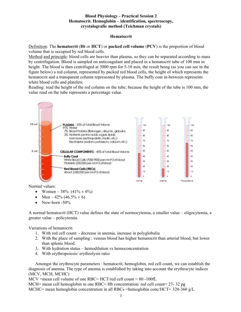
Blood Physiology - Practical Session 2 Hematocrit. Hemoglobin – identification, spectroscopy, crystalografic method (Teichman crystals) Hematocrit Definition: The hematocrit (Ht or HCT) or packed cell volume (PCV) is the proportion of blood volume that is occupied by red blood cells. Method and principle: blood cells are heavier than plasma, so they can be separated according to mass by centrifugation. Blood is sampled on anticoagulant and placed in a hematocrit tube of 100 mm in height. The blood is then centrifuged at 3000 rpm for 5-10 min, the result being (as you can see in the figure below) a red column, represented by packed red blood cells, the height of which represents the hematocrit and a transparent column represented by plasma. The buffy coat in-between represents white blood cells and platelets. Reading: read the height of the red column on the tube; because the height of the tube is 100 mm, the value read on the tube represents a percentage value. Normal values: • Women – 38% (41% ± 6%) • Men – 42% (46.5% ± 6) • New-born -50% A normal hematocrit (HCT) value defines the state of normocytemia, a smaller value – oligocytemia, a greater value – policytemia. Variations of hematocrit: 1. With red cell count: - decrease in anemia, increase in polyglobulia 2. With the place of sampling : venous blood has higher hematocrit than arterial blood, but lower than splenic blood. 3. With hydration status – hemodilution vs hemoconcentration 4. With erythropoiesis/ erythrolysis ratio Amongst the erythrocyte parameters : hematocrit, hemoglobin, red cell count, we can establish the diagnosis of anemia. The type of anemia is established by taking into account the erythrocyte indices (MCV, MCH, MCHC) MCV =mean cell volume of one RBC= HCT/red cell count = 80 -100fL MCH= mean cell hemoglobin in one RBC= Hb concentration/ red cell count= 27- 32 pg MCHC= mean hemoglobin concentration in all RBCs =hemoglobin conc/HCT= 320-360 g/L 1 With regard to these indices we can classify anemia into the following categories: I. normocromic, normocitic anemia: o acute hemorrhaging o aplastic/hypoplastic anemia o leukemia o renal/ hepatic disease II. hipocromic, microcytic anemia: o Iron deficient anemia o Cronic loss of blood o Hemolytic anemia o Cronic inflamation anemia III. normocromic, megalocytic anemia: o Anemia through B12deficit o Anemia through folic acid deficit Respective to the quantity of hemoglobin present in the blood there are several degrees of anemia: • Mild : 8-11g/100ml • Moderate : 6-8 g/100ml • Severe :<6g/100ml HEMOGLOBIN Hemoglobin (Hb) – major function in transporting blood gases. Hemoglobin: a cromoprotein Molecular weight = 68000da has 4 subunits, each having 17000da : - HEME (prosthetic group): ` - Fe2+ - tetrapirolic nucleus (same as in chlorophyll and B12 Vitamin) - GLOBIN (polypeptide) α, β, γ, δ = types of polypeptide chains. 2 Hb types Structure HbA α2β2 HbF α2γ2 HbA2 α2δ2 % 96-98 0,5-0,8 1,5-3,2 The conversion from HbF to HbA occurs 3-6 month after birth. 65% of Hb is synthesized in erythroblasts 35% of Hb is synthesized in reticulocytes. Schematic structure of hemoglobin Heme and Hb synthesis I. Krebs cycle -> succinil coenzyme A (SucCoA) II. SucCoA + glicin -> δ amino-levulinic acid (ALA) III. 2 ALA ->Porfobilinogen (pirolic nucleus) IV. 4 Pirols -> Protoporfirine IX (PP IX) V. PP IX + Fe++ -> HEM (Fe++ is inserted in the center of PP IX) VI. 4 Hem + polypeptide (globin) -> hemoglobin chain (α or β) VII. 2 α chains + 2 β chains -> Hb A Globin synthesis - dimeric structure - 2 polypeptide pairs There are: 2 α chains having 141 aa 2 β chains having 146 aa. Changing the sequence of aa leads to - type γ (146 aa, 37 different from type β) - type δ (146 aa, 10 different) - tip ε 3 The protein has an α helix spatial structure. The synthesis takes place in ribosomes, where the polypeptide α, β, γ, δ chains are formed. The 2 dimmers (α2 & β2 ) form together with hem HbA. There are also A1, A2, A3 types. HbA3 can be found in elder, having modified β chains. HbF (fetal) has a high binding capacity for oxygen. HbU (embryo type) – Gowers – has α2, ε2 chains. Iron metabolism Total iron amount in the body = 4+0,5g. It is divided in the following compartments: - hemoglobin – 67% - iron stored as feritine and hemosiderin – 27%. - myoglobin –3,5% - oxidative enzymes – 0,2% - plasma Iron absorption Minimum requirements: - newborn: 10 mg/24h - children: 5 mg/24h - young women: 20mg/24h - pregnant women: 30mg/24h - women after menopause: 10mg/24h - men: 10mg/24h 1 Fe 2+ binds 1O2 => 1Hb binds 4 O2 => 1g Hb binds 1.34ml O2 At a Hb concentration in the blood of 15g/100ml, there are 20 ml of O2 in 100 ml of blood. Identification of hemoglobin Principle: comparing clorhemine obtained from the tested blood with the Sahli hemoglobinometer clorhemine standard solution. Necessary material: - Sahli hemoglobinometer: - 2 lateral tubes with the standard solution – brown color. - 1 central tube used for the test – it has a gradation from 10 to 140 Sahli units (100th degree equals 16g Hb%) - annexed: a capillary pipette for blood maneuvering (with a 20 µl sign) + 1 distilled water pipette + 1 glass stirring stick. - HCl N/10 solution - Distilled water Protocol: • Put in the test tube HCl N/10 until 10th division. • Add with the capillary pipette 20 µl blood. • Brownish Clorhemine results. • Dilute the resulted solution with distilled water till it gets the same color with the standard probe. • Read the digits where the level has raised – meaning the HB amount in Sahli units. • In order to transform it in grams, calculate as follows: 100..................................................16g read value…………………..........X g X = 16 x read value / 100 Normal values: men – 15-17g% women – 13,5-15,5g% newborn – 17-20g. 4 Hb Spectroscopy Principle: Spectroscopy is based on the difference between the absorbtion spectra of hemoglobin and its related compounds. Necessary material: - spectroscope with direct vision - tubes - hemoglobin & derived solutions - crystals of sodium hydrosulfite - saturated potassium fericyanide. Protocol: • oxyhemoglobin spectra (OHb) - diluted blood solution (2-3 drops in 20 ml distilled water) - in contact with the air the blood turns bright red (Hb gets saturated with oxygen.) - put the tube in front of the spectroscope - the oxyhemoglobin absorbtion spectra has 2 bands: - a wide yellow one (577 nm) - a narrow green one (536 – 556 nm) • Reduced hemoglobin spectra - put in the HbO2 – 10 ml solution + a couple of crystals of sodium hydrosulfite. - the color turns violet red, due to the reduced Hb. - one can see a single green yellow band = Stokes band (530-548 nm) • Carboxihemoglobin spectra (HbCO) - Carboxihemoglobin is formed by the coordinative binding of CO to hem’s iron. It is a reversible reaction. The affinity of Hb for CO is 210 times higher then for O2. If we breathe CO – intoxication– 40% carboxihemoglobin = lethal value. - there are 2 bands: - A yellow one (564-579 nm) - A green one (530-548 nm) - both of them have a violet trend. • Met hemoglobin spectra - put 5-6 blood drops in 20 ml water. - add 8-10 drops of saturated potassium fericyanide solution. The solution gets dark brown due to the formed met hemoglobin. - it has 4 absorption bands: - A narrow red one (620-630 nm) – specific for met hemoglobin. - 2 pale yellow green bands - a wide blue one (486-578 nm) - met hemoglobin is improper for oxygen transport – a certain degree o hypoxia is reached. - Normally, erythrocytes contain 0,1-0,4 met hemoglobin;. The crystallographic method (Teichman crystals) Principle: Hemoglobin treated with “arising” HCl (resulted from the reaction of NaCl + CH 3 COOH ) -> clorhemine Clorhemine/hematin can be identified as yellow or brownish rhombic/diamond shaped crystals in clusters = Teichman crystals Necessary material: - a microscope slide, a glass slide cover, gas burner, microscope, blood, glacial acetic acid to which NaCl 1% has been added. 5 Protocol: - Pour 1 blood drop on the slide. - dry it by heating, but not over 40-45 degrees C – to avoid protein denaturation. Test it on the back of your hand. - after add 1 drop of glacial acetic acid and NaCl 1% and cover it with the glass slide cover. - heat it until it boils. Due to clorhemine the probe gets a brown color. - cool it and put it under the microscope. It is suggested to cool it slower in order to get bigger crystals. Discussion: - the method is used in forensic medicine in order to identify blood stains. - the crystals show the presence of blood without giving any information about the species. 6
