How does polyspermy happen in mammalian oocytes?
advertisement
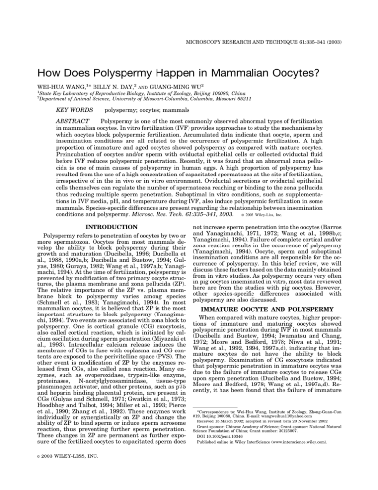
MICROSCOPY RESEARCH AND TECHNIQUE 61:335–341 (2003) How Does Polyspermy Happen in Mammalian Oocytes? WEI-HUA WANG,1* BILLY N. DAY,2 1 2 AND GUANG-MING WU2 State Key Laboratory of Reproductive Biology, Institute of Zoology, Beijing 100080, China Department of Animal Science, University of Missouri-Columbia, Columbia, Missouri 65211 KEY WORDS polyspermy; oocytes; mammals ABSTRACT Polyspermy is one of the most commonly observed abnormal types of fertilization in mammalian oocytes. In vitro fertilization (IVF) provides approaches to study the mechanisms by which oocytes block polyspermic fertilization. Accumulated data indicate that oocyte, sperm and insemination conditions are all related to the occurrence of polyspermic fertilization. A high proportion of immature and aged oocytes showed polyspermy as compared with mature oocytes. Preincubation of oocytes and/or sperm with oviductal epithelial cells or collected oviductal fluid before IVF reduces polyspermic penetration. Recently, it was found that an abnormal zona pellucida is one of main causes of polyspermy in human eggs. A high proportion of polyspermy has resulted from the use of a high concentration of capacitated spermatozoa at the site of fertilization, irrespective of in the in vivo or in vitro environment. Oviductal secretions or oviductal epithelial cells themselves can regulate the number of spermatozoa reaching or binding to the zona pellucida thus reducing multiple sperm penetration. Suboptimal in vitro conditions, such as supplementations in IVF media, pH, and temperature during IVF, also induce polyspermic fertilization in some mammals. Species-specific differences are present regarding the relationship between insemination conditions and polyspermy. Microsc. Res. Tech. 61:335–341, 2003. © 2003 Wiley-Liss, Inc. INTRODUCTION Polyspermy refers to penetration of oocytes by two or more spermatozoa. Oocytes from most mammals develop the ability to block polyspermy during their growth and maturation (Ducibella, 1996; Ducibella et al., 1988, 1990a,b; Ducibella and Buetow, 1994; Gulyas, 1980; Guraya, 1982; Wang et al., 1997a,b; Yanagimachi, 1994). At the time of fertilization, polyspermy is prevented by modification of two primary oocyte structures, the plasma membrane and zona pellucida (ZP). The relative importance of the ZP vs. plasma membrane block to polyspermy varies among species (Schmell et al., 1983; Yanagimachi, 1994). In most mammalian oocytes, it is believed that ZP is the most important structure to block polyspermy (Yanagimachi, 1994). Two events are associated with zona block to polyspermy. One is cortical granule (CG) exocytosis, also called cortical reaction, which is initiated by calcium oscillation during sperm penetration (Miyazaki et al., 1993). Intracellular calcium release induces the membrane of CGs to fuse with ooplasma and CG contents are exposed to the perivitelline space (PVS). The other event is modification of ZP by the enzymes released from CGs, also called zona reaction. Many enzymes, such as ovoperoxidase, trypsin-like enzyme, proteinases, N-acetylglycosaminidase, tissue-type plasminogen activator, and other proteins, such as p75 and heparin binding placental protein, are present in CGs (Gulyas and Schmell, 1971; Gwatkin et al., 1973; Hoodbhoy and Talbot, 1994; Miller et al., 1993; Pierce et al., 1990; Zhang et al., 1992). These enzymes work individually or synergistically on ZP and change the ability of ZP to bind sperm or induce sperm acrosome reaction, thus preventing further sperm penetration. These changes in ZP are permanent as further exposure of the fertilized oocytes to capacitated sperm does © 2003 WILEY-LISS, INC. not increase sperm penetration into the oocytes (Barros and Yanagimachi, 1971, 1972; Wang et al., 1998b,c; Yanagimachi, 1994). Failure of complete cortical and/or zona reaction results in the occurrence of polyspermy (Yanagimachi, 1994). Oocyte, sperm and suboptimal insemination conditions are all responsible for the occurrence of polyspermy. In this brief review, we will discuss these factors based on the data mainly obtained from in vitro studies. As polyspermy occurs very often in pig oocytes inseminated in vitro, most data reviewed here are from the studies with pig oocytes. However, other species-specific differences associated with polyspermy are also discussed. IMMATURE OOCYTE AND POLYSPERMY When compared with mature oocytes, higher proportions of immature and maturing oocytes showed polyspermic penetration during IVF in most mammals (Ducibella and Buetow, 1994; Iwamatsu and Chang, 1972; Moore and Bedford, 1978; Niwa et al., 1991; Wang et al., 1992, 1994, 1997a,d), indicating that immature oocytes do not have the ability to block polyspermy. Examination of CG exocytosis indicated that polyspermic penetration in immature oocytes was due to the failure of immature oocytes to release CGs upon sperm penetration (Ducibella and Buetow, 1994; Moore and Bedford, 1978; Wang et al., 1997a,d). Recently, it has been found that the failure of immature *Correspondence to: Wei-Hua Wang, Institute of Zoology, Zhong-Guan-Cun #19, Beijing 100080, China. E-mail: wangweihua11@yahoo.com Received 15 March 2002; accepted in revised form 20 November 2002 Grant sponsor: Chinese Academy of Science; Grant sponsor: National Natural Science Foundation of China; Grant number: 30125007. DOI 10.1002/jemt.10346 Published online in Wiley InterScience (www.interscience.wiley.com). 336 W.-H. WANG ET AL. oocytes to release CGs upon sperm penetration results from incomplete oocyte activation. A few hypotheses can explain the incomplete oocyte activation in mammals. The first is less calcium transients in immature oocytes (Machaty et al., 1997; Mehlmann and Kline, 1994). When compared with mature oocytes, immature oocytes had low calcium transients upon sperm penetration, calcium injection, and injection of inositol 1,4,5-trisphosphate (IP3). Immature oocytes are not sensitive to activation stimulation and such sensitivities increased during oocyte maturation (Machaty, 1997; Mehlmann and Kline, 1994). The second is incomplete migration of CGs under the ooplasma in immature oocytes (Wang et al., 1997a,d). CGs originate cortex cytoplasm during the oocyte’s early growth and development. Migration of CGs to ooplasma is necessary for subsequent exocytosis upon sperm penetration, and such a migration takes place during the final oocyte maturation. Gonadotropin stimulation and polymerization of microfilaments play important roles for CG migration to the cortex of oocytes (Ducibella et al., 1990b; Kim et al., 1996b). The third is reduced cortical endoplasmic reticulum (Charbonneau and Grey, 1984; Ducibella et al., 1988; Mehlmann et al., 1995). The number of cortical endoplasmic reticulum increases significantly during final oocyte maturation, which is thought to be calcium stores (Ducibella et al., 1988). In immature oocytes, there are fewer such calcium stores in the cortex of oocytes (Ducibella et al., 1988). And the fourth is a reduced amount of IP3 receptors (Mehlmann et al., 1996). IP3 receptors were significantly increased in the cortex of oocytes during final oocyte maturation. Immature oocytes have less IP3 receptors in the cortex of oocytes, thus the oocytes have less ability to respond to activators. Dramatic changes in these events mentioned above happen during the final maturation of oocytes. Thus, full oocyte cytoplasmic maturation including the ability to block polyspermy does not develop until oocytes reach metaphase II. AGED OOCYTES AND POLYSPERMY The lifetime for the oocytes to functionally block polyspermy is short (Ducibella, 1996). An increasing incidence of polyspermy occurs in most mammalian oocytes with increasing age of the oocytes. This could happen either in vivo (Hunter, 1991) or in vitro (Chian et al., 1992). Examination of CGs and ZP in the mice indicated that partial and unfunctional CG exocytosis is the main reason for polyspermic penetration in aged oocytes (Ducibella, 1996). It is also possible that changes in the cytoplasm or ooplasma during aging are related to the incomplete block to polyspermy in the aged oocytes. Reduced CG density in aged oocytes was observed in some mammalian oocytes (Ducibella, 1996; Gulyas, 1980; Guraya, 1982; Longo, 1974a,b; Szollosi, 1967; Wang et al., 1997a). Although we do not know if high polyspermic penetration in aged oocytes is due to reduced CG density, reduced enzymes activity in CGs, or reduced responsibility of ZP to CG contents, it is believed that degeneration in cytoplasm during aging makes all functions of oocytes associated with oocyte activation and development become low, thus the cortical reaction may be non-functional. Chian et al. (1992) found that some aged oocytes were fertilized normally, but their developmental competence significantly reduced, indicating that all cell functions are affected during aging. ZONA ABNORMALITY AND POLYSPERMY The ZP is a semi-transparent glycoprotein matrix that surrounds the oocyte and consists of ZP1, ZP2, and ZP3 (Bauskin et al., 1999; Bleil and Wassarman, 1980; Nakano et al., 1990; Shabanowitz and O’Rand, 1988). Although the mechanism by which the ZP blocks polyspermy is not completely understood, modification of the ZP by CG contents released during oocyte activation prevents subsequent sperm binding to and penetration of ZP are the most accepted mechanism for oocytes to block polyspermy. These modifications of ZP include changes in the zona composition and structure as revealed by electrophoresis (Bauskin et al., 1999; Bleil and Wassarman, 1980; Nakano et al., 1990; Shabanowitz and O’Rand, 1988) and other molecular techniques (Aviles et al., 1996; Barros and Yanagimachi, 1971, 1972; Miller et al., 1993). Zona hardening, which is measured by the time the oocyte’s ZP was digested by enzyme treatment, is also a parameter to detect zona reaction (Nakano et al., 1987). However, studies of zona reaction have always involved a group of oocytes, ranging from about 10 to more than a few hundred because of the low sensitivity of the methods used. It has also involved both reacted and non-reacted ZP because it is difficult to quantify zona components in single ZP. Recently, a modified technique that can reveal zona protein changes in single ZP in the mice was reported (Moos et al., 1994), but it has not been widely used in other animals due to the technique problem. Under differential interference contrast or Hoffman optics, the zona appears relatively homogeneous. However, when zona of living oocytes is imaged with an orientation-independent polarized light microscope, the polscope, three distinct layers, which differ in their retardance and azimuth, can be discerned (Keefe et al., 1997). Using the polscope, the protein matrix of internal layer of hamster zona was observed as oriented radially while the external layer was seen as oriented tangentially. An intermediate layer was randomly oriented (Keefe et al., 1997). With the polscope, ZP of the human oocytes exhibit a similar trilaminar structure (Fig. 1). Moreover, in one case, we found that abnormal zona in human oocytes exhibited minute discontinuities in inner zona layer (Fig. 1). All oocytes with this type of ZP were polyspermy. The zona of all normally fertilized oocytes from the same patient exhibited the normal trilaminar appearance of the zona layers. From these studies, it would appear that imaging ZP with the polscope might provide an easy, non-invasive method to diagnose abnormality in the block to polyspermy in human oocytes undergoing IVF. Abnormal ZP is always found in human oocytes during infertility treatment, which may be due to hypostimulation (Familiari et al., 1992). Familiari et al. (1992) speculated that the inner layer of ZP might play an important role in the block to polyspermy in human oocytes, as sperm was rarely found to penetrate this layer. Presumably, a defective ZP did not establish a functional zona block. Several studies have demonstrated that the thickness and integrity of the ZP are related to patient’s estradiol levels during stimulation. Oocytes POLYSPERMY IN MAMMALIAN OOCYTES Fig. 1. Zona pellucida imaged by Polscope in living human oocytes. A: An oocyte with normal zona pelluicda. The normal zona of oocytes shows the compact and smooth inner layer and the lower density and thinner outer layer; B: An oocyte with abnormal zona pelluicda. The zona pellucida of this oocyte shows fractured inner layer. This egg was polyploid. Scale bar ⫽ 10 m. from the patients with relatively good stimulation and response had optimal ZP thickness (Bertrand et al., 1996). However, how stimulation induces zona abnormality in the human is still unclear. It is necessary to study more details about this phenomenon in the human; perhaps, some animal models can help address the answer. NUMBER OF CAPACITATED SPERM AT THE SITE OF FERTILIZATION AND POLYSPERMY Both in vivo and in vitro studies indicated that the number of sperm at the site of fertilization is directly related to polyspermy even when mature oocytes were inseminated either in vivo or in vitro. A series of in vivo studies were conducted in porcine oocytes (Hunter, 1991). According to Hunter (1991), the incidence of polyspermy in pigs in vivo is less than 5%. However, higher rates, up to 60%, could be artificially induced under different conditions, such as oocytes ovulated after gonadotropin stimulation during the luteal phase of the cycle, delayed insemination, progesterone local microinjection and administration, and tubal insemination. Delayed insemination and progesterone also induced polyspermy in rabbits (Chang and Hunt, 1968). Under all of these conditions, it was found that number of sperm at the site of fertilization was significantly increased. Under physiological conditions, the isthmus of oviducts regulates the numbers of sperm released to the site of fertilization, thus avoids occurrence of polyspermic penetration. These results suggest that increased polyspermy of eggs in vivo occurs primarily because abnormally high numbers of competent spermatozoa reach the egg surface more or less simultaneously even if the oocytes have the ability to respond the first sperm penetration and release CGs to block further sperm penetration. When the fertilized oocytes pass through the isthmus and other parts of the oviduct where capacitated sperm are stored, it has been found that sperm can bind to the outer layer of ZP, but all of the sperm are blocked by the inner layer of zona. Thus, sperm are seldom seen in the PVS, as shown in Figure 2. In vitro studies also indicated that polyspermic fertilization was increased as sperm number was increased (Abeydeera and Day, 1997; Nagai et 337 Fig. 2. Confocal micrograph of a porcine eight-cell stage embryo collected from oviduct and image was taken at the equatorial section of the embryo (only 5 nuclei were shown in the image) The embryo was fixed and stained with propidium iodide. Nuclei in the blasomeres (arrows) inside the zona and sperm heads binding to the zona pellucida are shown. Note that there is no sperm head in the PVS, and all sperm were blocked at the innermost of the zona pellucida. al., 1984; Wang et al., 1991) regardless of the oocytes being collected from oviducts (in vivo matured-ovulated) or matured in vitro. Some in vitro conditions associated with polyspermic fertilization in pig oocytes have been examined and it has been found that CG exocytosis occurred during IVF (Wang et al., 1997a– c), but a delayed zona reaction was observed (Wang et al., 1999). From these studies, it seems that a simultaneous sperm penetration is the most possible reason for polyspermy of porcine oocytes inseminated in vitro. Much higher sperm concentrations are used for IVF and it is a few hundred times higher than that in the oviducts during natural conception. However, reduced sperm concentration could reduce polyspermy but it always reduces penetration rates (Abeydeera and Day, 1997). It is still unclear why a high concentration of sperm is necessary to obtain satisfactory fertilization rates during IVF. Probably, the conditions of sperm capacitation are not optimized in vitro, thus many sperm are not capacitated or capacitated simultaneously, then quickly undergo spontaneous acrosome reaction. A condition that regulates a high rate of sperm capacitation at different time, as in the oviduct, may reduce simultaneous sperm penetration. Thus, a low sperm concentration could be used for fertilization of oocytes in vitro. OVIDUCT SECRETION REDUCES POLYSPERMY Higher rates of polyspermic penetration are observed during IVF than natural insemination in the oviducts. When in vivo and in vitro fertilized porcine oocytes were compared, it was found that delayed and incomplete CG exocytosis existed in in vitro fertilized oocytes (Cran and Cheng, 1986). In vivo fertilization in the oviducts also allowed the dispersal of CG contents in the PVS after exocytosis (Cran and Cheng, 1986). However, released CG contents during IVF remained adjacent to the outside of the plasma membrane, suggest- 338 W.-H. WANG ET AL. ing that the enzymes from CGs do not immediately act on ZP during IVF. Thus, a delayed zona reaction happens. The dispersion of CG contents in PVS depends upon the concentration of calcium in medium (Cran and Cheng, 1986). In order to clarify whether it is maturation or exposure of oocytes to oviduct secretion that modulate polyspermy, a more direct comparison of sperm penetration and CG exocytosis between in vitro matured oocytes and in vivo oviduct oocytes was conducted recently (Wang et al., 1998a). A higher rate of polyspermy was observed in in vitro matured oocytes as compared to ovulated/oviduct oocytes. Oviduct secretory proteins have been identified in the ZP, PVS, and plasma membrane of oviduct oocytes in many mammals (Broermann et al., 1989; Brown and Cheng, 1986; Buhi et al., 1993). Some materials binding to ZPs were positively labeled by a lectin from archis hypogaea, which is specific for -D-Gal(1-3)-D-GalNAc (Fig. 2). ZPs were also modulated in the oviducts. Thus, the oocytes collected from oviducts showed thicker ZPs and larger PVS than in vitro mature oocytes. These changes in the extracellular matrix of oocytes while in the oviduct may help establish a functional block to polyspermy in pig oocytes. Polyspermic penetration was also reduced after in vitro matured oocytes were cultured in the collected oviductal fluid and oviductal epithelium cells before IVF (Kano et al., 1994; Kim et al., 1996a). It is suggested that some glycoproteins from oviductal fluid may enter PVS or plasma membrane, to facilitate the synchronous CG exocytosis and promote the reaction of ZP to CG exudates. The effects of oviductal epithelium cells on sperm function have also been noticed by in vivo and in vitro studies. In vivo, sperm are conveyed up to the ampulla from the uterus by way of the isthmus. Before fertilization, sperm stay in the isthmus to undergo capacitation. Only capacitated sperm move out of the isthmus sperm reservoir to the site of fertilization. Moreover, contact with the apical plasma membrane of oviduct epithelial cells can enhance sperm motility, inhibit spontaneous acrosome reaction of sperm, so even if only a few hundred sperm are released to move to the site of fertilization, almost all oocytes can be fertilized (Hunter, 1991, 1994, 1996). In vitro studies indicate that coculture of sperm with oviductal epithelium cells can control sperm movement thereby preventing polyspermic fertilization (Anderson and Killian, 1994; Dubuc and Sirrad, 1995; Nagai and Moor, 1990; Suarez, 1998). Binding of sperm to epithelium cells also inhibits spontaneous acrosome reaction thus retaining fertilizing ability (Dubuc and Sirrad, 1995). It is also believed that oviduct affects the sperm plasma membrane, stabilizes and reduces the numbers becoming capacitated simultaneously. In turn, it would appear that the effects of oviductal epithelium cells or their secretion on preventing polyspermic penetration are the results from the interactions between epithelium cells together with their secretion and both sperm and oocytes. Coculture of sperm or oocyte with cultured oviductal epithelium cells before or during IVF may be a good approach to produce normal fertilized oocytes in those mammals in which polyspermic penetration has not been resolved and/or IVF conditions have not been optimized, such as in domestic animals. PROTEIN SUPPLEMENTS IN FERTILIZATION MEDIUM AND POLYSPERMY Insemination medium plays a significant role(s) for successful IVF. One of the most important components in insemination media is protein supplementation. Although recent studies indicated that chemically defined insemination medium without protein supplement supported sperm penetration of oocytes in some mammals (Tajik et al., 1994; Wang et al., 1995), protein supplementation is important for maintaining sperm motility, capacitation, and acrosome reaction (Yanagimachi, 1994). Bovine serum albumin (BSA) and fetal calf serum (FCS) are widely used in most IVF media except that human serum albumin or maternal serum is usually used in human IVF. Protein supplementation is related to polyspermic penetration, but species-specific differences are obviously present. For example, supplementation of FCS in IVF medium can significantly increase polyspermy in bovine and pig oocytes (Tajik et al., 1993; Wang et al., 1994), but supplementation of BSA did not. It has been found that FCS reduces the effective concentration of calcium in the medium, thus supplementation of FCS inhibits CG contents disperse after exocytosis and delays its reaction to ZP (Cran and Cheng, 1986). FCS also inhibits the zona reaction in oocytes of some mammals (Downs et al., 1986; Ducibella et al., 1990a; Niwa and Wang, 2001; Schroeder et al., 1990; Zhang et al., 1992). However, in human IVF, supplementation of maternal serum does not increase polyspermic penetration. It was found that it is fetuin in FCS that regulates the CG’s effect on ZP (Schroeder et al., 1990). When mouse oocytes were cultured in vitro without FCS, premature CG exocytosis can induce zona reaction of oocytes before fertilization, and the oocytes cannot be fertilized. However, when FCS is present in the medium, it preserved the fertilizability of the mature oocyte (Eppig and Schroeder, 1986). Serum from other animals also has such a function, but the serum from human fetal cord did not (Eppig and Schroeder, 1986). It seems that FCS is not a suitable protein source for IVF medium and some substitute should be used, such as serum albumin or a chemically defined medium. OTHER FACTORS AND POLYSPERMY As mentioned above, sperm concentration is one of the main factors that affect sperm penetration and poyspermic penetration. An increased sperm penetration rate was accompanied by an increased polysermic penetration, which was observed in most animals, suggesting that a optimal concentration should be determined based on sperm motility, morphology and the components in the fertilization medium. The pH of insemination medium is also a factor that regulates sperm capacitation and acrosome reaction (Brackett and Oliphant, 1975). It has been found that high pH in medium supported a higher fertilization rate (Cheng, 1985; Tajik et al., 1994, Wang et al., 1995), but activity of enzymes from CGs seems lower in high pH conditions (Miller et al., 1993). Whether this is related to polyspermy in some animals, in which high pH insemination medium was used, is still unclear. Bicarbonate is another important component in insemination me- 339 POLYSPERMY IN MAMMALIAN OOCYTES Fig. 3. Confocal micrographs of porcine oocytes collected from oviduct (A and A’) and matured in culture (B and B’). Note the oocyte from oviduct has thicker zona pellucida and large PVS (inset in A). The zona of in vivo oocyte (A), not in vitro matured oocyte (B), was labeled by fluorescen isothiocyanate-labeled peanut agglutinin, which is used to label the lectin present in the cortical granules. Cortical granules in oocytes are distributed in the cortex of oocytes and form a monolayer under the ooplasma (arrows in A and B). In the egg cortex, CG distribution was not different between in vivo oocytes and in vitro oocytes (A’ and B’). Pb: polar body, PVS: perivitelline space; ZP: zona pellucida; M: metaphase chromosomes. Bar ⫽ 10 m. dium and it is important for maintaining sperm motility, inducing sperm capacitation and acrosome reaction (Suzuki et al., 1994; Tajik et al., 1994; Wang et al., 1995). Recently a Tris-buffered medium was successfully used to support sperm penetration of porcine (Abeydeera and Day, 1997) and bovine (Wang and Niwa, unpublished data) oocytes, and reduced polyspermy was reported when such a medium was used (Abeydeera and Day, 1997). It is unknown how Tris regulates sperm function and/or oocyte function during IVF. In some mammals, sperm capacitation stimulators, such as heparin and/or caffeine, are also necessary for sperm to penetrate oocyte in vitro (Abeydeera and Day, 1997; Park et al., 1989, Wang et al., 1991). The relationship of these additives on polyspermy or oocyte function remains to be clarified. High temperature (38.5–39°C) has been used to perform IVF in some animals (Cheng, 1985; Chian et al., 1992; Wang et al., 1991), since a high proportion of sperm could be capacitated in a high temperature condition, but its relationship with polyspermy is not clear. CONCLUSIONS As summarized in Figure 4, monospermic fertilization takes place only when fully mature oocytes with normal ZP are inseminated with optimal sperm concentration and optimized conditions. Incomplete oocyte cytoplasmic maturation, abnormal ZP, high concentration of sperm, inappropriate supplementations in in- Fig. 4. Schematic diagram of the occurrence of monospermic and polyspermic fertilization in the mammalian oocytes. Oocyte, zona pellucida, sperm, and fertilization conditions are all related to polyspermic fertilization. For details, see text. semination medium, and other odd factors during fertilization are all related to polyspermic fertilization. Species-specific differences make it difficult to compare the results from the different animals under different conditions, but molecular basis associated with polyspermy block may be similar in most mammals. It is necessary to address more details of polyspermic fertilization in mammals by technology developed in morphological and molecular biology. ACKNOWLEDGMENTS This work was partially supported by Hundred Talent Program to Wei-Hua Wang from Chinese Academy of Sciences and Natural Science Fund for Distinguished Young Scholar to Wei-Hua Wang from National Natural Science Foundation of China (No: 30125007). REFERENCES Abeydeera LR, Day BN. 1997. In vitro penetration of pig oocytes in a modified Tris-buffered medium: effect of BSA, caffeine and calcium. Theriogenology 48:537–544. 340 W.-H. WANG ET AL. Anderson SH, Killian GJ. 1994. Effect of macromolecules from oviductal conditioned medium on bovine sperm motion and capacitation. Biol Reprod 51:795–799. Aviles M, Jaber L, Castells MT, Kan FK, Ballesta J. 1996. Modifications of the lectin binding pattern in the rat zona pellucida after in vivo fertilization. Mol Reprod Dev 44:370 –381. Barros C, Yanagimachi R. 1971. Induction of zona reaction in golden hamster eggs by cortical granule material. Nature 233:268 –269. Barros C, Yanagimachi R. 1972. Polyspermy-preventing mechanisms in the golden hamster eggs. J Exp Zool 180:251–265. Bauskin AR, Franken DR, Eberspaecher U, Donner P. 1999. Characterization of human zona pellucida glycoproteins. Mol Hum Reprod 5:534 –540. Bertrand E, Bergh MV, Englert Y. 1996. Clinical parameters influencing human zona pellucida thickness. Fertil Steril 66:408 – 411. Bleil JD, Wassarman PM. 1980. Structure and function of the zona pellucida: identification and characterization of the proteins of the mouse oocyte’s zona pellucida. Dev Biol 76:185–202 Brackett BG, Oliphant G. 1975. Capaciation of rabbit spermatozoain vitro. Biol Reprod 12:260 –274. Broermann DM, Xie S, Nephew KP, Pope WF. 1989. Effects of the oviduct and wheat germ agglutinin on enzymatic digestion of porcine zona pellucidae. J Anim Sci 67:1324 –1329. Brown CR, Cheng WKT. 1986. Changes in composition of the porcine zona pellucida during development of the oocyte to the 2- to 4-cell embryo. J Embryol Exp Morph 92:183–191. Buhi WC, O’Brien B, Alvarez IM, Erdos G, Dubois D. 1993. Immunogold localization of porcine oviduct secretory proteins within the zona pellucida, perivitelline space, and plasma membrane of oviduct and uterine oocytes and early embryos. Biol Reprod 48:1274 – 1283. Chang MC, Hunt DM. 1968. Attempts to induce polyspermy in the rabbit by delayed insemination and treatment with progesterone. J Exp Zool 167:419 – 425. Charbonneau M, Grey RD. 1984. The onset of activation responsiveness during maturation coincides with the formation of the cortical endoplasmic reticulum in oocytes of Xenopus Iaevis. Dev Biol 102: 90 –97. Cheng WKT. 1985. In vitro fertilization of farm animals. Ph.D. Thesis. Chian RC, Nakahana H, Niwa K, Funahashi H. 1992. Fertilization and early cleavage in vitro of aging bovine oocytes after maturation in culture. Theriogenology 37:665– 672. Cran DG, Cheng WTK. 1986. The cortical reaction in pig oocytes during in vivo and in vitro fertilization. Gamete Res 13:241–251. Downs SM, Schroeder AC, Eppig JJ. 1986. Serum maintains the fertilizability of mouse oocytes matured in vitro by preventing hardening of zona pellucida. Gamete Res 15:115–122. Dubuc A, Sirard MA. 1995. Effect of coculturing spermatozoa with oviduct cells on the incidence of polyspermy in pig in vitro fertilization. Mol Reprod Dev 41:360 –367. Ducibella T. 1996. The cortical reaction and development of activation competence in mammalian oocytes. Human Reprod Update 2:29 – 42. Ducibella T, Buetow J. 1994. Competence to undergo normal, fertilization-induced cortical activation develops after metaphase I of meiosis in mouse oocytes. Dev Biol 165:95–104. Ducibella T, Rangarajian S Anderson E. 1988. The development of mouse oocyte cortical reaction competence in accompanied by major changes in cortical vesicle and not cortical granule depth. Dev Biol 130:789 –792. Ducibella T, Kurasawa S, Rangarajan S, Kopf GS, Schultz RM. 1990a. Precocious loss of cortical granules during mouse oocyte meiotic maturation and correlation with an egg induced modification of the zona pellucida. Dev Biol 137:46 –55. Ducibella T, Duffy P, Reindollar R, Su B. 1990b. Changes in the distribution of mouse oocyte cortical granules and ability to undergo the cortical reaction during gonadotropin-stimulated meiotic maturation and aging in vivo. Biol Reprod 43:870 – 876. Eppig JJ, Schroeder AC. 1986. Culture system for mammalkian oocyte development: progress and prospects. Theriogenology 25:97– 106. Familiari G, Nottola SA, Macchiarelli G, Micara G, Aragona C, Motta PM. 1992. Human zona pellucida during in vitro fertilization: an ultra structural study using saponin, ruthenium red, and osmiumthiocarbohydrazide. Mol Reprod Dev 32:51– 61. Gulyas BJ. 1980. Cortical granules of mammalian eggs. Int Rev Cytol 63:357–392. Gulyas BJ, Schmell ED. 1971. Ova peroxidase activity in ionophore treated mouse egg. I. Electron microscopic location. Gamete Res 3:267–277. Guraya SS. 1982. Recent progress in the structure, origin, composition, and function of cortical granules in animal egg. Int Rev Cytol 78:257–360. Gwatkin RBL, Williams DT, Hartmann JF, Kniazuk M. 1973. The zona reaction of hamster and mouse eggs: Production in vitro by a trypsin-like protease from cortical granules. J Reprod Fertil 32: 259 –265. Hoodbhoy T, Talbot P. 1994. Mammalian cortical granules, contents, fate and function. Mol Reprod Dev 39:439 – 448. Hunter RHF. 1991. Oviduct function in pigs, with particular reference to the pathological condition of polyspermy. Mol Reprod Dev 29: 385–391. Hunter RHF. 1994. Modulation of gamete and embryonic microenvironments by oviduct glycoproteins. Mol Reprod Dev 39:176 –181. Hunter RHF. 1996. Ovarian control of very low sperm/egg ratios at the commencement of mammalian fertilization to avoid polyspermy. Mol Reprod Dev 44:417– 422. Iwamatsu T, Chang MC. 1972. Sperm penetration in vitro of mouse oocytes at various times after maturation. J Reprod Fertil 31:237– 247. Kano K, Miyano T, Kato S. 1994. Effect of oviductal epithelial cells on fertilization of pig oocytes in vitro. Theriogenology 42:1061–1068. Keefe D, Tran P, Pellegrini C, Oldenbourg T. 1997. Polarized light microscopy and digital image processing identify a multi-laminar structure of the hamster zona pellucida. Hum Reprod 12:1250 – 1252 Kim N.H, Funahashi H, Abeydeera LR, Moon SJ, Prather RS, Day BN. 1996a. Effects of oviductal fluid on sperm penetration and cortical granule exocytosis during fertilization of pig oocytes in vitro. J Reprod Fertil 107:79 – 86. Kim NH, Day BN, Lee HT, Chung KS. 1996b. Microfilament assembly and cortical granule distribution during maturation, aprthenogenetic activation and fertilization in the porcine oocytes. Zygote 4:145–149. Longo FJ. 1974a. Unltrastructural changes in rabbit eggs aged in vivo. Biol Reorid 11:22–39. Longo FJ. 1974b. Unltrastructural analysis of spontaneous activation of hamster eggs aged in vivo. Anat Rec 179:27–55. Machaty Z, Funahashi H, Day BN, Prather RS. 1997. Developmental changes in the intracellular Ca2⫹ release mechanisms in porcine oocytes. Biol Reprod 56: 921–930. Mehlmann LM, Kline D. 1994. Regulation of intracellular calcium in the mouse egg:calcium release in response to sperm or inositol trisphosphate is enhanced after meiotic maturation. Biol Reprod 51:1088 –1098. MehlmannLM, Terasaki M, Jaffe A, Kline D. 1995. Reorganization of the endoplasmic reticulum during meiotic maturation of the mouse oocytes. Dev Biol 170:607– 615. MehlmannLM, Mikoshiba K, Kline D. 1996. Redistribution and increase in cortical inositol 1,4,5,-trisphosphate receptors after meiotic maturation of the mouse oocytes. Dev Biol 180:489 – 498. Miller DJ, Gong X, Decker G, Shur BD. 1993. Egg cortical granule N-acetylglucosaminidase is required for the mouse zona block to polyspermy. J Cell Biol 123:1431–1441. Miyazaki S, Shirakawa H, Nakada K, Honda Y. 1993. Essential role of the inositol 1,4,5-trisphosphate receptor/Ca2⫹ release channel in Ca2⫹ waves and Ca2⫹ oscillations at fertilization of mammalian eggs. Dev Biol 158:62–78. Moore HDM, Bedford JM. 1978. Ultrastructure of the equatorial segment of hamster spermatozoa during penetration of oocytes. J Ultrastruct Res 62:110 –117. Moos J, Kalab P, Kopf GS, Schuitz RM. 1994. Rapid, nonradioactive, and quantitative method to analyze zona pellucida modification in single mouse eggs. Mol Reprod Dev 38:91–93. Nagai T, Moor RM. 1990. Effect of oviduct cells on the incidence of polyspermy in pig oocytes fertilized in vitro. Mol Reprod Dev 26: 377–382. Nagai T, Niwa K, Iritani A. 1984. Effect of sperm concentration during preincubation in a defined medium on fertilization in vitro of pig follicular oocytes. J Reprod Fertil 70:271–275. Nakano M, Hatanaka Y, Tobita T. 1987. Solubilization of porcine zonae pellucidae by trypsin and pronase. Biochem Int 14:425– 433. Nakano M, Hatanaka Y, Kobayashi N, Noguchi S, Ishikawa S, Tobita T. 1990. Further fractionation of the glycoprotein families of porcine zona pellucida by anion-exchanges HPLC and some characterization of the separated fractions. J Biochem 107:144 –150. Niwa K, Wang WH. 2001. Cortical granules and cortical reaction in pig oocytes. In: Miyamoto H, Manabe H, editors. Reproductive bioltechnology: reproductive biotechnology update and its related physiology. Kyoto: Hokudo Printing Co. p 27–34. POLYSPERMY IN MAMMALIAN OOCYTES Niwa K, Park C-K, Okuda K. 1991. Penetration in vitro of bovine oocytes during maturation by frozen-thawed spermatozoa. J Reprod Fertil 91:329 –336. Park CK, Ohgoda O, Niwa K. 1989. Penetration of bovine follicular oocytes by frozen-thawed spermatozoa in the presence of caffeine and heparin. J Reprod Fertil 86:577–582. Pierce KE, Siebert MC, Kopf GS, Schultz RM, Calarco PG. 1990. Characterization and location of a mouse egg cortical granules antigen prior to and following fertilization or egg activation. Dev Biol 141: 381–392. Schmell ED, Gulyas BJ, Hedrick JL. 1983. Egg surface changes during fertilization and the molecular mechanism of the block to polyspermy. In: Hartmann JF, editor. Mechanism and control of animal fertilization. New York: Academic Press. p 365– 413. Schroeder AC, Schultz RM, Kopf GS, Talor FR, Becker RB, Eppig JJ. 1990. Fetuin inhibits zona pellucida hardening and conversion of ZP2 to ZP2f during spontaneous mouse oocyte maturation in vitro in the absence of serum. Biol Reprod 43:891– 897. Shabanowitz RB, O’Rand MG. 1988. Characterization of the human zona pellucida from fertilized and unfertilized eggs. J Reprod Fertil 82: 151–161 Suarez SS. 1998. The oviductal sperm reservoir in mammals: mechanisms of formation. Biol Reprod 58:1105–1107. Suzuki K, Ebihara M, Nagai T, Clarke NGE, Harrison RAP. 1994. Importance of bicarbonate/CO2 for fertilization of pig oocytes in vitro and synergism with caffeine. Reprod Fertil Dev 6:221–227. Szollosi D. 1967. Development of cortical granules and cortical reaction in rat and hamster eggs. Anat Rec 159:431– 446. Tajik P, Niwa K, Murase T. 1993. Effects of different protein supplements in fertilization medium on in vitro penetration of cumulusintact and cumulus-free bovine oocytes matured in culture. Theriogenology 40:949 –958. Tajik P, Wang WH, Okuda K, Niwa K. 1994. In vitro fertilization of bovine oocytes in a chemically defined, protein-free medium varying the bicarbonate concentration. Biol Reprod 50:1231–1237. Wang WH, Niwa K, Okuda K. 1991. In-vitro penetration of pig oocytes matured in culture by cryopreserved ejaculated spermatozoa. J Reprod Fertil 93:491– 49. Wang WH, Uchida M, Niwa K. 1992. Effects of follicle cells on in-vitro penetration of pig oocytes by cryopreserved, ejaculated spermatozoa. J Reprod Dev 38:125–131. 341 Wang WH, Abeydeera LR, Okuda K, Niwa K. 1994. Penetration of porcine oocytes during maturation in vitro by cryopreserved, ejaculated spermatozoa. Biol Reprod 50:510 –515. Wang WH, Abeydeera LR, Fraser LR, Niwa K. 1995. Functional analysis using chlortetracycline fluorescence and in vitro fertilization of frozen-thawed ejaculated spermatozoa incubated in a protein-free chemically defined medium. J Reprod Fertil 104:305–313. Wang WH, Hosoe M, Shioya Y. 1997a. Induction of cortical granule exocytosis of pig oocytes by spermatozoa during meiotic maturation. J Reprod Fertil 109:247–255. Wang WH, Sun QY, Hosoe M, Shioya Y, Day BN. 1997b. Quantified analysis of cortical granule distribution and exocytosis of porcine oocytes during meiotic maturation and activation. Biol Reprod 56: 1376 –1382. Wang WH, Abeydeera LR, Cantley TC, Day BN. 1997c. Effects of oocyte maturation media on development of pig embryos produced by in vitro fertilization. J Reprod Fertil 111:101–108. Wang WH, Hosoe M, Li RF, Shioya Y. 1997d. Development of the competence of bovine oocytes to release cortical granules and block polyspermy after meiotic maturation. Dev Growth Differ 39:607– 615. Wang WH, Abeydeera LR, Prather RS, Day BN. 1998a. Morphologic comparison of ovulated and in vitro-matured porcine oocytes, with particular reference to polyspermy after in vitro fertilization. Mol Reprod Dev 49:308 –316. Wang WH, Machanty Z, Abeydeera LR, Prather RS, Day BN. 1998b. Parthenogenetic activation of pig oocytes with calcium ionophore and the block to sperm penetration after activation. Biol Reprod 58:1357–1366. Wang WH, Abeydeera LR, Prather RS, Day BN. 1998c. Functional analysis of activation of porcine oocytes by spermatozoa, calcium ionophore and electrical pulse. Mol Reprod Dev 51:346 –353. Wang WH, Machaty Z, Abeydeera LR, Prather RS, Day BN. 1999. Time course of cortical and zona reactions of pig oocytes activated by thimerosal. Zygote 7:79 – 86. Wolf DP. 1978. The block to sperm penetration in zonal-free mouse eggs. Dev Biol 64:1–10. Yanagimachi R. 1994. Mammalian Fertilization. In: Knobil E, Neill JD, editors. The physiology of reproduction. New York: Raven Press. p 261–268. Zhang X, Rutledge J, Khamsi F, Armstrong DT. 1992. Release of tissue-type plasminogen activator by activated rat eggs and its possible role in the zona reaction. Mol Reprod Dev 32:28 –32
