Promega Notes Number 65, 1998, p. 14
The Chemistry of Bioluminescent Reporter
Assays
By Keith V. Wood
Promega Corporation
E-mail to: kwood@promega.com
Firefly luciferase is the most widely used bioluminescent reporter because its enzyme activity is closely coupled to protein synthesis, and
the luminescence assay is rapid, convenient and sensitive. Although various assay formulations for firefly luciferase have been
described, the most widely used contains coenzyme A in addition to beetle luciferin and ATP. In a 1-10 second measurement, this assay
provides linearity over a 100 million-fold concentration range with sensitivity greater than 10-20 moles of enzyme. Recently, Renilla
luciferase has also become widely used as a genetic reporter, although primarily as a co-reporter to firefly luciferase. Assay of Renilla
luciferase is also rapid and linear, but the sensitivity is limited somewhat by autoluminescence. An assay format called the DualLuciferaseTM Reporter (DLRTM) Assay*(a) has been designed to sequentially quantitate both firefly and Renilla luciferases from a
single sample. The integration of the two luciferase assays provides an efficient means for incorporating an internal control into
reporter measurements, or for analyzing two separate events in the same system. Bacterial luciferase, although the first luciferase to be
used as a reporter, is generally used to provide autonomous luminescence in bacterial systems through expression of the lux operon.
Ordinarily it is not useful for analysis in eukaryotic systems.
*Patent Pending.
(a)U.S.
Pat. Nos. 5,283,179, 5,641,641 and 5,650,289 have been issued to Promega Corporation for a firefly luciferase assay method, which affords greater light
output with improved kinetics as compared to the conventional assay.
INTRODUCTION
Bioluminescence as a natural phenomenon is widely experienced with amazement and delight at the prospect of living organisms
creating their own light. Basic research into this phenomenon has also led to practical applications, particularly in molecular biology
where bioluminescent enzymes have been widely used as genetic reporters. Moreover, the value of this application has grown
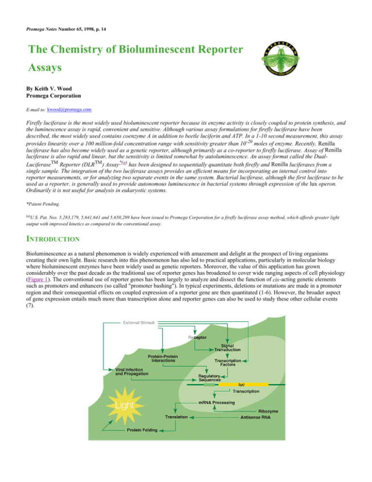
considerably over the past decade as the traditional use of reporter genes has broadened to cover wide ranging aspects of cell physiology
(Figure 1). The conventional use of reporter genes has been largely to analyze and dissect the function of cis-acting genetic elements
such as promoters and enhancers (so called "promoter bashing"). In typical experiments, deletions or mutations are made in a promoter
region and their consequential effects on coupled expression of a reporter gene are then quantitated (1-6). However, the broader aspect
of gene expression entails much more than transcription alone and reporter genes can also be used to study these other cellular events
(7).
Figure 1. Schematic diagram demonstrating the range of applications for reporter genes in
cellular biology.
Events upstream of transcription include receptor function (8-10), intracellular signal transduction (11,12) and the actions of specific
transcription factors (13-15). Events downstream of transcription include RNA processing (16,17), translation (18) and protein folding
(19,20). For monitoring of upstream events, the reporter gene is coupled to an "indicator sequence" specific for the event under study.
For example, coupling the reporter to a cAMP response element (CRE) permits regulated reporter expression in response to changing
intracellular cAMP levels (21,22). Coupling indicator sequences to reporter genes is particularly important in pharmaceutical research to
rapidly screen for new molecules acting as agonists or antagonists of receptor function. Reporter genes also support the development of
potentially important clinical methodologies, such as gene therapy or targeted genetic suppression, using antisense RNA or ribozymes.
The infection and propagation of viruses may be studied either by incorporating a reporter gene into the viral genome (23), or through
transactivation of a reporter gene in transgenic cells (24,25). For example, expression of Tat by infecting HIV particles can induce
transactivation of a reporter gene coupled to a second copy of the HIV LTR in the host cells (26).
Of the many different strategies available for using as genetic reporters, bioluminescence offers the most nearly ideal situation, because
the reporter measurements are nearly instantaneous, they are exceptionally sensitive and quantitative, and typically there is no
endogenous activity in the host cells to interfere with quantitation. Within bioluminescence, however, there are a large variety of distinct
chemistries. Bioluminescence is found in many phyla, particularly in the marine environment, including bacteria, dinoflagellates,
coelenterates, mollusks, echinoderms, annelids, crustaceans, fish, mushrooms and insects. Each of these groups has an independently
evolved biochemical system for light production and some have multiple systems (27). However, because only a few of these
biochemical systems have been characterized in detail, the practical choices for use as reporter genes are limited.
Luciferase genes have been cloned from bacteria, beetles (including firefly), Renilla, Aequorea, Vargula and Gonyaulax (a
dinoflagellate). Of these, only the luciferases from bacteria, firefly and Renilla have found general use as indicators of gene expression.
Hence, this overview of the biochemistry of bioluminescent reporters focuses on these three systems. Vargula luciferase has been used
in a few instances and is notable because it is secreted from the cells (28,29). However, the substrate for Vargula luciferase is difficult to
synthesize and is not available commercially, a fact that may contribute to its limited use. Aequorin, often referred to as a photoprotein
because it lacks catalytic turnover, is used primarily as a luminescent indicator of calcium (30,31). Finally, the dinoflagellate luciferase
has only recently been cloned and has yet to support any practical applications (32).
BACTERIAL LUCIFERASE
Bacterial luciferase is a dimeric enzyme of 80kDa found in several marine bacteria and one species of terrestrial bacteria (33,34).
Although probably most dramatic as the luminous symbionts of many species of bioluminescent fish and mollusks, the biochemical
studies and research applications have come mostly from free living marine species. The luminescence is generated from an oxidation
reaction involving FMNH 2 and an aliphatic aldehyde to yield FMN, carboxylate and blue light of 490nm (Figure 2). Although catalytic
turnover of the luciferase would be expected to yield a steady luminescence intensity, in practice the enzyme assay generates only a
brief burst of light upon addition of substrates; the FMNH2 is rapidly auto-oxidized in aqueous solutions and thus is not available for
sustained catalysis. This is one of the reasons that bacterial luciferases are not generally preferred as bioluminescent reporter genes over
the alternatives, most notably firefly luciferase.
Figure 2. The bioluminescence chemistries of bacterial, firefly and Renilla luciferases. The actions of
luciferase from three genera on the substrates aldehyde (bacterial), luciferin (firefly) and coelenterazine
(Renilla) and the resulting end products, carboxylate, oxyluciferin and coelenteramide, respectively.
The genes encoding the bacterial luciferases are called lux; the luxA and luxB genes encode the alpha and beta subunits of the enzyme.
Because they were the first to be cloned, bacterial luciferases were the first to be applied as genetic reporters (35). This was first
demonstrated in bacterial hosts and continues to be the dominant application for bacterial luciferases. The primary advantage for
working in bacterial hosts is the ability to express the complete lux operon including the luxC, luxD and luxE genes, which encode the
fatty acid reductase needed to recycle the reaction product back to the aldehyde substrate. Recycling of the FMN product is achieved
through the normal redox homeostasis of the bacterial cells. Thus, autonomous expression of luminescence can be attained in bacterial
cells with expression of the lux operon. All other bioluminescent reporter systems require the exogenous addition of luciferin.
The multiple genes comprising the lux operon make this system unsuitable for expression in eukaryotic hosts. In addition, the dimeric
structure of the luciferase itself is limiting, although luxAB fusion genes have been created to overcome this dilemma (36-38).
Nevertheless, those who have compared bacterial luciferase to firefly luciferase have generally found the latter to be more sensitive and
convenient as a reporter molecule (39). This is true even for bacterial hosts in cases where expression of the entire operon is not a
consideration. It should also be noted that in situ luminescence measurements using the entire lux operon are complicated by the
expression of multiple genes and the interactions of their gene products with cellular biochemistry and energetics. Thus, although
expression of the lux operon works well for labeling bacterial cells with autonomous luminescence, alternative bioluminescent reporter
genes are better suited for the majority of reporter applications.
FIREFLY LUCIFERASE
Firefly luciferase is by far the most commonly used of the bioluminescent reporters (40-42). This monomeric enzyme of 61kDa
catalyzes a two-step oxidation reaction to yield light, usually in the green to yellow region, typically 550-570nm (Figure 2) (43). The
first step is activation of the luciferyl carboxylate by ATP to yield a reactive mixed anhydride. In the second step, this activated
intermediate reacts with oxygen to create a transient dioxetane that breaks down to the oxidized products, oxyluciferin and CO 2. Upon
mixing with substrates, firefly luciferase produces an initial burst of light that decays over about 15 seconds to a low level of sustained
luminescence. This kinetic profile is thought to be caused by slow release of the enzymatic product, thus limiting catalytic turnover after
the initial reaction.
Various strategies to generate a stable luminescence signal have been tried to make the assay more convenient for routine laboratory
use. The most successful of these incorporates coenzyme A to yield maximal luminescence intensity that slowly decays over several
minutes (44,45). The mechanism of action for coenzyme A in the luminescent reaction is unclear, although it probably stems from the
evolutionary ancestry of firefly luciferase. The amino acid sequence of firefly luciferase is related to a diverse family of acyl-CoA
synthetases. By analogy to the catalytic mechanism of these related enzymes, formation of a thiol ester between CoA and luciferin
seems likely (43). An optimized assay containing coenzyme A generates steady-state luminescence in less than 0.3 seconds with
linearity to enzyme concentration over a 100 million-fold range. The assay sensitivity allows quantitation to less than 10-20 moles of
enzyme (46).
The popularity of firefly luciferase as a genetic reporter is due both to the sensitivity and convenience of the enzyme assay, and to the
tight coupling of protein synthesis with enzyme activity. The gene encoding firefly luciferase, luc, is actually a cDNA and thus contains
no introns. Because the luciferase is a monomer that does not require any post-translational modifications, it is available as a mature
enzyme directly upon translation from its mRNA. It has been shown that catalytic competence is attained immediately after release from
the ribosome (40). Hence, the luciferase assay provides a nearly instantaneous measure of total reporter expression in the cell. Although
assay design is important to the effective use of firefly luciferase, the design of the reporter vectors should also be considered (47,48).
One consideration is the subcellular compartmentalization of luciferase when expressed in eukaryotic cells. Luciferase is normally
directed into peroxisomes, but with high levels of expression the peroxisomes become saturated resulting in enzyme remaining in the
cytoplasm (49-51). Not surprisingly, the relative enzyme stability can differ in these two compartments, leading to concentrationdependent biases in the correlation between protein synthesis and quantifiable luminescence (19,47). Also, there is some evidence that
impairment of normal peroxisomal activity can lead to destabilization of transgenic cell lines expressing the luciferase gene. To
overcome these problems, a modified form of the luciferase gene called luc+ was developed, in which the peroxisome targeting
sequence was deleted. The luc+ gene has additional modifications, such as the addition of a consensus Kozak sequence, improved codon
usage and removal of several consensus regulatory sequences (52).
The genetic context of the luciferase gene is also important, and one aspect in particular should be noted. Although incorporation of the
SV40 small t intron downstream of luc is a common practice, this is a small intron, which is inappropriately processed in many
mammalian cells (53,54). Incorrect splicing can lead to truncation of the reporter gene and thus reduced expression of luminescence.
The pGL3 Vectors(b,c) developed at Promega do not contain the SV40 intron and also have several other features designed to optimize
reporter performance, such as an improved polyadenylation sequence. These new vectors can support 20- to 200-fold greater expression
of luminescence than the earlier pGL2 Vectors(c) (48).
(b)U.S.
Pat. No. 5,670,356 has been issued to Promega Corporation for a modified luciferase technology.
(c)The method of recombinant expression of Coleoptera luciferase is covered by U.S. Pat. No. 5,583,024 assigned to The Regents of the University of California.
RENILLA LUCIFERASE
Renilla luciferase is a 31kDa monomeric enzyme that catalyzes the oxidation of coelenterazine to yield coelenteramide and blue light of
480nm (Figure 2). The host organism, Renilla reniformis, is a coelenterate that creates bright green flashes upon tactile stimulation,
apparently to ward off potential predators. The green light is created through association of the luciferase with a green fluorescent
protein. Although Renilla and Aequorea are both luminous coelenterates based on coelenterazine oxidation, and both have a green
fluorescent protein, their respective luciferases are structurally unrelated. In particular, the Renilla luciferase does not require calcium in
the luminescent reaction. Like firefly luciferase, the reporter gene for Renilla luciferase is a cDNA called Rluc(d) (55,56).
(d)The
cDNA encoding luciferase from Renilla reniformis is covered by U.S. Pat. No. 5,292,658 assigned to the University of Georgia Research Foundation, Inc.,
and sublicensed from SeaLite Sciences, Inc., Norcross, GA. The pRL family of Renilla luciferase cDNA vectors is for research use only.
Even though as a reporter the Renilla luciferase provides many of the same benefits as firefly luciferase, it offers no particular
advantages and its assay chemistry is somewhat more limited. The primary limitation is the presence of a low level of nonenzymatic
luminescence, termed autoluminescence, which reduces the assay sensitivity. The level of this autoluminescence can be greatly
influenced by the availability of hydrophobic microenvironments to protect the luminescent excited state from quenching by water.
Thus, the presence of hydrophobic proteins, cellular debris and detergent micelles can diminish assay sensitivity. The firefly luciferase
assay does not suffer from autoluminescence, probably because the two-step mechanism would be impossible without coordination by
the catalyst.
To enable efficient cell lysis with minimal autoluminescence, a detergent-based reagent called Passive Lysis Buffer (PLB) was
developed by Promega. This reagent suppresses autoluminescence roughly 20-fold compared to a previously developed lysis reagent for
firefly luciferase (Cell Culture Lysis Reagent; CCLR). The PLB also provides a uniform level of autoluminescence between samples of
differing composition. The PLB name is derived from its ability to quantitatively extract both the firefly and Renilla luciferases from
most mammalian cells without requiring freeze/thaw or scraping. The assay sensitivity of firefly luciferase is approximately 10-fold
greater than that of Renilla luciferase using PLB.
Although Renilla luciferase is not generally preferred over firefly luciferase, it has recently become popular as a companion reporter for
experiments where two different reporters are needed. An integrated assay format, called the Dual-LuciferaseTM Reporter (DLRTM)
Assay, allows rapid sequential quantification of both the firefly and Renilla luciferases from the same sample (57). The firefly luciferase
is measured first by adding a luciferase assay reagent to generate a "glow-type" luminescent signal. After quantifying the firefly
luminescence, this reaction is quenched and the Renilla luciferase reaction is initiated by adding a proprietary reagent to the same tube.
This reagent also produces a "glow-type" signal from the Renilla luciferase that decays slowly over the course of the measurement. With
the DLRTM Assay System, both reporters yield linear assays over a 10 million-fold concentration range with subattomole sensitivities,
and with no endogenous activity of either reporter in the experimental host cells.
As a companion reporter, Renilla luciferase is most commonly used as an internal control to compensate for experimental variables such
as transfection efficiency. A typical example is an experiment to identify the promoter region of the 27-hydroxylase gene (Figure 3,
Panels A and B). Differing amounts of DNA taken from upstream of the gene were cloned into the pGL3-Basic Vector(b,c). Each of
these new luc vectors was transformed into duplicate cultures of mammalian cells along with the pRL-TK Vector(d) containing the Rluc
gene coupled to the thymidine kinase promoter as an internal control. Each data point is the calculated average of the duplicate cultures,
and the entire experiment was done twice, once each on two different days. The results show that using the firefly luciferase reporter
alone yielded noninterpretable and nonreproducible results. However, normalizing the measurements of firefly luciferase by the Renilla
luciferase revealed a predictable and reproducible pattern of reporter expression. Moreover, the additional reporter measurements
needed to salvage this experiment required only a few additional seconds per sample.
Figure 3. Examples showing the combined use of the firefly and Renilla luciferases. To map a promoter
region, cells were transformed with three different deletion fragments coupled to the firefly luciferase gene.
The deletion fragments were pXP1Rsal (700bp), pXB1 (413bp) and pXP1EcoRI (257bp), as indicated on the xaxis. All transformations were performed in duplicate and all experiments were repeated on a different day.
Panel A shows measurements of the firefly luciferase reporter alone. Panel B shows the same data normalized
to measurements of the Renilla luciferase, expressed from a cotransfected control vector. Panels C and D: The
effects of ethanol and rifampicin on transcription/translation in cell-free S30 extracts were tested for different
promoters. The firefly luciferase reporter was coupled to a tac promoter and the Renilla luciferase reporter was
coupled to a T7 promoter (T7 RNA polymerase was added to the extract). RLU, relative light units.
The firefly and Renilla luciferases can also be used to monitor two separate events within the same system. An example is the
simultaneous expression of both luciferases under different promoters in E. coli S30 extracts (Figure 3, Panels C and D). The firefly
luciferase was coupled to a tac promoter and thus was subjected to transcription by the host polymerase; the Renilla luciferase was
coupled to a T7 promoter and thus subjected to transcription by added T7 RNA polymerase. The results show that titrating increasing
amounts of ethanol into the extract suppressed expression of both luciferases roughly equally. In contrast, the addition of rifampicin,
which is a specific inhibitor of the E. coli polymerase, suppressed expression of only the firefly luciferase, as expected. These examples
illustrate how the use of two reporters can increase the value and reliability of experimental data based on reporter measurements.
Obtaining such data is made rapid and convenient by integrating the two luciferase assays into the DLRTM Assay.
CONCLUSIONS
Although it would be preferable to measure any intracellular process with quantitative precision, the reality is that in most cases this is
difficult, if not impossible. Even where direct quantitation is possible, acquiring accurate and reproducible data is typically labor
intensive and thus costly. Hence, the role of genetic reporters is to enable rapid quantitative evaluation of physiological events without
extraordinary effort or cost. Firefly luciferase is nearly ideal for this because of the close association between protein synthesis and
enzyme activity, and because the assay is rapid, sensitive and convenient. Although luminescence measurements in most research
applications are 5-10 seconds per sample, in some cases assays of less than one second are performed allowing for thousands of assays
per hour. Because of these general performance features and its applicability to virtually any host system, firefly luciferase has been
used as a reporter in hundreds of published studies.
Renilla luciferase is generally not recommended as an alternative to firefly luciferase, although it shares many of the same benefits.
Nevertheless, Renilla luciferase serves as an excellent companion to firefly luciferase in cases where two independent reporters are
needed. The integration of both reporter assays into a single format allows rapid co-reporter analysis to be achieved with little additional
effort. With the availability of genes and assays for both the firefly and Renilla luciferases, virtually any experimental design using
reporters is achievable with the quantitative performance afforded by bioluminescence. As noted in the introduction, a wide range of
applications much beyond the conventional analysis of promoter and enhancer activity is possible. Furthermore, with the convenience of
bioluminescent reporter assays, it is likely that their broad application in cellular biology research will become increasingly
commonplace. Bacterial luciferase, although the first to be cloned, plays a much lesser role due to its more complex protein structure
and assay chemistry. Its primary applications will be to confer autonomous luminescence to bacterial cells through expression of the lux
operon.
ACKNOWLEDGEMENTS
The author would like to thank Hany Segev and Alik Honigman of the Hebrew University for their example of promoter mapping of the
27-hydroxylase gene (Figure 3, Panels A and B) and Robin Hurst and Scott Lesley of Promega Corporation for their example of the
differential luciferase expression in S30 extracts (Figure 3, Panels C and D).
REFERENCES
1.
2.
3.
4.
5.
6.
7.
8.
9.
10.
11.
12.
13.
14.
15.
16.
17.
18.
19.
20.
21.
22.
23.
24.
25.
26.
27.
28.
29.
30.
31.
32.
33.
34.
35.
36.
37.
38.
39.
40.
41.
42.
43.
44.
45.
46.
47.
48.
49.
50.
Yingling, J.M. et al. (1997) Mol. Cell. Biol. 17, 7019.
Brannon, M. et al. (1997) Gen. Dev. 11, 2359.
Geltinger, C., Hörtnagel, K. and Polack, A. (1996) Gen. Express. 6, 113.
Lin, M.C.M. et al. (1997) FASEB J. 11, 1145.
Iwasaki, Y. et al. (1997) Endocrinology 138, 5266.
Hinshelwood, M.M., Michael, M.D. and Simpson, E.R. (1997) Endocrinology 138, 3704.
Wood, K.V. (1995) Curr. Opin. Biotech. 6, 50.
von Bülow, G. and Bram, R. (1997) Science 278, 138.
Croston, G.E. et al. (1997) Endocrinology 138, 3779.
Ye, P. et al. (1997) Endocrinology 138, 5466.
Martínez-Martínez, S. et al. (1997) Mol. Cell. Biol. 17, 6437.
Müller, H. et al. (1997) Mol. Cell. Biol. 17, 5508.
Durocher, D. et al. (1997) EMBO 16, 5687.
Woronicz, J.D. et al. (1997) Science 278, 866.
Bian, J. and Sun, Y. (1997) Mol. Cell. Biol. 17, 6330.
Tarun, S.Z. and Sachs, A.B. (1997) Mol. Cell. Biol. 17, 6876.
Gallie, D.R. and Kobayashi, M. (1994) Gene 142, 159.
Kollmus, H. et al. (1994) J. Virol. 68, 6087.
Pinto, M., Morange, M. and Bensaude, O. (1990) J. Biol. Chem. 266, 13941.
Schroder, H. et al. (1993) EMBO J. 12, 4137.
Himmler, A., Stratowa, C. and Czernilofsky, A.P. (1993) J. Recept. Res. 13, 79.
Castanon, M.J. and Spevak, W. (1994) Biochem. Biophys. Res. Comm. 198, 626.
Chen, B.K. et al. (1994) J. Virol. 68, 654.
Olivo, P.D. (1994) J. Virol. Meth. 47, 117.
Olivo, P.D., Frolov, I. and Schlesinger, S. (1994) Virology 198, 381.
Aguilar-Cordova, E. et al. (1994) AIDS Res. Hum. Ret. 10, 295.
Hastings, J.W. (1983) J. Mol. Evol. 19, 309.
Thompson, E.M., Nagata, S. and Tsuji, F.I. (1990) Gene 96, 257.
Inouye, S. et al. (1992) Proc. Natl. Acad. Sci. USA 89, 9584.
Prasher, D., McCann, R.O. and Cormier, M.J. (1985) Biochem. Biophys. Res. Comm. 126, 1259.
Tanahashi, H. et al. (1990) Gene 96, 249.
Li, L., Hong, R. and Hastings, J.W. (1996) In: Bioluminescence and Chemiluminescence: Molecular Reporting with Photons,
Hastings, J.W., Kricka, L.J. and Stanley, P.E., eds., John Wiley & Sons, New York.
Meighen, E.A. (1993) FASEB J. 7, 1016.
Dunlap, P.V. (1991) Photochem. Photobiol. 54, 1157.
Engebrecht, J. et al. (1985) Science 227, 1345.
Boylan, M., Pelletier, J. and Meighan, E.A. (1989) J. Biol. Chem. 264, 1915.
Kirchner, G. et al. (1989) Gene 81, 349.
Hill, P.J., Throup, J.P. and Stewart, G.S.A.B. (1993) In: Proceedings of the VIIth International Symposium on Bioluminescence
and Chemiluminescence, Szalay, A.A., Kricka, L.J. and Stanley, P., eds.
Gelmini, S. et al. (1993) In: Proceedings of the VIIth International Symposium on Bioluminescence and Chemiluminescence,
Szalay, A.A., Kricka, L.J. and Stanley, P., eds.
de Wet, J.R. et al. (1985) Proc. Natl. Acad. Sci. USA 82, 7870.
Ow, D. et al. (1986) Science, 234, 856.
de Wet, J.R. et al. (1987) Mol. Cell. Biol. 7, 725.
Wood, K. (1995) Photochem. Photobiol. 62, 662.
Airth, R.L., Rhodes, W.C. and McElroy, W.D. (1958) Biochim. Biophys. Acta 27, 519.
Wood, K.V. (1991) In: Bioluminescence and Chemiluminescence: Current Status, Stanley, P.E. and Kricka, L.J., eds., John Wiley
& Sons, Chichester, 11.
Dual LuciferaseTM Reporter 1000 Assay System Technical Manual #TM046, Promega Corporation.
Sherf, B.A. and Wood, K.V. (1994) Promega Notes 49, 14.
Groskreutz, D.J. et al. (1994) Promega Notes 50, 2.
Gould S.J., Keller, G.-A. and Subramani, S. (1987) J. Cell Biol. 105, 2923.
Sommer, J.M. et al. (1992) Mol. Biol. Cell 3, 749.
51.
52.
53.
54.
55.
56.
Keller, G.-A. et al. (1987) Cell Biol. 84, 3264.
pGL3 Luciferase Reporter Vectors Technical Manual #TM033, Promega Corporation.
Huang, M.T.F. and Gorman, C.M. (1990) Mol. Cell. Biol. 10, 1805.
Evans, M.J. and Scarpulla, R.C. (1989) Gene 84, 135.
Lorenz, W.W. et al. (1991) Proc. Natl. Acad. Sci. USA 88, 4438.
Lorenz, W.W. et al. (1993) In: Bioluminescence and Chemiluminescence: Status Report, Szalay, A., Kricka, L.J. and Stanley, P.,
eds., John Wiley & Sons, Chicester, 191.
57. Sherf, B.A. et al. (1996) Promega Notes 57, 2.
Editor's Note: Promega offers an extensive family of luciferase-based assays for the reporting of genetic elements in eukaryotic
systems. Please see the article on Rapid Luciferase Reporter Assay Systems that begins on page 9 and the Product Highlights section,
which covers luciferase mammalian reporter vectors, luciferase assay systems and Turner Designs luminometers offered by Promega.
We also carry a complementary line of transfection reagents for mammalian cells.
© 1998 Promega Corporation. All Rights Reserved.
DLR and Dual-Luciferase are trademarks of Promega Corporation.
Product claims are subject to change. Please contact Promega Technical Services or access the Promega online catalog for the most
up-to-date information on Promega products.

