Atopic eczema
advertisement
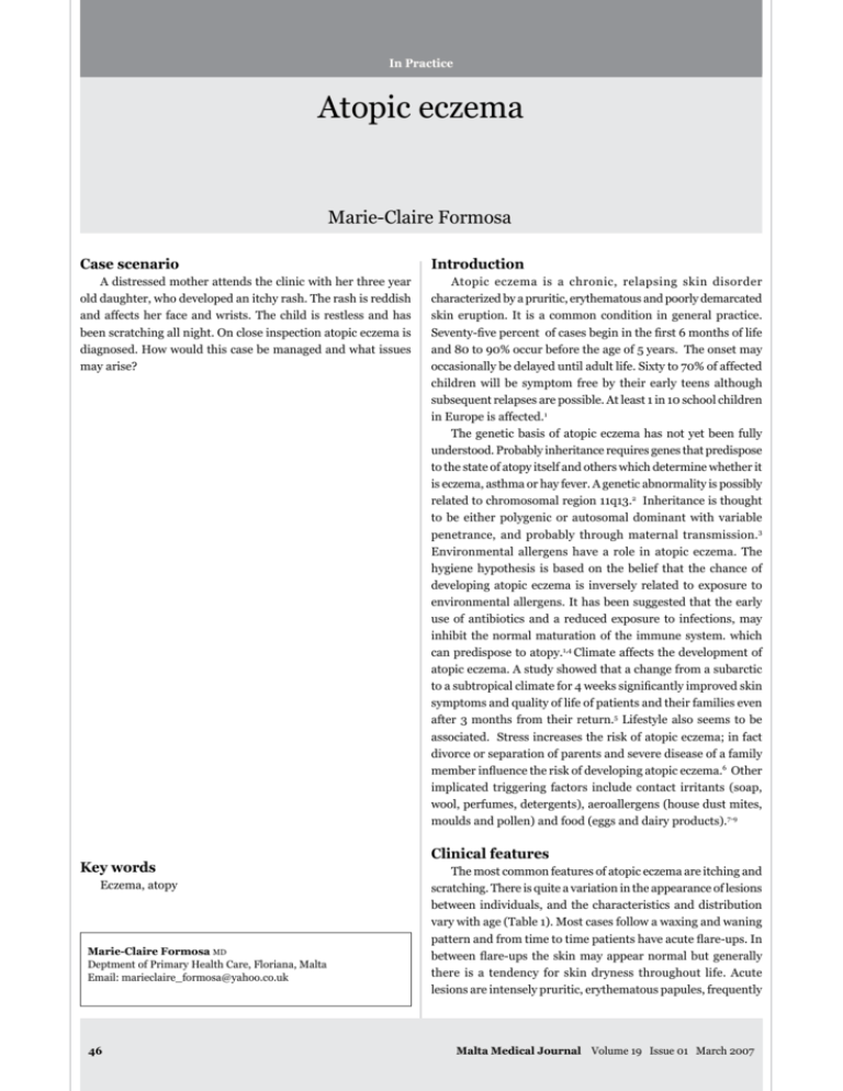
In Practice Atopic eczema Marie-Claire Formosa Case scenario Introduction A distressed mother attends the clinic with her three year old daughter, who developed an itchy rash. The rash is reddish and affects her face and wrists. The child is restless and has been scratching all night. On close inspection atopic eczema is diagnosed. How would this case be managed and what issues may arise? Atopic eczema is a chronic, relapsing skin disorder characterized by a pruritic, erythematous and poorly demarcated skin eruption. It is a common condition in general practice. Seventy-five percent of cases begin in the first 6 months of life and 80 to 90% occur before the age of 5 years. The onset may occasionally be delayed until adult life. Sixty to 70% of affected children will be symptom free by their early teens although subsequent relapses are possible. At least 1 in 10 school children in Europe is affected.1 The genetic basis of atopic eczema has not yet been fully understood. Probably inheritance requires genes that predispose to the state of atopy itself and others which determine whether it is eczema, asthma or hay fever. A genetic abnormality is possibly related to chromosomal region 11q13.2 Inheritance is thought to be either polygenic or autosomal dominant with variable penetrance, and probably through maternal transmission.3 Environmental allergens have a role in atopic eczema. The hygiene hypothesis is based on the belief that the chance of developing atopic eczema is inversely related to exposure to environmental allergens. It has been suggested that the early use of antibiotics and a reduced exposure to infections, may inhibit the normal maturation of the immune system. which can predispose to atopy.1,4 Climate affects the development of atopic eczema. A study showed that a change from a subarctic to a subtropical climate for 4 weeks significantly improved skin symptoms and quality of life of patients and their families even after 3 months from their return.5 Lifestyle also seems to be associated. Stress increases the risk of atopic eczema; in fact divorce or separation of parents and severe disease of a family member influence the risk of developing atopic eczema.6 Other implicated triggering factors include contact irritants (soap, wool, perfumes, detergents), aeroallergens (house dust mites, moulds and pollen) and food (eggs and dairy products).7-9 Key words Eczema, atopy Marie-Claire Formosa MD Deptment of Primary Health Care, Floriana, Malta Email: marieclaire_formosa@yahoo.co.uk 46 Clinical features The most common features of atopic eczema are itching and scratching. There is quite a variation in the appearance of lesions between individuals, and the characteristics and distribution vary with age (Table 1). Most cases follow a waxing and waning pattern and from time to time patients have acute flare-ups. In between flare-ups the skin may appear normal but generally there is a tendency for skin dryness throughout life. Acute lesions are intensely pruritic, erythematous papules, frequently Malta Medical Journal Volume 19 Issue 01 March 2007 Table 1: Age related distribution of atopic eczema Age group Distribution Pattern Infant Face/scalp Non specific distribution elsewhere Itchy/erythematous papules Vesicular/weeping lesions Occasionally hypo pigmentation Small scratch marks Older child Leathery, dry and excoriated Scratching may alter pattern Elbow/knee flexures Wrists/ankles Tends to be symmetrical Mid-teens Limb flexures Hands Remains clear or Localized hand eczema or Generalized low grade eczema Adults as in childhood +/- trunk/face/hands Tendency towards lichenification More widespread but low grade White demographism striking associated with extensive excoriations, erosions and exudates. A sub-acute state, characterized by erythema, excoriations and scaling, is also recognized. Chronic eczema leads to thickened plaques of skin and prominent skin markings.10,11 Atopic eczema also has a non-dermatological clinical scenario, since it greatly affects the quality of life of patients and their families.12 These disturbances range from minor problems to major handicaps. It is essential that the general practitioner recognizes these symptoms since the patients’ mental health and social and emotional functioning seem to be more affected than the physical functioning.13 Sleep disturbances are common. Over 60% of parents participating in a study reported that eczema affected how well they or their child slept. This was directly associated with the severity of the eczema.14 These disturbances can lead to delayed growth and development due to the fact that growth hormone levels rise during stages 3 and 4 of sleep, which may not be reached if sleep is recurrently disturbed. Other sources of anxiety to both the child and the parents include school absences and bullying due to the dermatological features. These may lead to under performance of the child which would generate further anxiety. The emotional impact can become even more significant in the preteen and teen years. This spectrum of symptoms should be considered when offering treatment. Diagnosing atopic eczema Diagnosis is based on assessment and clinical findings. Table 2 illustrates the Williams Criteria for diagnosing atopic eczema.10 Diagnosis is based on identifying one major criterion and three or more minor criteria. Lab tests which could aid diagnosis especially in difficult cases include increased serum levels of IgE and peripheral blood eosinophilia. Skin prick tests are positive for most common allergens. Occasionally a skin biopsy is required. This reveals a state of chronic inflammation, which involves mast cells, lymphocytes, and eosinophils.15 Malta Medical Journal Volume 19 Issue 01 March 2007 Treatment Managing atopic eczema requires a holistic approach. Explanation, reassurance and encouragement to both the patients and the parents are crucial. The pathology and course of the disease should be explained, avoiding scientific jargon which might confuse the patients. It is important to explain that eczema is not contagious and that there is no need to isolate the child from other peers or siblings. Parents should also know what clinical features to look for which would need prompt referral to the doctor, including fever, erythema and warmth around the affected areas, and formation of pus-filled blisters. The concept of allergen and trigger factors avoidance should be explained. Significant improvement can be obtained by simple measures such as wearing cotton underclothes instead of wool, keeping nails short, avoiding extremes of temperature and avoiding direct contact with detergents and other irritant chemicals. Household pets especially cats and dogs should be discouraged.11,16 Patients also benefit from reducing exposure to dust mites. Mattress, pillow and duvet covers and washing bedding at 55oC are effective methods of reducing exposure.17 The role of diet in atopic eczema is conflicting. It is believed that food allergy is a trigger in 10% of cases. A milk free diet is however recommended. Breast feeding is also advised but there is little evidence that it modifies or prevents eczema.15 Follow up visits are of utmost importance to monitor the progress of the disease and to discuss any worries and difficulties encountered. Since eczema is associated with a psychological burden, the family doctor should also be on the look out for symptoms of anxiety and depression in both the children and their parents and offer the necessary treatment. Another important aspect in follow up is keeping a growth chart for children with chronic severe eczema, to detect any growth delays as early as possible. 47 Table 3: Skin type and recommended product18 Table 2: Williams Criteria 10 Skin Type Product Hairy skin Weeping eczema Mildly dry skin Moderately to severe dry skin lotion cream cream or lotion ointment or cream Major Criteria 1) Patient must have an itchy condition or parental report of scratching/rubbing in a child Medical treatment Medical treatment focuses on adequate skin hydration, reducing inflammation and treating secondary infections. Patients and parents of younger patients should be made aware of the wide array of products available. The family doctor should discuss the expectations and concerns associated with the treatment offered. Emollients The most fundamental step in fighting atopic eczema is re-hydration. The principle mode of action of emollients is improving the depleted lipid barrier of the skin thus reducing skin water loss. This reduces dryness and pruritus and increases the skin resistance to irritants. If used regularly, emollients reduce eczema flare ups and the need of topical steroids.18 Emollients are available as lotions, creams and ointments. The lipid content is lowest in lotions and highest in ointments. The higher the lipid content, the greasier and stickier the emollient feels. Choice of emollient should be based on individual preference, body site and level of skin dryness (Table 3).18 Initially the emollient should be applied generously 3 times a day, preferably after bath or showering. Frequency might be increased to every hour if skin is very dry. Topical steroids Topical steroids have been used to control atopic eczema for over 40 years. They are available in different potencies (Table 4). The weakest steroid that controls the eczema should be used. Patients should be reviewed regularly and checked for local and systemic side effects. Elderly people and infants tend to have thinner skin, therefore weaker preparations should be used. The National Institute for Clinical Excellence recommends using a once daily application of topical steroids. This has a lower risk of adverse effects, greater convenience for the patient and lower costs.19 The use of ‘finger tip units’ (FTUs) is recommended. One FTU weighs 500mg and is roughly equivalent to the amount of cream or ointment that can be squeezed from a tube with a standard (5mm) nozzle onto an adult index finger from the finger tip to the first crease. One FTU is sufficient to treat a skin area about twice that of the flat of the hand with the fingers together. The FTUs required depend on the area to be treated and the age of the patient (Table 5).20 48 Plus 3 or more of the following 1) History of itchiness in skin creases such as folds of the elbows, behind knees, front of ankles or around neck (or cheeks if <4years) 2) Personal history of asthma, or hay fever or other atopic disease in a first-degree relative in patients younger than 4 years 3) General dry skin in the last year 4) Visible flexural eczema (or eczema effecting the cheeks or forehead and outer limbs in children younger than 4 years old) 5) Onset in first 2 years of life (not used if child is <4) How should emollients be applied with topical steroids? Inflamed skin should be treated with corticosteroids, whilst the rest of the skin should be treated with emollient. The two products should not be mixed since this would dilute the effect of the corticosteroid. A 30 minutes gap is recommended before applying the emollient to the inflamed skin previously treated with corticosteroids.18 Antibiotics If there is clinical evidence of a bacterial infection, flucloxacillin (or erythromycin if allergic) is the preferred oral antibiotic. Microbiological investigations are recommended if there is no response to the antibiotic but practicality in general practice is debatable. There is no evidence that topical antibiotic and corticosteroid preparations are superior to topical steroids alone. Topical antibiotics should be avoided or reserved for single small lesions only. Also, there is no evidence that bath oils containing antimicrobials are more effective and their routine use is not recommended.21 Table 4: Examples of topical corticosteroids of different potency Potency Example Weak Moderate Strong Very strong 1% Hydrocortisone Clobetasone butyrate (Eumovate®) Bethamethasone 17- valerate (Betnovate®) Hydrocortisone butyrate (Locoid® ) Clobetasol propionate (Dermovate®) Malta Medical Journal Volume 19 Issue 01 March 2007 Table 5: FTUs related to age and area to be treated 20 FTUs/Age 3-6 months 1-2 years 3-5 years Entire area Face and neck 1 1.5 1.5 Arm and hand 1 1.5 2 Leg and foot 1.5 2 3 Chest and abdomen 1 2 3 Back and buttocks 1.5 3 3.5 Calcineurin inhibitors – pimecrolimus and tacrolimus Pimecrolimus and Tacrolimus are topical steroid-free medications with immune modulating and anti-inflammatory properties. They act by down-regulating the mediator release of cytokine expression of various cells which contribute to the inflammation. The National Institute for Clinical Excellence has issued guidelines in August 2004, regarding their use. Calcineurin inhibitors are recommended as an option in adults and children two years and older that have not been controlled by topical corticosteroids and when there is a serious risk of important adverse effects from further use of topical corticosteroids. 22 Preparations are costly, but seem to be safe, with occasional transient burning sensation being the most common side effect. This decreases with usage. Other less common side effects include headache, cough, fever, flu-like symptoms, muscle Table 6: Type of referral related to clinical scenario 34 Emergency referral • Infection with disseminated HSV (Eczema herpeticum) Urgent referral • Severe disease not responding to treatment in primary care • Bacterial infection unresponsive to oral antibiotics +/- topical steroid Referral as soon as possible • Severe social and psychological problems arising from atopic eczema (e.g. sleeplessness and school absenteeism ) • Treatment requiring excessive amounts of potent topical steroids Routine referral • Failure to control symptoms in primary care • If patient or family might benefit from additional advice on application of treatment (e.g. bandaging techniques) • Patch testing if contact dermatitis is suspected • Dietary factors are suspected (Routine referral to a dietician) Malta Medical Journal Volume 19 Issue 01 March 2007 6-10 years 2 2.5 4.5 3.5 5 adults 2.5 4 8 7 7 aches, folliculitis and acne. The risk of developing skin cancer and lymphoma when using these products appears to be very low but it is carefully being monitored. Systemic absorption is low and skin atrophy is not a problem, hence these preparations are suitable for face, neck and flexures.23,24 Antihistamines Oral antihistamines are effective as systemic anti-pruritics, sedatives and mild anxiolytics. Histamine release is not the main cause of the itching, so the newer non-sedating antihistamines help less than might be expected.1 Other treatment options Wet wrap dressing During the last two decades wet wrap dressing has been advocated as a relatively safe and effective treatment option in children with severe and refractory atopic eczema, best used at night time. Large amounts of emollient and sometimes a mild topical steroid are applied under a damp layer of bandage. A second dry layer is then applied on top. As the bandage dries, the skin cools, reducing pruritus. This technique needs to be demonstrated to the patient’s parents. It is contraindicated in the presence of secondary infection. The use of wet wrap dressing with diluted topical corticosteroids for up to 14 days is a safe intervention treatment in children, with temporary systemic bioactivity of the corticosteroid as the only reported serious side effect. 25 Coal tar and ichthammol Coal tar and ichthammol are used for chronic lichenified eczema. They may be applied as crude coal tar, tar-containing creams or in combination with zinc paste. This is not suitable for facial use and side effects include skin irritation, folliculitis and staining. Prebiotics Orally administered prebiotics e.g. Lactobacillus and Bifido bacterium species might help prevent and treat atopic disorders. 26 This is based on the hypothesis that atopic individuals have alterations in their intestinal micro flora as compared to unaffected individuals. Restoring these imbalances would therefore decrease the symptoms. Prebiotics modulate the post-natal development of the immune system via the gut 49 flora. Such modulation could have consequences on reduction of allergies and infections later in life.27 The prebiotic Lactobacillus rhamnosus GG when taken by a woman during pregnancy and then either continued during breast feeding or given to the baby, may help prevent the occurrence of atopic eczema in children with a family history of atopy. 28,29 Experimental treatments Experimental treatments include trials of gamma interferon and interleukin-2.30 These are inhibitors of TH2 cell function, which appear to be hyperactive in atopic eczema. Results when using these treatments are promising. Oral mycophenolate mofetil, an inhibitor of purine synthesis, has also been shown in two small studies to be an effective alternative treatment.31 Complications and associated conditions Patients suffering from atopic eczema have an increased propensity to develop dermatological infections. This is secondary to decreased immunity and reduced barrier function of the skin. Ninety percent of atopic eczema patches are colonized by Staphylococcus areus. It is being evaluated whether this is a cause rather than an effect of atopic eczema. There is also evidence that Malasezzia species are a cause of eczema with head and neck distribution. Patients are also prone to viral infections, including Molluscum contagiosum, warts and most dangerously widespread herpes simplex (eczema herpeticum). Viral infections are not as yet perceived as aetiological factors.32 There are various skin changes associated with atopic eczema. About 20% of patients develop icthyosis vulgaris with associated keratosis piliaris and hyper-linear palms.10 Keratosis piliaris produces a stippled skin appearance on thighs, legs, buttocks and trunks. Hyperlinear palms consist of increased number of fine lines and accentuated markings on the palms. Lichenification of the wrists, ankles, and popliteal fossa is characteristic of chronic eczema. This is observed as thickened, leather, hyper-pigmented skin.10 Atopic cataracts can occur in patients with very severe atopic eczema. These occur much earlier in life than senile cataracts. They are typically bilateral and central. Incidence appears to be unrelated to the use of topical steroids.10 Atopic eczema is also associated with other atopic conditions. Asthma or hay fever is found in about 50% of children with peak onset at 2 and 8 years respectively.33 Referral criteria A number of patients may need specialist care. The National Institute for Clinical Excellence has issued guidelines when this should occur (Table 6).34 50 Prognosis Good prognostic factors include early age of onset and involvement of flexural surfaces. It is believed that more adults will be suffering from atopic eczema in the future, considering that the prevalence amongst children has increased and the fact that atopic eczema in most adults continues for many years.35,36 Conclusion The clinical scenario illustrates a typical presentation of atopic eczema. The family doctor dealing with this case should start by explaining to the mother what atopic eczema is. Other important points to be mentioned during this first encounter include the concept of triggering factors, and what treatments are available. The child would benefit from a mild topical steroid applied daily in conjunction with an emollient cream, applied three times daily preferably after a bath or shower. A sedating anti-histamine administered before bed time is also indicated to help the child sleep better at night. A follow-up appointment should be organized after one week of treatment to assess the clinical improvement and act accordingly. The mother should also be advised to consult the doctor, should any difficulties arise. Atopic eczema is a common chronic dermatological condition which has a considerable psychological burden. It affects not only the suffering child but also other family members. A holistic approach based on both medical and non-medical treatment should be offered by the caring family practitioner to improve the quality of life and make atopic eczema a bearable condition. References 1. Hunter, J.A.A., Savin, J.A., Dahl ,M.V. Clinical Dermatology. 3rd ed. Oxford: Blackwell Science Ltd, 2002, Chapter 7. 2. Coleman, R., Trembath, R.C., Harper, J.I. Chromosome 11q13 and atopy underlying atopic eczema. Lancet 1993;341:1121-22. 3. Millington, G.W. Genetic imprinting and dermatological disease. Clinical and Experimental Dermatology 2006;31: 681-8. 4. Flohr, C., Pascoe, D., Williams, H.C. Atopic Dermatitis and the ‘Hygiene Hypothesis’: Too clean to be true?. British Journal of Dermatology 2005;152:206-16. 5. Byremo, G., Rod, G., Carlsenk, H. Effect of climatic change in children with atopic eczema. Allergy 2006;61:1403-10. 6. Bockelbrink, A., Heinrich, J., Scafer, I., et al. for LISA Study Group. Atopic eczema in children; another harmful sequel of divorce. Allergy 2006;61:1397-402. 7. Aberg, N., Familial occurrence of atopic disease: genetic versus environmental factors. Clinical and Experimental Allergy 1993;23: 829-34. 8. Mc Morran, J., Crowther, D.C., Mc Morran, S., Prince, C., YoungMin, S., Pleat, J., Wacogne, I., Atopic Eczema – Pathogenesis. [Online]. (URL http://www.gpnotebook.co.uk/ simplepage.cfm?ID=496631814&linkID=13808&cook=yes). (Accessed 9 December 2006). 9. Langan, S.M., Williams, H.C. What causes worsening of eczema? A systemic review. British Journal of Dermatology 2006; 155:504-14. Malta Medical Journal Volume 19 Issue 01 March 2007 10.Krafchik B.R., Atopic Dermatits. [Online]. (URL www.emedicine. com/derm/topic38.htm). (Accessed 26 November 2006). 11. Spagnola, C., D Korb, J. Atopic Dermatits. [Online]. (URL www. emedicine.com/ped/topic2567.htm ). (Accessed 9 December 2006). 12.Chamlin, S.L., Frieden, I.J., Williams, M.L., Chren, M.M. Effect of atopic dermatitis on young American children and their families. Paediatrics 2004;114:607-11. 13.Holm, E.A., Wulf, H.C., Stegmann, H., Jemec, G.B.E. Life quality assessment among patients with atopic eczema. British Journal of Dermatology 2006;154: 819-25. 14.Chamlin, S.L., Mattson, C.L., Frieden, I.J. et al. The price of pruritus: sleep disturbance and co sleeping in atopic dermatitis. Archives of Peadiatric and Adolescent Medicine 2005;159:74550. 15.Mc Morran, J., Crowther, D.C., Mc Morran, S., Prince, C., YoungMin, S., Pleat, J., Wacogne, I., Atopoic EczemaInvestigations. [Online]. (URL www.gpnotebook.co.uk/ simplepage.cfm?ID=1376124960 ). (Accessed 9 December 2006). 16.Mc Morran, J., Crowther, D.C., Mc Morran, S., Prince, C., YoungMin, S., Pleat, J., Wacogne, I., Atopic Eczema – General Education. [Online]. (URL www.gpnotebook.co.uk/simplepage. cfm?ID=1456472096). (Accessed 9 December 2006). 17.Charman, C., Clinical evidence: Atopic Eczema. British Medical Journal 1999;318:1600-4. 18.Prodigy – Quick Reference Guide. Emollients. [Online]. (URL www.prodigy.nhs.uk/Eczema-atopic). (Accessed 11 December 2006). 19.National Institute for Clinical Excellence. Quick Reference Guide. Frequency of application of topical corticosteroids for atopic eczema. August 2004. 20. Patient UK – Fingertip Units for Topical Steroids. [Online]. (URL www.patient.co.uk/showdoc/27000762). (Accessed 12 February 2007). 21.Prodigy - Quick Reference Guide. Flare-up in infants and Children. [Online]. (URL www.prodigy.nhs.uk/Eczema-atopic). (Accessed 11 December 2006). 22. National Institute for Clinical Excellence. Quick Reference Guide. Tacrolimus and pimecrolimus for atopic eczema. August 2004. Malta Medical Journal Volume 19 Issue 01 March 2007 23.Wahn, U., Bos, J.D., Goodfield, M. Efficacy and safety of pimecorlimus cream in the long-term management of atopic eczema in children. Paediatirics 2002;110:2-9. 24.Pustisek, N., Lipozenci, J., Gubgevi, S. Tacrolimus ointment: a new therapy for atopic dermatitis - review of literature. Acta Dermatovenerologica Croatica 2002;10:25-32. 25.Devillers, A.C.A., Oranje, A.P. Efficacy and safety of ‘wet wrap’ dressing as an intervention treatment in children with severe and/ or refractory atopic dermatitis: A critical review of the literature. British Journal of Dermatology 2006;154:579-85. 26.Drugs and Therapeutics Bulletin 2005;43:6-8. 27.Rizzo, M. Prebiotics reduce the risk of atopic dermatitis. Archives of Disease in Childhood 2006;91:814-9. 28.Rautava, S., Kalliomaki, M., Isolauri, E. Prebiotics during pregnancy and breast-feeding might confer immunomudulatory protection against atopic disease in infants. Journal of Allergy and Clinical Immunology 2002;109:119-21. 29.Kalliomaki, M., Salminen, S., Poussa, T., Arvilommi, H., Isolauri E. Prebiotics and prevention of atopic disease: 4 year follow up of a randomised placebo-controlled trial. Lancet 2003:361:1869-71. 30.Chang, T.T., Stevens, S.R. Atopic dermatitis: the role of recombinant interferon-gamma therapy. American Journal of Clinical Dermatology 2002:;3:175-83. 31.Grundmann-Kollmann, M., Kaufmann, R., Zollner, T.M. Treatment of atopic dermatitis with mycophenolate mofetil. British Journal of Dermatology 2001; 145:351-2. 32.Lubbe, J. Secondary infections in patients with atopic dermatitis. American Journal of Clinical Dermatology 2003;4:641-54. 33.Mc Morran, J., Crowther, D.C., Mc Morran, S., Prince, C., YoungMin, S., Pleat, J., Wacogne, I., Atopic Eczema – Associated conditions. [Online]. (URL www.gpnotebook.co.uk/simplepage. cfm?ID=1355808800). (Accessed 9 December 2006). 34.National Institute of Clinical Excellence. Referral Practice A guide to appropriate referral from general practice to specialist services. May 2000. 35.Williams, H.C., Strachan, D..P. The Natural history of childhood eczema from the British 1958 birth cohort study. British Journal of Dermatology 1998;139:834-9. 36.Sandstrom Falk, M.H., Faergermann, J. Atopic dermatitis in adults; does it disappear with age? Acta Dermato-Venereologica 2006;86:135-9. 51
