REVIEWS
The origin of extracellular fields and
currents — EEG, ECoG, LFP and spikes
György Buzsáki1,2,3, Costas A. Anastassiou4 and Christof Koch4,5
Abstract | Neuronal activity in the brain gives rise to transmembrane currents that can be
measured in the extracellular medium. Although the major contributor of the extracellular
signal is the synaptic transmembrane current, other sources — including Na+ and Ca2+
spikes, ionic fluxes through voltage- and ligand-gated channels, and intrinsic membrane
oscillations — can substantially shape the extracellular field. High-density recordings of
field activity in animals and subdural grid recordings in humans, combined with recently
developed data processing tools and computational modelling, can provide insight into
the cooperative behaviour of neurons, their average synaptic input and their spiking
output, and can increase our understanding of how these processes contribute to the
extracellular signal.
Center for Molecular and
Behavioural Neuroscience,
Rutgers, The State University
of New Jersey,
197 University Avenue,
Newark, New Jersey 07102,
USA.
2
New York University
Neuroscience Institute, New
York University Langone
Medical Center, New York,
New York 10016, USA.
3
Center for Neural Science,
New York University, New
York, New York 10003, USA.
4
Division of Biology,
California Institute of
Technology, 1200 East
California Boulevard,
Pasadena, California 91125,
USA.
5
Allen Institute for Brain
Science, 551 North 34th
Street, Seattle, Washington
98103, USA.
Correspondence to G.B.
e-mail: gyorgy.buzsaki@
nyumc.org
doi:10.1038/nrn3241
1
Electric current contributions from all active cellular
processes within a volume of brain tissue superimpose
at a given location in the extracellular medium and
generate a potential, Ve (a scalar measured in Volts),
with respect to a reference potential. The difference in
Ve between two locations gives rise to an electric field
(a vector whose amplitude is measured in Volts per
distance) that is defined as the negative spatial gradient
of Ve. Electric fields can be monitored by extracellularly
placed electrodes with submillisecond time resolution
and can be used to interpret many facets of neuronal
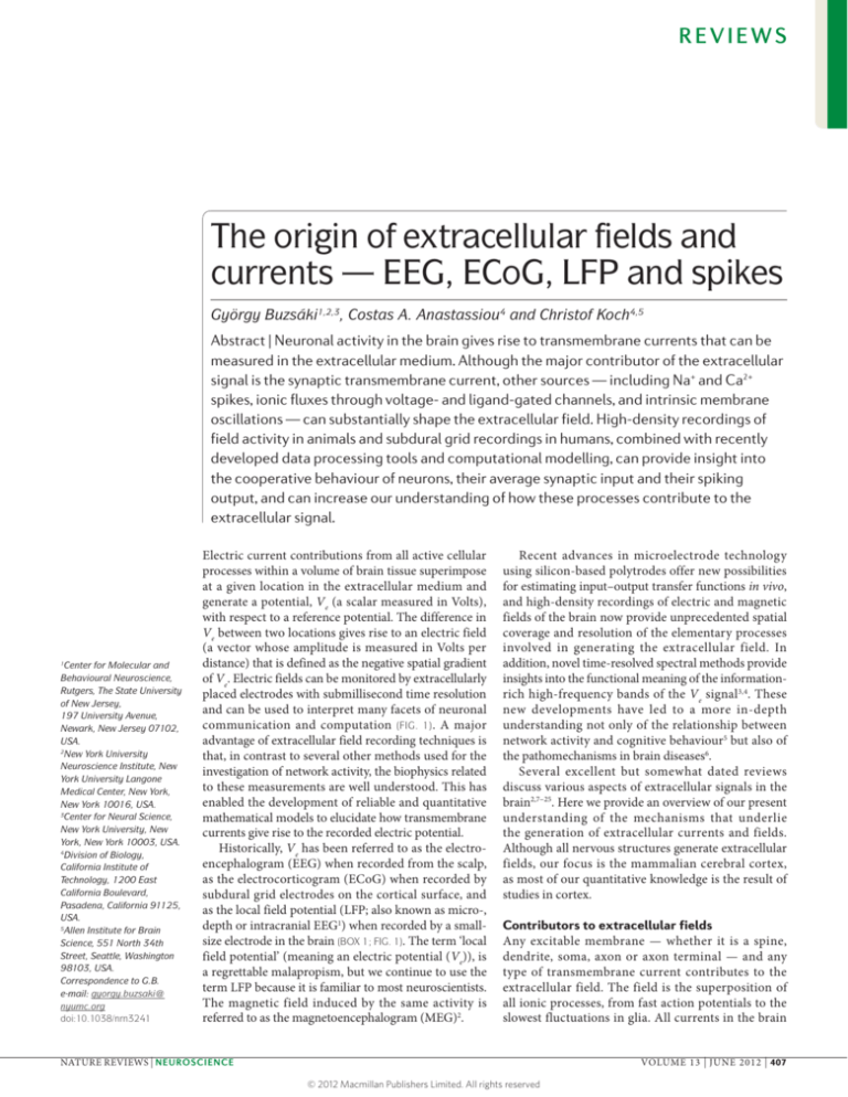
communication and computation (FIG. 1). A major
advantage of extracellular field recording techniques is
that, in contrast to several other methods used for the
investigation of network activity, the biophysics related
to these measurements are well understood. This has
enabled the development of reliable and quantitative
mathematical models to elucidate how transmembrane
currents give rise to the recorded electric potential.
Historically, Ve has been referred to as the electroencephalogram (EEG) when recorded from the scalp,
as the electrocorticogram (ECoG) when recorded by
subdural grid electrodes on the cortical surface, and
as the local field potential (LFP; also known as micro-,
depth or intracranial EEG1) when recorded by a smallsize electrode in the brain (BOX 1; FIG. 1). The term ‘local
field potential’ (meaning an electric potential (Ve)), is
a regrettable malapropism, but we continue to use the
term LFP because it is familiar to most neuroscientists.
The magnetic field induced by the same activity is
referred to as the magnetoencephalogram (MEG)2.
Recent advances in microelectrode technology
using silicon-based polytrodes offer new possibilities
for estimating input–output transfer functions in vivo,
and high-density recordings of electric and magnetic
fields of the brain now provide unprecedented spatial
coverage and resolution of the elementary processes
involved in generating the extracellular field. In
addition, novel time-resolved spectral methods provide
insights into the functional meaning of the informationrich high-frequency bands of the Ve signal3,4. These
new developments have led to a more in-depth
understanding not only of the relationship between
network activity and cognitive behaviour5 but also of
the pathomechanisms in brain diseases6.
Several excellent but somewhat dated reviews
discuss various aspects of extracellular signals in the
brain2,7–25. Here we provide an overview of our present
understanding of the mechanisms that underlie
the generation of extracellular currents and fields.
Although all nervous structures generate extracellular
fields, our focus is the mammalian cerebral cortex,
as most of our quantitative knowledge is the result of
studies in cortex.
Contributors to extracellular fields
Any excitable membrane — whether it is a spine,
dendrite, soma, axon or axon terminal — and any
type of transmembrane current contributes to the
extracellular field. The field is the superposition of
all ionic processes, from fast action potentials to the
slowest fluctuations in glia. All currents in the brain
NATURE REVIEWS | NEUROSCIENCE
VOLUME 13 | JUNE 2012 | 407
© 2012 Macmillan Publishers Limited. All rights reserved
REVIEWS
800 µV 150 µV
b
a
Cz
Depth
(LFP)
SM
EC
Grid
(ECoG)
Strip
(ECoG)
HC
Strip
(ECoG)
Am
Fz
Scalp
EEG
O2
1s
1s
c
100
500
0
0
–500
d
–100
1s
–1000
iEEG (µV)
MEG (fT)
1000
LFP surface
I
II
III
Intracellular
V
20 mV
IV
LFP depth
VI
Figure 1 | Extracellular traces using different recording methods are
fundamentally similar. a | Simultaneous recordings from
three Reviews
depth electrodes
(two
Nature
| Neuroscience
selected sites each) in the left amygdala and hippocampus (measuring the local field
cortex (measuring the electrocorticogram (ECoG); two four-contact strips placed under
the inferior temporal surface (measuring the ECoG); an eight-contact strip placed
over the left orbitofrontal surface (measuring the ECoG); and scalp electroencephalography (EEG) over both hemispheres (selected sites are the Fz and O2) in a patient with
drug-resistant epilepsy. The amplitude signals are larger and the higher-frequency
patterns have greater resolution at the intracerebral (LFP) and ECoG sites compared to
scalp EEG. b | A 6 s epoch of slow waves recorded by scalp EEG (Cz, red), and LFP (blue)
recorded by depth electrodes placed in the deep layers of the supplementary motor area
(SM) and entorhinal cortex (EC), hippocampus (HC) and amygdala (Am). Also shown are
multiple-unit activity (green) and spikes of isolated neurons (black ticks). c | Simultaneously
recorded magnetoencephalogram (MEG; black) and anterior hippocampus depth EEG
(red) from a patient with drug-resistant epilepsy. Note the similar theta oscillations
recorded by the depth electrode and the trace calculated by the MEG, without any phase
delay. d | Simultaneously recorded LFP traces from the superficial (‘surface’) and deep
(‘depth’) layers of the motor cortex in an anaesthetized cat and an intracellular trace
from a layer 5 pyramidal neuron. Note the alternation of hyperpolarization and
depolarization (slow oscillation) of the layer 5 neuron and the corresponding changes in
the LFP. The positive waves in the deep layer (close to the recorded neuron) are also
known as delta waves. iEEG, intracranial EEG. Part a courtesy of G. Worrell, Mayo Clinic,
Minneapolis, Minnesota, USA, and S. Makeig, University of California at San Diego, USA.
Part b is reproduced, with permission, from REF. 157 © (2011) Cell Press. Part c courtesy of
S. S. Dalal, University of Konstanz, Germany, and J.-P. Lachaux and L. Garnero, Université
de Paris, France. Part d is reproduced, with permission, from REF. 158 © (1995) Society
for Neuroscience.
superimpose at any given point in space to yield Ve
at that location. Thus, any transmembrane current,
irrespective of its origin, leads to an intracellular as
well as an extracellular (that is, LFP) voltage deflection.
The characteristics of the LFP waveform, such as the
amplitude and frequency, depend on the proportional
contribution of the multiple sources and various
properties of the brain tissue. The larger the distance
of the recording electrode from the current source, the
less informative the measured LFP becomes about the
events occurring at the location(s) of the source(s). This
is mainly owing to the fact that the Ve amplitude scales
with the inverse of the distance r between the source
and the recording site, and to the inclusion of other
(interfering) signals (leading to ‘spatial averaging’). In
addition to the magnitude and sign of the individual
current sources, and their spatial density, the temporal
coordination of the respective current sources (that
is, their synchrony) shapes the extracellular field.
Thus, extracellular currents can emerge from multiple
sources, and these are described below.
Synaptic activity. In physiological situations,
synaptic activity is often the most important source
of extracellular current flow. The idea that synaptic
currents contribute to the LFP stems from the
recognition that extracellular currents from many
individual compartments must overlap in time to induce
a measurable signal, and such overlap is most easily
achieved for relatively slow events, such as synaptic
currents7,10,23. The dendrites and soma of a neuron form
a tree-like structure with an electrically conducting
interior that is surrounded by a relatively insulating
membrane, with hundreds to tens of thousands of
synapses located along it. Neurotransmitters acting on
synaptic AMPA and NMDA receptors mediate excitatory
currents, involving Na + or Ca 2+ ions, respectively,
which flow inwardly at the synapse. This influx of
cations from the extracellular into the intracellular
space gives rise to a local extracellular sink. To achieve
effective electroneutrality within the time constants of
relevance for systems neuroscience, the extracellular
sink needs to be ‘balanced’ by an extracellular source,
that is, an opposing ionic flux from the intracellular to
the extracellular space, along the neuron; this flux is
termed passive current or return current. Depending on
the location of the sink current(s) and its distance from
the source current(s), a dipole or a higher-order n-pole
is formed (FIG. 2a). The contribution of a monopole to
Ve scales as 1/r, whereas the contribution of a dipole
decays faster, as 1/r2; this steeper decay is due to the two
opposing charges that comprise the dipole cancelling
each other out to first order.
Notably, GABA subtype A (GABA A) receptormediated inhibitory currents are typically assumed
to add very little to the extracellular field as the Cl –
equilibrium potential is close to the resting membrane
potential26,27. However, in actively spiking neurons the
membrane is depolarized, and therefore inhibitory (and
often hyperpolarizing) currents can generate substantial
transmembrane currents28–30 (FIG. 2b,c).
408 | JUNE 2012 | VOLUME 13
www.nature.com/reviews/neuro
© 2012 Macmillan Publishers Limited. All rights reserved
REVIEWS
Fast action potentials. Fast (Na+) action potentials generate the strongest currents across the neuronal membrane and can be detected as ‘unit’ or ‘spike’ activity in
Box 1 | Recordings methods of extracellular events
Electroencephalography
Electroencephalography (EEG) is one of the oldest and most widely used methods for
the investigation of the electric activity of the brain10,16. The scalp electroencephalogram, recorded by a single electrode, is a spatiotemporally smoothed version of the local
field potential (LFP), integrated over an area of 10 cm2 or more. Under most conditions, it
has little discernible relationship with the firing patterns of the contributing individual
neurons16, and this is largely due to the distorting and attenuating effects of the soft and
hard tissues between the current source and the recording electrode. The recently
introduced ‘high-density’ EEG recordings, in combination with source-modelling that
can account for the gyri and sulci (as inferred from structural MRI imaging) of the
subject, have substantially improved the spatial resolution of EEG16,146,147.
Magnetoencephalography
Magnetoencephalography (MEG) uses superconducting quantum interference devices
(SQUIDs) to measure tiny magnetic fields outside the skull (typically in the 10–1,000 fT
range) from currents generated by the neurons2. Because MEG is non-invasive and has a
2
, it has
become a popular method for monitoring neuronal activity in the human brain. An
advantage of MEG is that magnetic signals are much less dependent on the
conductivity of the extracellular space than EEG. The scaling properties (that is, the
frequency versus power relationship) of EEG and MEG often show differences, typically
in the higher-frequency bands. These differences may be partly explained by the
capacitive properties of the extracellular medium (such as skin and scalp muscles) that
distort the EEG signal but not the MEG signal148.
Electrocorticography
Electrocorticography (ECoG) is becoming an increasingly popular tool for studying
various cortical phenomena in clinical settings149. It uses subdural platinum–iridium or
stainless steel electrodes to record electric activity directly from the surface of the
cerebral cortex, thereby bypassing the signal-distorting skull and intermediate tissue.
The spatial resolution of the recorded electric field can be substantially improved
(<5 mm2)102 by using flexible, closely spaced subdural grid or strip electrodes (FIG. 1).
Local field potential
EEG, MEG and ECoG mainly sample electrical activity that occurs in the superficial
layers of the cortex. Electrical events at deeper locations can be explored by inserting
metal or glass electrodes, or silicon probes into the brain to record the LFP (also known
contains both action potentials and other membrane potential-derived fluctuations in a
studying cortical electrogenesis. Many observation points, with short distances
between the recording sites and with minimal impact on brain tissue, are needed to
achieve high spatial resolution. In principle, the spiking activity of nearly all or at least a
representative fraction of the neuron population in a small volume can be monitored
with a sufficiently large density of recording sites. Additional clues about the
intracellular dynamics can be deduced from the waveform changes of the
extracellular action potentials99,150. Progress in this field has been accelerated by the
availability of micro-machined silicon-based probes with ever-increasing numbers of
recording sites ,151,152.
Voltage-sensitive dye imaging
Voltage changes can also be detected by membrane-bound voltage-sensitive dyes or
by genetically expressed voltage-sensitive proteins
. Using the voltage-sensitive
dye imaging (VSDI) method, the membrane voltage changes of neurons in a region of
interest can be detected optically, using a high-resolution fast-speed digital camera, at
the peak excitation wavelength of the dye. A major advantage of VSDI is that it directly
potential. A second advantage is that the provenance of the signal can be identified if a
known promoter is used to express the voltage-sensitive protein. Limitations are
inherent in all optical probe-based methods156, and for VSDI these include interference
with the physiological functions of the cell membrane, photoxicity, a low
signal-to-noise ratio and the fact that it can only measure surface events.
the extracellular medium27. Although Na+ spikes generate large-amplitude Ve deflections near the soma (FIG. 2d),
until recently they were thought not to contribute substantially to the traditionally considered LFP band
(<100 Hz) or to the scalp-recorded EEG10,16, because
the strongest fields they generate are of short duration
(<2 ms) and nearby neurons rarely fire synchronously
in such short time windows under physiological conditions31. However, synchronous action potentials from
many neurons can contribute substantially to highfrequency components of the LFP. Therefore, with
appropriate methods, valuable information can be
extracted from the LFP about the temporal structure of
spiking neuronal populations (see below).
Calcium spikes. Other non-synaptic events that can
contribute prominently to the extracellular field are
the long-lasting (10–100 ms) Ca2+-mediated spikes32.
Because voltage-dependent regenerative Ca2+ spikes are
often triggered by NMDA receptor-mediated excitatory
postsynaptic potentials (EPSPs) 33 – 36 , separating
them from EPSPs in extracellular recordings is not
straightforward. A potential differentiating factor is that,
in contrast to EPSPs, Ca2+ spikes can actively propagate
within the cell and can therefore generate fields across
the laminar boundaries of afferent inputs. Ca2+ spikes
can also be triggered by back-propagating somatic
action potentials37, in which case they are independent
of synaptic activity. Because dendritic Ca2+ spikes are
large (10–50 mV) and long lasting37–39, their share in the
measured extracellular events can be substantial under
certain circumstances (FIG. 3). Unfortunately, very little
is known about Ca2+ spikes in vivo40.
Intrinsic currents and resonances. I h currents and
I T currents are prominent examples of intrinsic,
v o l t a g e - d e p e n d e nt m e m b r a n e r e s p o n s e s 3 9 .
Although synaptically induced voltage changes are a
prerequisite for the activation of voltage-dependent
hyperpolarization-activated cyclic nucleotide (HCN)gated and T-type calcium channels, the large membrane
and extracellular currents that these channels generate
are not synaptic events. These and other voltagegated currents contribute to intrinsic resonance and
oscillation of the membrane potential. Several neuron
types possess resonant properties; that is, they respond
more effectively to inputs of a particular frequency
range39. When intracellular depolarization is sufficiently
strong, the resonant property of the membrane can
give way to a self-sustained oscillation of the voltage.
Voltage-dependent resonance and oscillations at theta
frequency have been described in principal neurons of
several cortical regions39,41–44. By contrast, perisomatic
inhibitory interneurons have a preferred resonance in
the gamma frequency (30–90 Hz) range45,46. Because
resonance is both voltage- and frequency-dependent39,41,
its impact on the magnitude of the extracellular field can
vary in a complex manner. To contribute substantially to
the LFP, resonant membrane potential fluctuations must
occur synchronously in nearby neurons, a feature that
most often occurs in inhibitory interneurons.
NATURE REVIEWS | NEUROSCIENCE
VOLUME 13 | JUNE 2012 | 409
© 2012 Macmillan Publishers Limited. All rights reserved
REVIEWS
a
d
By convention, a site on the
neuronal membrane where
positive charges enter the
neuron.
Normalized power
1 nA
Electroneutrality
The phenomenon that, owing
to charge conservation, at any
given point in time the total
charge entering and leaving the
cell across all of its membrane
equals zero.
Dipole
An ideal electric dipole is
defined by two charges of
opposite polarity with infinitely
small separation, such that the
product of the charge times the
distance r separating them
remains finite. The electric
potential of a dipole falls off
as 1/r2.
Equilibrium potential
The voltage difference between
intracellular and extracellular
space of a neuron when the net
ionic flux across the membrane
equals zero.
Ih currents
Currents flowing through
hyperpolarization deinactivated
cyclic nucleotide-gated channels.
IT currents
Low-threshold
(hyperpolarization-induced)
transient Ca2+ currents, which
often lead to burst firing.
Resonance
A property of the neuronal
membrane to respond to some
input frequencies more
strongly than others. At the
resonant frequency, even weak
periodic driving can produce
large-amplitude oscillations.
Silicon probes
Multiple-site recording
electrodes for high spatial
density monitoring of the
extracellular field. The
recordings sites can record Ve
along one, two or even three
orthogonal axes.
µV
1
0.1
10–2
10–3
101
102
Frequency (Hz)
103
400
200
0
–30 –15
–70.5
0
ms
15
30
300
200
100
0
–100
–200
SLM
10 µV
SR
100 µm
–70.0
400
SP
SO
–0.4 0.0 0.4 0.8
40 mV
5 ms
50 V
600
A loop current that flows in the
opposite direction to an active
sink or source.
10
c
b
Return current
100
10–1
10–4 0
10
Vm
Locations along the neuronal
membrane where positive
charge flows out of the neuron.
For negative charge, the
location of sinks and sources is
inverted.
0.1 µV
Count
Sources
Membrane potential
100
Distance from SP (µm)
Sink
Amplitude (normalized)
Figure 2 | Excitatory and inhibitory postsynaptic currents are the most ubiquitous contributors to Ve.
Nature Reviews | Neuroscience
a | Computer-simulated local field potential (LFP) traces (left panel; grey) in response to an excitatory synaptic current
input (a sink, shown by the blue circle) injected into the distal apical dendrite of a purely passive layer 5 pyramidal model
neuron. The waveform of the injected current is illustrated in the box. Red and blue contour lines correspond to positive
and negative values for the LFP amplitude, respectively. The calculated double logarithmic power spectra of the
transmembrane potential are also shown (right panel), following injection of current into the apical dendrite near the
injection site (blue trace), mid-apical dendrite (green trace) and soma (orange trace). Note that high-frequency activity
decreases with the distance from the active synaptic site (that is, the sink). b | A monosynaptic inhibitory connection
between a putative layer 3 entorhinal cortical interneuron (red circle) and intracellularly recorded pyramidal cell (blue
triangle). Below it, a cross-correlogram between the spikes of the reference interneuron (at time 0, red line) and the
pyramidal cell and, superimposed on it, the spike-triggered average of the membrane potential (Vm) of the pyramidal cell
(in blue). Note the small, short-latency hyperpolarization (the dip) superimposed on the rising phase of the intracellular
theta oscillation and the corresponding decreased spike discharge of the pyramidal cell. c | Inhibition-induced LFPs. LFPs
were generated in the vicinity of a pyramidal neuron (bottom cell) by intracellularly induced action potentials in a nearby
basket cell (top cell), and were recorded extracellularly at six sites in multiple layers of the hippocampus. The mean LFP
amplitude at each site is shown by the blue squares. Example LFP traces (blue) from six sites and the action potential of the
basket cell (red trace) are shown on the right. Note that the largest positive response by inhibition-induced
hyperpolarization occurs near the soma. d | Extracellular contribution of an action potential (‘spike’) to the LFP in the
vicinity of the spiking pyramidal cell. The magnitude of the spike is normalized. The peak-to-peak voltage range is
indicated by the colour of the traces. Note that the spike amplitude decreases rapidly with distance from the soma,
without a change in polarity within the pyramidal layer (the approximate area of which is shown by the box), in contrast to
the quadrupole (that is, reversed polarity signals both above and below the pyramidal layers) formed along the
somatodendritic axis. The distance-dependence of the spike amplitude within the pyramidal layer is shown (bottom left
panel) with voltages drawn to scale, using the same colour identity as the traces in the boxed area in d. The same traces are
shown normalized to the negative peak (bottom right panel). Note the widening of the spike with distance from the soma,
owing to greater contributions from dendritic currents and intrinsic filtering of high-frequency currents by the cell
membrane. SLM, stratum lacunosum moleculare; SO, stratum oriens; SP, stratum pyramidale; SR, stratum radiatum. Part a
is reproduced, with permission, from REF. 83 © (2010) Springer. Part b is reproduced, with permission, from REF. 137 ©
(2010) Society for Neuroscience. Part c is reproduced from REF. 29 © (2009) Macmillan Publishers Ltd. All rights reserved.
Part d courtesy of E. W. Schomburg, California Institute of Technology, USA.
Spike afterhyperpolarizations and ‘down’ states.
Elevation of the intracellular concentration of a certain
ion may trigger influx of other ions through activation of
ligand-gated channels, and this will in turn contribute to
Ve. For example, bursts of fast spikes and associated dendritic Ca2+ spikes are often followed by hyperpolarization
of the membrane, owing to activation of a Ca2+-mediated
increase of K+ conductance in the somatic region47.
As the amplitude and duration of such burst-induced
afterhyperpolarizations (AHPs) can be as large (and last
as long as) synaptic events, AHPs also contribute to the
extracellular field48, particularly when bursting of nearby
neurons occurs in a temporally coordinated fashion: for
example, following hippocampal sharp-wave events49.
In the intact brain, responses to unexpected stimuli or
movement initiation are often associated with relatively
long-lasting (0.5–2 s) LFP shifts, which might be mediated by synchronized AHPs. This slow LFP is often
410 | JUNE 2012 | VOLUME 13
www.nature.com/reviews/neuro
© 2012 Macmillan Publishers Limited. All rights reserved
REVIEWS
A
Ba
Extra
0.2 mV
50 mV
Bb
50 ms
2s
Bc
Bd
–50 mV Intra
–70 mV
C Vdend
Agar
ECoG
Pia
1
2/3 Vdend
4
5
D
D1
D2
D3
10 mV
∆ F/F
D4
50%
10 ms
ECoG spike
amplitude (mV)
Cover-glass
1.0
0.5
0.0
0
25
50
Slow potential
amplitude (mV)
E
473 nm; 140 µW
593 nm; 220 µW
10 ms
100 ms
200 µV
Figure 3 | Non-synaptic contributions to the LFP. Ca spikes, disfacilitation and
disinhibition contribute to the local field potential (LFP). A | Voltage-dependence of a
theta-frequency oscillation in a hippocampal pyramidal cell dendrite
. A continuous
recording of extracellular (extra) and intradendritic (intra) activity in a hippocampal CA1
pyramidal cell is shown. The holding potential was manually shifted to progressively more
depolarized levels by intradendritic current injection. The recording electrode contained
QX-314 to block Na+ spikes. Note the large increase in the amplitude of the intradendritic
theta oscillation upon depolarization. Arrows, putative high-threshold Ca2+ spikes
phase-locked to the LFP theta oscillation. Ba | Dendritic Ca2+ spikes (shown by an arrow)
have a large amplitude and are long-lasting
. Bb–Bd | The response of a CA1 pyramidal
cell to ventral hippocampal commissural stimulation (vertical arrows) paired with dendritic
depolarization. Such inhibition can delay (Bb), prevent (Bc) or abort (Bd) the dendritic Ca2+
spike. LFPs recorded from a nearby electrode in the pyramidal layer show the timing and
magnitude of the stimulation (lower traces in Bb–Bd). Note that the number of Na2+ spikes
remains approximately the same, irrespective of the presence or absence of the Ca2+ spike.
C | Whisker stimulation-evoked dendritic Ca2+ spikes correlate with surface cortical LFP
changes. The setup for recording the electrocorticogram (ECoG), intradendritic potential
(Vdend) and Ca2+ fluorescence is shown in the left panel. The relationship between the
intradendritic potential amplitude (horizontal arrows) and simultaneously measured Ca2+
F/F) is shown in the middle panel. The ECoG response as a function of the Ca2+ spike
(‘slow potential’) amplitude is shown in the right panel. D | ‘Down’ states in cortical
pyramidal cells during sleep produce extracellular LFP ‘delta’ waves. Shown are
simultaneously recorded LFP (top) and unit activity (bottom) at three layer 5 intracortical
2+
down states (shaded areas), reflected as positive waves (delta waves) in the LFP, can be
either strongly localized (in D2 and D3) or more widespread (in D1 and D4). E | Generation
of extracellular potentials by depolarization or hyperpolarization of a limited number of
CA1 neurons that express both channelrhodopsin 2 (ChR2) and halorhodopsin, in response
to blue (top) and yellow (bottom) light
. Note the depolarization-induced negative
LFP (top) and the hyperpolarization-induced positive LFP (bottom) in the pyramidal layer.
Part A is reproduced, with permission, from REF. 159 ©
B is reproduced,
with permission, from REF. 160 © (1996) National Academy of Sciences. Part C is
reproduced from REF. 161 © (1999) Macmillan Publishers Ltd. All rights reserved. Part D is
reproduced, with permission, from REF. 56 © (2005) Cambridge Journals. Part E courtesy of
E. Stark, New York University, Langone Medical Center, USA.
referred to as Bereitschaftspotential50, readiness potential
or contingent negative variation51.
During non-rapid eye movement (non-REM) sleep,
the membrane potential of cortical neurons periodically
shifts (0.5–1.5 Hz) between a hyperpolarized ‘down’
state and a more depolarized ‘up’ (that is, spiking)
state52 (FIGS 1d,3D). At least part of the cessation of
spiking during the down states can be explained by
AHPs of the synchronously bursting pyramidal cells
in the up state48,53. The temporally coordinated silent
down state of nearby neurons is associated with a
positive Ve in infragranular layers and a negative Ve
in the supragranular layers (these down states are also
known as delta waves 48,54–56). Various mechanisms
contribute to these state transitions, including a
gradual decrease in extracellular Ca 2+ concentration
and a corresponding decrease in synaptic transmission,
inactivation of I h channels 53,57, and other network
effects52. As the largest-amplitude up–down shifts of
the membrane voltage occur in large layer 5 pyramidal
neurons53,58, it has been suggested that the large voltage
shifts in the somata of the synchronously active–silent
neurons induce the formation of an extracellular
dipole between deep (infragranular) and superficial
(supragranular) layers48,58. Neither interneurons nor the
thalamocortical inputs are active during the down state,
so that the down state (characterized by delta waves) is a
disfacilitatory, non-synaptic event that can be mimicked
by synchronous hyperpolarization of nearby pyramidal
neurons (FIG. 3E).
Gap junctions and neuron–glia interactions. Direct
electric communication between neurons through
gap junctions (also known as electrical ‘synapses’)59–61
can enhance neuronal synchrony49,62,63. Although gap
junctions allow ionic movement across neurons and,
therefore, do not involve any extracellular current flow,
they can affect neuronal excitability and contribute
indirectly to the extracellular field.
Membrane potential changes in non-neuronal cells,
such as glia, may also give rise to Ve. Recent studies on
neuron–glia interactions have indicated that the glial
syncytium may contribute to slow and infraslow (<0.1 Hz)
field patterns1,64,65. These slow LFPs may arise from glia,
glia–neuron interactions or from vascular events66–68.
Ephaptic effects. Neurons are surrounded by a
conducting medium — the extracellular space —
and can therefore ‘sense’ the electric gradients they
generate during neuronal processing. In fact, the
effect of gradients brought about by synchronous
population activity along cable-like dendrites can
be mimicked by appropriate intracellular current
injections69,70. This raises the question of whether the
spatiotemporal field fluctuations in the brain are merely
an epiphenomenon of coordinated cellular activity or
whether they also have a functional ‘feedback’ (or even
amplification) role by affecting the discharge properties
of neurons71. That is, do they serve any function for
the organism or are they like the heartbeat, a useful
diagnostic epiphenomenon? Given the resistivity of
NATURE REVIEWS | NEUROSCIENCE
VOLUME 13 | JUNE 2012 | 411
© 2012 Macmillan Publishers Limited. All rights reserved
REVIEWS
the extracellular medium in the mammalian brain
and the highly transient nature of spikes, it is unlikely
that spikes from individual neurons greatly affect the
excitability of nearby neurons through ephaptic coupling.
However, the situation is very different when many
neurons are simultaneously active, as such synchrony
can generate strong spatial gradients in the extracellular
voltage. Experiments have shown that small-amplitude,
slow-frequency application of extracranial currents
(trans-cranial electrical stimulation) has a detectable
effect on neuronal activity72 and cognitive function73;
the small but effective voltage gradients brought about
in brain tissue by such external fields are comparable to
the voltage gradients produced by population patterns
in vivo under physiological conditions70,74–76. Ephaptic
coupling has been shown to affect population activity
during hypersynchronous epileptic discharges 77,78.
Furthermore, ephaptic feedback may enhance spike–
field coherence and bias the preferred spiking phases
with respect to the LFP also under physiological
conditions75,76,79–81; for example, during hippocampal
sharp waves or theta waves70,76,77.
Ephaptic coupling
The effect of the extracellular
field on the transmembrane
potential of a neuron.
Open field
When the sink (or the source) is
substantially spatially
separated from the return
currents of the dipole.
Closed field
When the sink (or the source) is
minimally spatially separated
from the return currents of the
dipole.
Power law (of LFP)
The power law of LFP describes
a relationship between the
amplitude of the extracellular
signal and its temporal
frequency. A descending
straight line on the log–log plot
(power versus frequency)
would be an indication of a
power law that scales as 1/fn.
Low-pass frequency filtering
A process by which the
frequency components of a
signal beyond a cutoff
frequency are increasingly
attenuated, typically owing to
a serial capacitance (for
example, the bi-lipid
membrane).
Neuronal geometry and architecture
All neuron types contribute to the extracellular field,
but their relative contribution depends in part on the
shape of the cell. Pyramidal cells are the most populous
cell type. They have long, thick apical dendrites that
can generate strong dipoles along the somatodendritic
axis. Such dipoles give rise to an open field, as there is
considerable spatial separation of the active sink (or
the source) from the return currents. This induces
substantial ionic flow in the extracellular medium
(FIG. 2). Therefore, neurons that generate open fields,
such as pyramidal cells, make a sizeable contribution
to the extracellular field. By contrast, spherically
symmetric neurons — such as thalamocortical cells
— that emanate dendrites of relatively equal size in all
directions, can give rise to a closed field82. However, a
strictly closed field only occurs when several dendrites
are simultaneously activated. As this is rarely the case,
depolarization of a single dendrite generates a small
dipole even in spherically symmetric cells83.
Assuming a homogeneous medium, the two most
important determinants of the extracellular field strength
are the spatial alignment of neurons and the temporal
synchrony (discussed in the next section) of the dipole
moments they generate13,22,84. In cytoarchitecturally
regular structures, such as the cortex, the apical dendrites
of pyramidal neurons lie parallel to each other and the
afferent inputs run perpendicular to the dendritic
axis. This geometry is ideal for the superposition
of synchronously active dipoles and is the primary
reason why LFPs are largest in cortex. In the rodent
hippocampus, the somata of pyramidal cells occupy only
a few rows. By contrast, in the human hippocampus the
cell bodies are vertically shifted relative to each other
and form a wider somatic layer85. As a result, the source
currents from the soma flow in the opposite direction
to the sink currents from the dendrites of neighbouring
neurons, effectively cancelling each other. This partly
explains why the amplitude of the LFP decreases from
rat to cat, and from cat to primate86,87. Another reason
why brain size affects the magnitude of the extracellular
current is that mammals with smaller brains have smaller
pyramidal neurons, which are therefore more densely
packed compared to mammals with larger brains88,
leading to a smaller conductivity σ. Indeed, all LFP
patterns have larger amplitude in the mouse brain than
in the rat brain89.
Another important geometric factor that affects
the magnitude of the extracellular current flow is the
highly folded nature of the cortex in higher mammals10.
When the cortical sheet bends to form a gyrus, the
apical dendrites are pushed closer to each other on
the concave side, and current density becomes higher
compared to when the apical dendrites occupy the
convex side of the curve16. The influence of tissue
curving on the LFP is particularly striking in the dentate
gyrus–hippocampus–subiculum axis, where concave
and convex bends alternate90. In subcortical structures,
spatial regularity of neurons and afferents is much less
prominent. Nevertheless, afferent fibres from one source
may have some asymmetric distribution on spherically
symmetric neurons (for example, cortical afferents to
the medium spiny neurons of the striatum91), whose
temporally synchronous activity can generate spatially
distinct sinks and sources.
Temporal scaling properties
Geometric factors alone cannot fully explain the
magnitude of the extracellular current. For example,
the cerebellum is a perfectly ordered structure with
stratified inputs and a single layer of giant Purkinje
neurons, but it generates very small extracellular
fields 92. This is because cerebellar computation is
mainly local and therefore does not require the
cooperation of large numbers of neurons. However,
when synchrony is imposed on the cerebellar cortex
from the outside, large-amplitude LFP signals can
emerge from cerebellar circuits93. Thus, in addition to
cytoarchitecture, a second critical factor in determining
the magnitude of the extracellular current is the
temporally synchronous fluctuations of the membrane
potential in large neuronal aggregates. Synchrony,
which is often brought about through network
oscillations, explains why different brain states are
associated with dramatically different magnitudes of
LFP9–14. A consistent quantitative feature of the LFP is
that the magnitude of LFP power (that is, the square of
the Fourier amplitude) is inversely related to temporal
frequency f, that is, there is 1/fn scaling with n = 1–2
(the exact value of n depends on various factors)94,95.
These features have given rise to much speculation
regarding the relationship between network features
of the brain and the extracellular signal (see below),
although a strict power law behaviour of the LFP is still
being debated94,96–98.
The 1/fn scaling of the LFP power can be primarily
attributed to the low-pass frequency filtering property
of dendrites83,99,100. Simulations have shown that in
layer 5 pyramidal neurons (FIG. 2a) the effect of a
412 | JUNE 2012 | VOLUME 13
www.nature.com/reviews/neuro
© 2012 Macmillan Publishers Limited. All rights reserved
REVIEWS
Phase–amplitude coupling
The power of a faster
oscillation is phase-modulated
by a slower oscillation.
Ohmic
Electrical current flow through
a purely resistive milieu. The
extracellular cytoplasm is
primarily ohmic in the
1–10,000 kHz frequency
range.
Current source density
(CSD). The current source
density reflects the rate of
current flow in a given direction
through the unit surface (unit,
A m–2) or volume (unit, A m–3).
Anisotropic
Ansiotropic tissue can conduct
electricity in a directiondependent manner.
high-frequency local input (100 Hz) to the distal
dendrite can be detected extracellularly near the
distal dendritic segment, whereas the signal is
attenuated approximately 100-fold near the soma.
Slower signals (for example, 1 Hz) are attenuated much
less. The low-pass filtering effect of a purely passive
neuron depends on the distance between the soma and
the location of the input, and on the membrane time
constant27. This suggests that dendritic morphology
is an important factor in frequency filtering and
that pyramidal cells, with their long dendrites, are
particularly effective low-pass filters. However, as the
electrotonic length and input resistance of neurons can
be effectively altered by synaptically induced excitatory
and inhibitory conductance changes26,101, the frequency
filtering performance of neurons depends not only on
the geometric characteristics of the neurons but also
on their physiological state. Another frequently cited
cause of high-frequency attenuation of the LFP is the
capacitive nature of the extracellular medium itself 96,102,
although the capacitive and inductive properties of the
brain tissue remain a subject of debate16,24,103.
Network mechanisms also contribute to the 1/fn
feature of the power spectrum. In a brief time window,
only a limited number of neurons can be recruited in
a given volume, whereas in longer time windows the
activity of many more neurons can contribute to the LFP,
therefore generating larger amplitude LFP at slower
frequencies. This frequency dependence is also
reflected in the phase coherence–distance relationship,
with lower-frequency signals having higher coherence
compared to high-frequency signals. Provided that
neuronal recruitment occurs within the time constant
of an integrating mechanism (for example, NMDA or
GABAB receptors have a slow time constant, whereas
AMPA or GABAA receptors have a fast time constant),
the amplitude of low-frequency LFP components will
be larger than the amplitude of high-frequency LFP
components. Finally, the different network oscillations
generated in the cerebral cortex show a hierarchical
relationship5,104,105, often expressed by cross-frequency
coupling between the various rhythms 106–111. As the
phase of the slower oscillations modulates the power
of higher-frequency events (a phenomenon known as
phase–amplitude coupling), the duration of the faster
events is limited by the ‘allowable’ phase of the slower
event. In summary, multiple mechanisms can contribute
to the 1/fn power scaling.
Although the phenomenological 1/fn relationship
may capture various statistical aspects of brain dynamics
at longer timescales, it should be emphasized that
most neuronal computation takes place in short time
windows (from tens to hundreds of milliseconds).
The spectral properties of such short time windows
strongly deviate from the scale-free frequency–power
distribution and are often dominated by oscillations or
sensory input-triggered ‘evoked’ or ‘induced’ events.
These stimulus-driven, transient LFP events are the
physiologically relevant time windows from which one
aims to infer neuronal computation from the mean field
behaviour of neuronal populations13.
The role of volume conduction in Ve
The electric field specifies the forces acting upon a
charged particle. The field is defined at every point
of space from which one can measure a force ‘felt’ by
an electric charge, and it can be transmitted through
volume (for example, through brain tissue); a
phenomenon known as volume conduction. The origin
of the volume-conducted field is the return currents of
the dipoles 18,22,83. The extent of volume conduction
depends on the intricate relationships between the
current dipole and the features of the conductive
medium84,112. Consequently, some LFP patterns can
be recorded far away from the source, whereas others
remain relatively local. The most robust demonstration
of the importance and extent of volume conduction is
that return currents from active dipoles in brain tissue
can be measured on the scalp by electric recording
methods (BOX 1).
Assuming that conductivity in the brain is purely
ohmic, the Ve induced by a current dipole depends on
the magnitude and location of the current source, and
on the conductivity of the extracellular medium. In turn,
conductivity in the medium depends on the degree of
isotropy and homogeneity of the medium and is therefore a function of a number of factors, including the
geometry of the extracellular space. The relationship
between Ve and the current source density (CSD) J (measured in A m–2) at a particular point of brain tissue is
given by Maxwell’s equations of electromagnetism, that
in their simplified form (that is, when the magnetic con→
tributions can be neglected) dictate ∇(σ→ Ve) = –∇ J ,
→
–1
where σ (amplitude measured in S m ) is the extracellular conductivity tensor. The properties of σ→ crucially
affect the waveform and functionality of the spatiotemporal Ve deflections. Assuming that the extracellular
milieu can be satisfactorily described by a purely homogeneous and isotropic ohmic conductivity σ, Ve is governed by Laplace’s equation ∇2Ve = 0, with the boundary
condition along a cable-like source described by σ Ve = J
(with J as the transmembrane current density). For a
single point source in an unbounded isotropic volume
conductor, the solution is Ve = I/4πσr, in which I (unit,
A) is the current amplitude of the point source and r
(unit, m) is the distance from the source to the measurement. Multiple current sinks and sources then combine
linearly by the superposition principle. Conceptually,
the point-source equation is key to computing the
extracellular potential in response to any transmembrane current. It also follows that the transmembrane
voltage, often used in intracellular versus extracellular
comparisons, is a relatively poor estimator of the LFP,
whereas the transmembrane current is a more reliable
estimator99. The above calculations assume that the
extracellular medium is homogeneous and isotropic
(that is, a constant σ). Measurements of the extracellular
medium in the relevant frequency range (<10 kHz) have
not yet fully resolved this issue, with some experiments
concluding that the extracellular medium is anisotropic
and homogeneous24,113, and others suggesting that it is
strongly anisotropic, inhomogeneous68,103,114 and may
even possess capacitive features91,96,97.
NATURE REVIEWS | NEUROSCIENCE
VOLUME 13 | JUNE 2012 | 413
© 2012 Macmillan Publishers Limited. All rights reserved
REVIEWS
Striking examples of volume-conducted events have
been described in hemispherectomized patients over
the missing hemisphere 115. Furthermore, auditoryevoked brain stem responses recorded over the scalp
are a clinically used diagnostic tool that is based on
volume conduction 116. Volume conduction clearly
poses problems for the interpretation of the functional
meaning of the relationship between signals recorded
from different brain locations. For example, two nearby
dipoles with different orientations can produce volumeconducted fields at distant sites. When the coherence
between signals recorded at these distant sites increases
(for example, as a function of behaviour), this may
be falsely interpreted to reflect some ‘dynamic’ or
‘functional coupling’ between the circuits residing at
the sites of the recording electrodes, even though the
coherence increase was brought about by the temporal
shifts between the two close dipoles117. For these reasons,
verification of the local nature of the signal always
requires the demonstration of a correlation between the
LFP and local neuronal firing.
The inverse problem of LFP
Extracellular signals provide information about
the collective behaviour of aggregates of neurons,
particularly with regard to the temporal scales of their
activity. However, the same macroscopic extracellular
signal can be generated by diverse cellular events.
Thus, a seemingly similar theta oscillation in the
hippocampus and neocortex may be brought about
by different elementary mechanisms. A common
obstacle in interpreting the ‘mean field signal’ is the
‘inverse problem’ 16,118. The inverse problem arises
when attempting to infer the microscopic variables
from the macroscopic ones — in this case, inferring
the characteristics of the primary current dipoles
from the spatiotemporal profile of the volumeconducted field. The inverse problem is commonly
dealt with by first solving the ‘forward problem’ —
deriving macroscopic variables from their elementary,
causal constituents — and then using the established
relationships between microscopic and macroscopic
variables to gain insight into the microscopic events
from the macroscopic patterns. The first step in this
process is to identify the contribution of the suspected
synaptic and non-synaptic mechanisms of the LFP by
correlating the macroscopic events (that is, the LFP)
and the microscopic events119,120,122. The second step is
to experimentally recreate the LFP from its primary
constituents, such as synaptic currents and the spiking
patterns of various neuron types. The technical
means required to create such LFP patterns are now
available (FIG. 3E). Alternatively, synthetic mean fields
can be generated in network models of neurons in
which events in the different domains of the neurons
are timed on the basis of experimentally observed
temporal patterns.
Localizing the current sinks and sources: CSD analysis.
In deciphering the location of the current sources (that
is, cations flowing from the intracellular space to the
extracellular space) and sinks (that is, cations flowing
into the cell) that give rise to the LFP, the concept of
CSD is useful. CSD is a quantity that represents the
volume density of the net current entering or leaving
the extracellular space113,121. Consider a distant current
source relative to three linearly and equally spaced
recording sites in a homogenous volume (FIG. 4). Each
electrode will measure some contribution to the field
from the distant source, and the voltage difference
between the middle and side electrodes will be small.
As a consequence, the difference between the ‘voltage
differences per distance’ (that is, the second spatial
derivative of Ve, a vector with units of V m–2) between
the middle and side electrodes is small; an indication
that the field can be attributed to a distant source. By
contrast, if the three electrodes span the location of
the current-generating synapse or neuron group, the
voltage at the three recording sites will be unequal
and the difference magnitude of this derivative will be
large; an indication of the local origin of the current.
The current flow between two recording sites can be
calculated from the voltage difference and resistivity
using Ohm’s law, provided that information about
the conductance (which is inversely proportional to
resistivity) of the tissue is available (0.15–0.35 Ω m
in brain tissue68,103,113). The conductance is a factor of
both conductivity and the specific geometry of volume.
Using high-density recording probes to monitor the
LFP, it is possible to precisely determine the maximum
CSD and therefore the exact location of the current
sink (or source).
Interpreting current density. Unfortunately, it is not
possible to conclude using CSD measurement alone
whether, for example, an outward current close to
the cell body layer is due to active inhibitory synaptic
currents or reflects the passive return current of active
excitatory currents impinging along the dendritic arbor.
The missing information may be obtained by selectively
stimulating the various anatomically identified inputs
to the recorded circuit (FIG. 4). This process helps to
attribute the sinks (and sources) to the known sources
of synaptic inputs 106,122. In addition to anatomical
knowledge, simultaneous intracellular recordings from
representative neurons within the population responsible
for the generation of the LFP may be required.
Alternatively, it is possible to record extracellularly from
identified pyramidal cells and inhibitory interneurons
in the same volume of tissue and use the spike–field
correlations to determine whether, for example, a local
current is an active hyperpolarizing current or a passive
return current from a more distant depolarizing event.
Unfortunately, ambiguity may still remain if the sinks
and sources are generated by a non-synaptic mechanism
rather than by a synaptic mechanism.
Somatic hyperpolarization brought about by
the activity of perisomatic basket neurons44,123 also
generates a voltage gradient between the soma
and dendrites (inhibitory dipole; FIGS 2b,c,4a,b). As
dendritic excitation and somatic inhibition result in
the same direction of current flow, the excitatory and
414 | JUNE 2012 | VOLUME 13
www.nature.com/reviews/neuro
© 2012 Macmillan Publishers Limited. All rights reserved
REVIEWS
a
b
o
Recordings close to source
CSD close to source
Recordings far from source
CSD far from source
p
r
hf
0.5 mV
100 ms
c
150 mV mm–2
100 ms
d
o
p
CA1
Source
r
hf
Sink
CA3
2 mV
100 ms
Nature Reviews
Neuroscience
Figure 4 | Identifying current sources. a | A current source–sink dipole, embedded in a homogeneous
and |isotropic
conductive medium, that is induced by barrage-like inhibitory input (shown by the red symbol) impinging on the perisomatic
region. Lines show the iso-potentials (red, positive; blue, negative). A triplet of linearly and equally spaced recording
electrodes (shown in yellow) is located near the soma (top), that is, close to the current source, and another is located far from
the current source. b | Ve traces (left panels) measured at the three equally spaced locations relative to an ideal infinite
(reference) site. The middle trace in the top panel is from the electrode positioned closest to the soma. The voltage
contribution induced by the active dipole decays in the medium as the inverse square of the distance (compare with FIG. 2a).
The current source density (CSD) traces (right panels) are calculated from the voltage traces. Although dipole-induced Ve can
be measured far from the source, CSD is spatially confined and can therefore help to identify the anatomical location of the
dipole. c | Simultaneous recordings from 96 sites (six shanks (represented by columns in the figure) with 16 recording sites
each (LFP traces shown in grey)) in a behaving rat. Simultaneously recorded evoked field responses in the CA1–dentate gyrus
axis of the rat hippocampus (black lines show the outline of the layers) in response to electrical stimulation of entorhinal
afferents are shown. Such trisynaptic activation of CA1 pyramidal cells is reflected as negative LFP (and sink, blue) in the
apical dendritic layer (stratum radiatum, r). The black rectangle indicates missing channels. d | A CSD map of average
spontaneously occurring sharp waves. Note the nearly identical distribution of sinks and sources in CA1 during the evoked
responses and sharp waves, supporting the idea that sharp waves reflect CA3-induced depolarization of the apical dendrites
of CA1 neurons. Selective activation of known afferents thus can be used to ‘calibrate’ the locations of sinks and sources, and
relate them to the CSD distribution of spontaneously occurring LFP events. hf, stratum lacunosum-moleculare; o, stratum
oriens; p, pyramidal layer. Parts c and d courtesy of J. Csicsvari, Institute of Science and Technology, Austria, and D. Sullivan,
New York University, Langone Medical Center, USA.
inhibitory return currents will superimpose in the
extracellular space, resulting in large-amplitude LFPs.
Although strong somatic inhibition can enhance the
magnitude of the LFP, it may at the same time ‘veto’ the
occurrence of action potentials in pyramidal cells. This
complex relationship is the reason why large-amplitude
extracellular current flow may be associated with
strong spiking, moderate spiking or no spike output
at all from the pyramidal neurons. As a result, the
measured correlation between LFP and spiking activity
can vary substantially even within a small volume.
Such variable coupling between LFP and unit firing
may be one of the sources of the controversy regarding
the contribution of LFP versus spikes to the functional
MRI (blood oxygen level-dependent (BOLD)) signal
because often there is a strong correlation between
LFP power in the gamma-frequency band and spiking
activity23,124.
NATURE REVIEWS | NEUROSCIENCE
VOLUME 13 | JUNE 2012 | 415
© 2012 Macmillan Publishers Limited. All rights reserved
REVIEWS
200
0.5
0
10
100
200 400
Time (ms)
5
0
0
0
15
0
200 400
0
200 400
0
200 400
Time (ms)
Power (dB)
1
–5
Power decrease (%)
b
Frequency (Hz)
Firing rate (normalized)
a
No interneurons
No pyramidal cells
No spikes
14
12
10
8
6
4
2
0
100
600
Frequency (Hz)
Figure 5 | Spike contribution to the LFP. a | Average multiunit recording of the visual cortex of a monkey during
Nature Reviews | Neuroscience
time–frequency–power difference plots demonstrating the difference between baseline power (in dB) and power in
and synchrony of units after stimulus onset. The arrow indicates sustained gamma frequency oscillation. b | The effect of
local field potential (LFP) ‘de-spiking’ on spectral power. The figure shows the percentage change of power at different
frequencies after de-spiking the LFP. Thick lines indicate the frequencies at which there was a significant difference
between the original LFP power and the power of the LFP after removing interneuron spikes (No interneurons), pyramidal
cell spikes (No pyramidal cells) or all spikes (No spikes). Part a is reproduced from REF. 162. Part b is reproduced, with
permission, from REF. 111 © (2012) Society for Neuroscience.
The CSD method described above is, in principle,
applicable to any other a priori identified rhythmic
or transient LFP event. However, it is important
to emphasize that conventional one-dimensional
(typically along the somatodendritic axis) estimation
of CSD is possible only in a situation in which the
LFP varies little in the lateral direction, that is, within
the same layer. The assumption is often not satisfied
when the layers curve. In this case, two-dimensional
estimation of the CSD, using equally spaced highdensity electrodes in both vertical and horizontal
directions, is required113,125. Further complications arise
when several dipoles are involved in the generation
of LFP patterns, particularly when these dipoles are
temporally disparate, as is the case in the generation
of most cortical patterns48,126,127. Nevertheless, the
above strategies have been successfully used in the
identification of evoked and spontaneous LFP patterns
in multiple brain regions121,122,128,129. The ever-increasing
density of recording sites on silicon-based recording
probes130 in combination with optogenetic tools131 will
help us to disentangle the contribution of multiple
dipoles.
Spike contribution to the LFP
As noted above, any transmembrane current
contributes to the LFP, including currents that are
generated by action potentials. The action potential
includes not only the ‘spike’ itself but also spikeinduced AHPs, which have durations and magnitudes
that vary for different neuron types and that can change
as a function of brain state132. The spike contribution
to the LFP has important implications. First, increased
spiking generates a broad-frequency spectrum with a
power distribution that depends on the composition of
the active cell types95,98,111,133,134. Second, both increased
spike frequency and synchrony increase spectral
power, particularly in the higher-frequency (>100 Hz)
bands 135,136 (FIG. 5). However, when spike AHPs are
also considered, the contribution of action potentials
may be substantial in the lower-frequency range as
well, even in the absence of synaptic transmission119.
Thus, increased power in the higher-frequency bands
can be regarded as an index of spiking synchrony.
Third, high-frequency power has a restricted spatial
component: it increases in layers with a high density
of cell bodies111,137 and axon terminals. Fourth, highfrequency power, which largely reflects spiking activity,
co-varies with LFP components that emanate from
postsynaptic potentials and other non-spike-related
membrane voltage fluctuations 18,22,23,86,98,100–112,133,136.
Fifth, the high-frequency power can be phase-locked
to lower-frequency oscillations; this occurs because it is
largely the phase-locked spiking neurons that generate
the rhythmic extracellular currents 22,23,86,111,112,133,136.
Last, the high-frequency power of extracellular
LFP provides indirect access to the spike outputs of
neurons4,111,124,138. Together, these aspects show that
spike ‘contamination’ of the LFP should be regarded
as good news, in that high-frequency LFP power
can provide a ‘proxy’ for the assessment of neuronal
outputs. The ‘mesoscopic’ information provided by
the high-frequency band of the LFP is therefore an
important link between the macroscopic-level EEG
and the microscopic-level spiking activity of neuronal
assemblies.
Conclusions and future directions
Electric currents from all excitable membranes
contribute to the extracellular voltage. These currents
emerge mainly from synaptic activity but often with
substantial contributions from Ca2+ spikes and other
voltage-dependent intrinsic events, as well as from
action potentials and spike afterpotentials. The two
most important factors contributing to the LFP are
the cellular-synaptic architectural organization of
416 | JUNE 2012 | VOLUME 13
www.nature.com/reviews/neuro
© 2012 Macmillan Publishers Limited. All rights reserved
REVIEWS
Theta
*
SPW-R
CA1
CA3
ori
pyr
*
CA1
ori
pyr
rad
rad
Im
Im
mol
mol
gc
gc
CA3
hil
hil
Figure 6 | Spikes are embedded in unique synapsembles and spatially distributed LFP. Spike-triggered averages of
the local field potential (LFP) in the hippocampus during exploration (left panel) and sleep Nature
(right panel).
During
Reviews | Neuroscience
exploration, spikes were sampled while the rat ran on a linear track for a water reward; during sleep, spikes were sampled
during sharp wave-ripples (SPW-R). Recordings were made by an eight-shank (300 m intershank distance), 256-site
silicon prove (32 recording sites on each shank, linerarly spaced 50 m apart). The LFP was smoothed both within and
across shanks. The LFP was triggered by the spikes of a fast-firing putative interneuron in CA1 stratum oriens (ori; shown
by a star). Both panels show a 100 s snapshot of the LFP map at the time of the spike occurrence. Note that during
exploration (left panel), the spike is associated with synaptic activity (negative wave, hot colours) mainly in the stratum
lacunosum-moleculare (lm; shown by an arrow) and the dentate molecular layer (mol), indicating entorhinal cortex
activation. During sleep (right panel), activity arises in CA3 and invades the CA1 stratum radiatum (rad; shown by an
arrow). We propose that such LFP ‘snapshots’ reflect unique constellations of cell assemblies responsible for the discharge
of the neuron. The LFP map changes characteristically with time (see Supplementary information S1 and S2 (movies)). We
suggest that the time-evolving constellation of the LFP map or vector reflects a unique distribution of postsynaptic
potentials (that is, synapsembles139) brought about by the evolving spike assemblies within and upstream of the
hippocampus. Sufficiently high-density LFP recordings can therefore be informative of the evolving cell assemblies that
the network and synchrony of the current sources.
The extracellular potential can be reconstructed from
simultaneous monitoring of several current source
generators across the neuronal membrane, provided
that sufficient details are known about the contributing
sources and the extracellular milieu. This forward
reconstruction is theoretically possible because the
physical processes underlying the generation of Ve are
mostly understood. The forward reconstruction of the
LFP is accelerated by advancements in microelectrode
technology and other new methods, and developments
in computational modelling. Reconstruction of the
LFP signal from the measured current sources and
sinks can, in turn, provide insights into resolving
the inverse problem, that is, the deduction of the
microscopic processes from the macroscopic LFP
measurements.
A practically important application of the forward–
inverse relationship would be the reconstruction of cell
assembly sequences from the constellation of the LFP.
Cell assemblies can be defined as a temporal coalition
of neurons — typically within gamma cycles — the
collective action of which can lead to the discharge of
a downstream ‘reader’ neuron139. Such assemblies (or
‘neural letters’) are organized into assembly sequences
(or ‘neural words’) by the slower rhythms. Although
the temporal organization of neuronal dynamics
can be effectively inferred from the cross-frequency
coupling of the various brain rhythms, additional
steps are required to reveal the spiking content of
the LFP patterns. In the intact brain, spiking neurons
are embedded in interconnected networks and may
be influenced by the local electric field through
ephaptic effects. Therefore, the output spikes of
the cell assemblies within and across networks are
transformed into spatially distributed transmembrane
events through synaptic activity (‘synapsembles’)139. Of
course, these transmembane events are responsible for
the LFP. We suggest that as the composition of spiking
assemblies varies over time, the spike patterns induce
unique patterns of LFPs, which vary from moment to
moment (for example, from one gamma cycle to the
next). Recording the LFP from a sufficiently large and
representative neuronal volume with sufficiently high
spatial density may therefore provide access to the
time-evolving synaptic currents brought about by the
spiking assemblies (FIG. 6; Supplementary information
S1 and S2 (movies)). Such synapsembles139, reflected
indirectly by the LFP vectors, can be as informative
about the encoded information as the spiking cell
assemblies themselves140–142. In support of this idea, it
has been shown that during cognitive tasks, the spatial
distribution of spectral power varies in a task-relevant
manner98,134,143–145. We foresee that the spatially resolved,
wide-band LFP signal, which contains information
about both afferent patterns and assembly outputs, may
turn out to be the most useful signal for understanding
neuronal computations11,13,135.
NATURE REVIEWS | NEUROSCIENCE
VOLUME 13 | JUNE 2012 | 417
© 2012 Macmillan Publishers Limited. All rights reserved
REVIEWS
1.
2.
3.
4.
5.
6.
7.
8.
9.
10.
11.
12.
13.
14.
15.
16.
17.
18.
19.
20.
21.
22.
23.
24.
25.
Petsche, H., Pockberger, H. & Rappelsberger, P. On
the search for the sources of the electroencephalogram.
Neuroscience 11, 1–27 (1984).
An excellent review of the sources of the local field.
Hämäläinen, M., Hari, R., Ilmoniemi, R. J., Knuutila, J.
& Lounasmaa, O. V. Magnetoencephalography —
theory, instrumentation, and applications to
noninvasive studies of the working human brain. Rev.
Mod. Phys. 65, 413–497 (1993).
An exhaustive review of and excellent tutorial on
the theory and methods of MEG.
Bokil, H., Andrews, P., Kulkarni, J. E., Mehta, S. &
Mitra, P. P. Chronux: a platform for analyzing neural
signals. J. Neurosci. Methods 192, 146–151 (2010).
Crone, N. E., Korzeniewska, A. & Franaszczuk, P. J.
Cortical gamma responses: searching high and low.
Int. J. Psychophysiol. 79, 9–15 (2011).
Buzsáki, G. & Draguhn, A. Neuronal oscillations in
cortical networks. Science 304, 1926–1929 (2004).
Uhhaas, P. J. & Singer, W. Neural synchrony in brain
disorders: relevance for cognitive dysfunctions and
pathophysiology. Neuron 52, 155–168 (2006).
Elul, R. The genesis of the EEG. Int. Rev. Neurobiol.
15, 227–272 (1971).
Pfurtscheller, G. & Lopes da Silva, F. H. Event-related
EEG/MEG synchronization and desynchronization:
basic principles. Clin. Neurophysiol. 110,
1842–1857 (1999).
Creutzfeldt, O. D., Watanabe, S. & Lux, H. D. Relations
between EEG phenomena and potentials of single
cortical cells. I. Evoked responses after thalamic and
epicortical stimulation. Electroencephalogr. Clin.
Neurophysiol. 20, 1–18 (1966).
Niedermayer, E. & Lopes da Silva, F. H.
Electroencephalography: Basic Principles, Clinical
Applications, And Related Fields 5th edn (Wolters
Kluwer, 2005).
A classical and comprehensive text, covering both
basic and clinical aspects of EEG.
Freeman, W. J. Origin, structure, and role of
background EEG activity. Part 1. Analytic amplitude.
Clin. Neurophysiol. 115, 2077–2088 (2004).
Freeman, W. J. Origin, structure, and role of
background EEG activity. Part 2. Analytic phase. Clin.
Neurophysiol. 115, 2089–2107 (2004).
Buzsáki, G. Rhythms Of The Brain (Oxford Univ. Press,
2006).
Olejniczak, P. Neurophysiologic basis of EEG. J. Clin.
Neurophysiol. 23, 186–189 (2006).
Destexhe, A. & Sejnowski, T. J. Interactions between
membrane conductances underlying thalamocortical
slow-wave oscillations. Physiol. Rev. 83, 1401–1453
(2003).
Nunez, P. & Srinivasan, R. Electric Fields Of The Brain
(Oxford Univ. Press, 2006).
A comprehensive review of the physical attributes
of the EEG.
Okada, Y. C., Wu, J. & Kyuhou, S. Genesis of MEG
signals in a mammalian CNS structure.
Electroencephalogr. Clin. Neurophysiol. 103,
474–485 (1997).
Nadasdy, Z., Csicsvari, J., Penttonen, M. & Buzsáki, G.
in Neuronal Ensembles: Strategies For Recording And
Decoding (eds Eichenbaum, H. & Davis, J. L.) 17–55
(Wiley-Liss, 1998).
Steriade, M. Corticothalamic resonance, states of
vigilance and mentation. Neuroscience 101,
243–276, (2000).
He, B. J., Snyder, A. Z., Zempel, J. M., Smyth, M. D. &
Raichle, M. E. Electrophysiological correlates of the
brain’s intrinsic large-scale functional architecture.
Proc. Natl Acad. Sci. USA 105, 16039–16044
(2008).
Srinivasan, R., Winter, W. R. & Nunez, P. L. Source
analysis of EEG oscillations using high-resolution EEG
and MEG. Prog. Brain Res. 159, 29–42 (2006).
Buzsáki, G., Traub, R. D. & Pedley, T. A. in Current
Practice of Clinical Encephalography (eds Ebersole,
J. S. & Pedley, T. A.) 1–11 (Lippincott-Williams and
Wilkins, 2003).
Logothetis, N. K. & Wandell, B. A. Interpreting the
BOLD signal. Annu. Rev. Physiol. 66, 735–769
(2004).
Logothetis, N. K., Kayser, C. & Oeltermann, A. In vivo
measurement of cortical impedance spectrum in
monkeys: implications for signal propagation. Neuron
55, 809–823 (2007).
Riedner, B. A., Hulse, B. K., Murphy, M. J., Ferrarelli, F.
& Tononi, G. Temporal dynamics of cortical sources
underlying spontaneous and peripherally evoked slow
waves. Prog. Brain Res. 193, 201–218 (2011).
26. Bartos, M., Vida, I. & Jonas, P. Synaptic mechanisms
of synchronized gamma oscillations in inhibitory
interneuron networks. Nature Rev. Neurosci. 8,
45–56 (2007).
27. Koch, C. Biophysics Of Computation (Oxford Univ.
Press, 1999).
28. Trevelyan, A. J. The direct relationship between
inhibitory currents and local field potentials.
J. Neurosci. 29, 15299–15307 (2009).
29. Glickfeld, L. L., Roberts, J. D., Somogyi, P. &
Scanziani, M. Interneurons hyperpolarize pyramidal
cells along their entire somatodendritic axis. Nature
Neurosci. 12, 21–23 (2009).
30. Bazelot, M., Dinocourt, C., Cohen, I. & Miles, R.
Unitary inhibitory field potentials in the CA3 region of
rat hippocampus. J. Physiol. 588, 2077–2090
(2010).
31. Andersen, P., Bliss, T. V. & Skrede, K. K. Unit analysis
of hippocampal polulation spikes. Exp. Brain Res. 13,
208–221 (1971).
32. Wong, R. K., Prince, D. A. & Basbaum, A. I.
Intradendritic recordings from hippocampal neurons.
Proc. Natl Acad. Sci. USA 76, 986–990 (1979).
33. Hirsch, J. A., Alonso, J.-M. & Reid, R. C. Visually
evoked calcium action potentials in cat striate cortex.
Nature 378, 612–616 (1995).
34. Schiller, J., Major, G., Koester, H. J. & Schiller, Y.
NMDA spikes in basal dendrites of cortical pyramidal
neurons. Nature 404, 285–289 (2000).
35. Polsky, A., Mel, B. W. & Schiller, J. Computational
subunits in thin dendrites of pyramidal cells. Nature
Neurosci. 7, 621–627 (2004).
36. Larkum, M. E., Nevian, T., Sandler, M., Polsky, A. &
Schiller, J. Synaptic integration in tuft dendrites of
layer 5 pyramidal neurons: a new unifying principle.
Science 325, 756–760 (2009).
37. Stuart, G., Spruston, N. & Hausser, M. Dendrites
(Oxford Univ. Press, 2008).
38. Schiller, J., Schiller, Y., Stuart, G. & Sakmann, B.
Calcium action potentials restricted to distal apical
dendrites of rat neocortical pyramidal neurons.
J. Physiol. 505, 605–616 (1997).
39. Llinas, R. R. The intrinsic electrophysiological
properties of mammalian neurons: insights into
central nervous system function. Science 242,
1654–1664 (1988).
40. Kamondi, A., Acsady, L. & Buzsáki, G. Dendritic spikes
are enhanced by cooperative network activity in the
intact hippocampus. J. Neurosci. 18, 3919–3928
(1998).
41. Storm, J. F. Temporal integration by a slowly
inactivating K+ current in hippocampal neurons.
Nature 336, 379–381 (1988).
42. Silva, L. R., Amitai, Y. & Connors, B. W. Intrinsic
oscillations of neocortex generated by layer five
pyramidal neurons. Science 251, 432–435
(1991).
43. Leung, L. S. & Yim, C. Y. Intrinsic membrane potential
oscillations in hippocampal neurons in vitro. Brain
Res. 553, 261–274 (1991).
44. Freund, T. F. & Buzsáki, G. Interneurons of the
hippocampus. Hippocampus 6, 347–470 (1996).
45. Cardin, J. A. et al. Targeted optogenetic stimulation
and recording of neurons in vivo using
cell-type-specific expression of Channelrhodopsin-2.
Nature Protoc. 5, 247–254 (2010).
46. Pike, F. G. et al. Distinct frequency preferences of
different types of rat hippocampal neurones in
response to oscillatory input currents. J. Physiol. 529,
205–213 (2000).
47. Hotson, J. R. & Prince, D. A. A calcium-activated
hyperpolarization follows repetitive firing in
hippocampal neurons. J. Neurophysiol. 43, 409–419
(1980).
48. Buzsáki, G. et al. Nucleus basalis and thalamic control
of neocortical activity in the freely moving rat. J.
Neurosci. 8, 4007–4026 (1988).
49. Ylinen, A. et al. Sharp wave-associated high-frequency
oscillation (200 Hz) in the intact hippocampus:
network and intracellular mechanisms. J. Neurosci.
15, 30–46 (1995).
50. Kornhuber, H. H., Becker, W., Taumer, R., Hoehne, O.
& Iwase, K. Cerebral potentials accompanying
voluntary movements in man: readiness potential and
reafferent potentials. Electroencephalogr. Clin.
Neurophysiol. 26, 439 (1969).
51. Walter, W. G., Cooper, R., Aldridge, V. J., McCallum,
W. C. & Winter, A. L. Contingent negative variation: an
electric sign of sensorimotor association and
expectancy in the human brain. Nature 203,
380–384 (1964).
418 | JUNE 2012 | VOLUME 13
52. Steriade, M., Nunez, A. & Amzica, F. A novel slow
(< 1 Hz) oscillation of neocortical neurons in vivo:
depolarizing and hyperpolarizing components.
J. Neurosci. 13, 3252–3265 (1993).
53. Sanchez-Vives, M. V. & McCormick, D. A. Cellular and
network mechanisms of rhythmic recurrent activity in
neocortex. Nature Neurosci. 3, 1027–1034 (2000).
54. Jasper, H. & Stefanis, C. Intracellular oscillatory
rhythms in pyramidal tract neurones in the cat.
Electroencephalogr. Clin. Neurophysiol. 18, 541–553
(1965).
55. Rappelsberger, P., Pockberger, H. & Petsche, H. The
contribution of the cortical layers to the generation of
the EEG: field potential and current source density
analyses in the rabbit’s visual cortex.
Electroencephalogr. Clin. Neurophysiol. 53, 254–269
(1982).
56. Sirota, A. & Buzsáki, G. Interaction between
neocortical and hippocampal networks via slow
oscillations. Thalamus Relat. Syst. 3, 245–259
(2005).
57. Luthi, A. & McCormick, D. A. H-current: properties of
a neuronal and network pacemaker. Neuron 21, 9–12
(1998).
58. Steriade, M. & Buzsáki, G. in Brain Cholinergic System
(eds Steriade, M. & Biesold, D.) 3–64 (Oxford Univ.
Press, 1989).
59. Bennett, M. V. & Zukin, R. S. Electrical coupling and
neuronal synchronization in the mammalian brain.
Neuron 41, 495–511 (2004).
60. Cruikshank, S. J., Landisman, C. E., Mancilla, J. G. &
Connors, B. W. Connexon connexions in the
thalamocortical system. Prog. Brain Res. 149, 41–57
(2005).
61. Katsumaru, H., Kosaka, T., Heizmann, C. W. & Hama, K.
Gap-junctions on GABAergic neurons containing the
calcium-binding protein parvalbumin in the rat
hippocampus (Ca1 region). Exp. Brain Res. 72,
363–370 (1988).
62. Barth, D. S. Submillisecond synchronization of fast
electrical oscillations in neocortex. J. Neurosci. 23,
2502–2510 (2003).
63. Traub, R. D., Bibbig, A., LeBeau, F. E., Buhl, E. H. &
Whittington, M. A. Cellular mechanisms of neuronal
population oscillations in the hippocampus in vitro.
Annu. Rev. Neurosci. 27, 247–278 (2004).
64. He, B. J., Snyder, A. Z., Zempel, J. M., Smyth, M. D. &
Raichle, M. E. Electrophysiological correlates of the
brain’s intrinsic large-scale functional architecture.
Proc. Natl Acad. Sci. USA 105, 16039–16044
(2008).
65. Kang, J., Jiang, L., Goldman, S. A. & Nedergaard, M.
Astrocyte-mediated potentiation of inhibitory
synaptic transmission. Nature Neurosci. 1, 683–692
(1998).
66. Vanhatalo, S. et al. Infraslow oscillations modulate
excitability and interictal epileptic activity in the
human cortex during sleep. Proc. Natl Acad. Sci. USA
101, 5053–5057 (2004).
67. Hughes, S. W., Lorincz, M. L., Parri, H. R. & Crunelli, V.
Infraslow (<0.1 Hz) oscillations in thalamic relay
nuclei basic mechanisms and significance to health
and disease states. Prog. Brain Res. 193, 145–162
(2011).
68. Poskanzer, K. E. & Yuste, R. Astrocytic regulation of
cortical UP states. Proc. Natl Acad. Sci. USA 108,
18453–18458 (2011).
69. Chan, C. Y. & Nicholson, C. Modulation by applied
electric fields of Purkinje and stellate cell activity in
the isolated turtle cerebellum. J. Physiol. 371,
89–114 (1986).
70. Anastassiou, C. A., Montgomery, S. M., Barahona, M.,
Buzsáki, G. & Koch, C. The effect of spatially
inhomogeneous extracellular electric fields on
neurons. J. Neurosci. 30, 1925–1936 (2010).
71. Faber, D. S. & Korn, H. Electrical field effects: their
relevance in central neural networks. Physiol. Rev. 69,
821–863 (1989).
72. Ozen, S. et al. Transcranial electric stimulation
entrains cortical neuronal populations in rats. J.
Neurosci. 30, 11476–11485 (2010).
73. Marshall, L., Helgadottir, H., Molle, M. & Born, J.
Boosting slow oscillations during sleep potentiates
memory. Nature 444, 610–613 (2006).
74. Bikson, M. et al. Effects of uniform extracellular DC
electric fields on excitability in rat hippocampal slices
in vitro. J. Physiol. 557, 175–190 (2004).
75. Radman, T., Su, Y., An, J. H., Parra, L. C. & Bikson, M.
Spike timing amplifies the effect of electric fields on
neurons: implications for endogenous field effects. J.
Neurosci. 27, 3030–3036 (2007).
www.nature.com/reviews/neuro
© 2012 Macmillan Publishers Limited. All rights reserved
REVIEWS
76. Anastassiou, C. A., Perin, R., Markram, H. & Koch, C.
Ephaptic coupling in cortical neurons. Nature
Neurosci. 14, 217–223 (2011).
77. Jefferys, J. G. Nonsynaptic modulation of neuronal
activity in the brain: electric currents and extracellular
ions. Physiol. Rev. 75, 689–723 (1995).
The most comprehensive text on the ephaptic
effects in the brain to date.
78. McCormick, D. A. & Contreras, D. On the cellular and
network bases of epileptic seizures. Annu. Rev.
Physiol. 63, 815–846 (2001).
79. Yim, C. C., Krnjevic, K. & Dalkara, T. Ephaptically
generated potentials in CA1 neurons of rat’s
hippocampus in situ. J. Neurophysiol. 56, 99–122
(1986).
80. Frohlich, F. & McCormick, D. A. Endogenous electric
fields may guide neocortical network activity. Neuron
67, 129–143 (2010).
81. Deans, J. K., Powell, A. D. & Jefferys, J. G. Sensitivity
of coherent oscillations in rat hippocampus to AC
electric fields. J. Physiol. 583, 555–565 (2007).
82. Lorente de Nó, R. A study of nerve physiology. Studies
from the Rockefeller Institute for Medical Research
Part I, Vol. 131 (The Rockefeller Institute for Medical
Research, 1947).
83. Linden, H., Pettersen, K. H. & Einevoll, G. T. Intrinsic
dendritic filtering gives low-pass power spectra of local
field potentials. J. Comput. Neurosci. 29, 423–444
(2010).
84. Linden, H. et al. Modeling the spatial reach of the LFP.
Neuron 72, 859–872 (2011).
85. Amaral, D. & Lavenex, P. in The Hippocampus Book
(eds Andersen, P. et al.) 37–114 (Oxford Univ. Press,
2007).
86. Buzsáki, G. Theta oscillations in the hippocampus.
Neuron 33, 325–340 (2002).
87. Kahana, M. J., Seelig, D. & Madsen, J. R. Theta
returns. Curr. Opin. Neurobiol. 11, 739–744
(2001).
88. Herculano-Houzel, S., Collins, C. E., Wong, P. & Kaas,
J. H. Cellular scaling rules for primate brains. Proc.
Natl Acad. Sci. USA 104, 3562–3567 (2007).
89. Buzsáki, G. et al. Hippocampal network patterns of
activity in the mouse. Neuroscience 116, 201–211
(2003).
90. Buzsáki, G. in Electrical Activity of the Archicortex
(eds Buzsáki, G. & Vanderwolf, C. H.) 143–167
(1985).
91. Graybiel, A. M. Habits, rituals, and the evaluative
brain. Annu. Rev. Neurosci. 31, 359–387 (2008).
92. Niedermeyer, E. The electrocerebellogram. Clin. EEG
Neurosci. 35, 112–115 (2004).
93. Kandel, A. & Buzsáki, G. Cerebellar neuronal activity
correlates with spike and wave EEG patterns in the rat.
Epilepsy Res. 16, 1–9 (1993).
94. Milstein, J., Mormann, F., Fried, I. & Koch, C.
Neuronal shot noise and Brownian 1/f2 behavior in the
local field potential. PLoS ONE 4, e4338 (2009).
95. Miller, K. J., Sorensen, L. B., Ojemann, J. G. & den
Nijs, M. Power-law scaling in the brain surface electric
potential. PLoS Comput. Biol. 5, e1000609 (2009).
96. Pritchard, W. S. The brain in fractal time: 1/f-like
power spectrum scaling of the human
electroencephalogram. Int. J. Neurosci. 66, 119–129
(1992).
One of the first papers discussing the fractal
nature of the EEG.
97. Bedard, C. & Destexhe, A. Macroscopic models of
local field potentials and the apparent 1/f noise in
brain activity. Biophys. J. 96, 2589–2603 (2009).
98. Manning, J. R., Jacobs, J., Fried, I. & Kahana, M. J.
Broadband shifts in local field potential power spectra
are correlated with single-neuron spiking in humans.
J. Neurosci. 29, 13613–13620 (2009).
99. Gold, C., Henze, D. A., Koch, C. & Buzsáki, G. On the
origin of the extracellular action potential waveform:
A modeling study. J. Neurophysiol. 95, 3113–3128
(2006).
The first simultaneous intra- and extracellular
modelling of action potentials.
100. Pettersen, K. H., Hagen, E. & Einevoll, G. T. Estimation
of population firing rates and current source densities
from laminar electrode recordings. J. Comput.
Neurosci. 24, 291–313 (2008).
101. Bernander, O., Douglas, R. J., Martin, K. A. C. &
Koch, C. Synaptic background activity influences
spatiotemporal integration in single pyramidal cells.
Proc. Natl Acad. Sci. USA 88, 11569–11573
(1991).
102. Bazhenov, M., Lonjers, P., Skorheim, S., Bedard, C. &
Dstexhe, A. Non-homogeneous extracellular resistivity
103.
104.
105.
106.
107.
108.
109.
110.
111.
112.
113.
114.
115.
116.
117.
118.
119.
120.
121.
122.
123.
124.
125.
126.
127.
affects the current-source density profiles of up-down
state oscillations. Philos. Transact. A Math. Phys. Eng.
Sci. 369, 3802–3819 (2011).
Goto, T. et al. An evaluation of the conductivity profile
in the somatosensory barrel cortex of Wistar rats.
J. Neurophysiol. 104, 3388–3412 (2010).
Lakatos, P. et al. An oscillatory hierarchy controlling
neuronal excitability and stimulus processing in the
auditory cortex. J. Neurophysiol. 94, 1904–1911
(2005).
Roopun, A. K. et al. Temporal interactions between
cortical rhythms. Front. Neurosci. 2, 145–154
(2008).
Bragin, A. et al. Gamma (40–100 Hz) oscillation in the
hippocampus of the behaving rat. J. Neurosci. 15,
47–60 (1995).
Chrobak, J. J. & Buzsáki, G. Gamma oscillations in the
entorhinal cortex of the freely behaving rat. J. Neurosci.
18, 388–398 (1998).
Canolty, R. T. & Knight, R. T. The functional role of
cross-frequency coupling. Trends Cogn. Sci. 14,
506–515 (2010).
Fell, J. & Axmacher, N. The role of phase
synchronization in memory processes. Nature Rev.
Neurosci. 12, 105–118 (2011).
Schroeder, C. E. & Lakatos, P. Low-frequency neuronal
oscillations as instruments of sensory selection. Trends
Neurosci. 32, 9–18 (2009).
Belluscio, M. A., Mizuseki, K., Schmidt, R., Kempter, R.
& Buzsáki, G. Cross-frequency phase-phase coupling
between theta and gamma oscillations in the
hippocampus. J. Neurosci. 32, 423–435 (2012).
Kajikawa, Y. & Schroeder, C. E. How local is the local
field potential? Neuron 72, 847–858 (2011).
Nicholson, C. & Freeman, J. A. Theory of current
source-density analysis and determination of
conductivity tensor for anuran cerebellum.
J. Neurophysiol. 38, 356–368 (1975).
A pioneering study of the physical basis of the
extracellular currents.
Hoeltzell, P. B. & Dykes, R. W. Conductivity in the
somatosensory cortex of the cat — evidence for
cortical anisotropy. Brain Res. 177, 61–82 (1979).
Cobb, W. & Sears, T. A. A study of the transmission of
potentials after hemispherectomy. Electroencephalogr.
Clin. Neurophysiol. 12, 371–383 (1960).
Jewett, D. L. & Williston, J. S. Auditory-evoked far
fields averaged from the scalp of humans. Brain 94,
681–696 (1971).
Sirota, A. et al. Entrainment of neocortical neurons
and gamma oscillations by the hippocampal theta
rhythm. Neuron 60, 683–697 (2008).
Alifanov, O. M. Solution of the inverse heat conduction
problems by iterative methods. J. Eng. Physics
(Russia) 26, 682–689 (1974).
Einevoll, G. T. et al. Laminar population analysis:
estimating firing rates and evoked synaptic activity
from multielectrode recordings in rat barrel cortex.
J. Neurophysiol. 97, 2174–2190 (2007).
Li, X. & Ascoli, G. A. Effects of synaptic synchrony on
the neuronal input-output relationship. Neural
Comput. 20, 1717–1731 (2008).
Mitzdorf, U. Current source-density method and
application in cat cerebral cortex: investigation of
evoked potentials and EEG phenomena. Physiol. Rev.
65, 37–100 (1985).
The most frequently cited text on the methods of
CSD analysis.
Buzsáki, G., Czopf, J., Kondakor, I. & Kellenyi, L.
Laminar distribution of hippocampal rhythmic slow
activity (RSA) in the behaving rat: current-source
density analysis, effects of urethane and atropine.
Brain Res. 365, 125–137 (1986).
Klausberger, T. & Somogyi, P. Neuronal diversity and
temporal dynamics: the unity of hippocampal circuit
operations. Science 321, 53–57 (2008).
Rasch, M. J., Gretton, A., Murayama, Y., Maass, W. &
Logothetis, N. K. Inferring spike trains from local
field potentials. J. Neurophysiol. 99, 1461–1476
(2008).
Pettersen, K. H., Devor, A., Ulbert, I., Dale, A. M. &
Einevoll, G. T. Current-source density estimation based
on inversion of electrostatic forward solution: effects of
finite extent of neuronal activity and conductivity
discontinuities. J. Neurosci. Methods 154, 116–133
(2006).
Steriade, M. Neuronal Substrates Of Sleep And
Epilepsy (Cambridge Univ. Press, 2003).
Steriade, M., McCormick, D. A. & Sejnowski, T. J.
Thalamocortical oscillations in the sleeping and
aroused brain. Science 262, 679–685 (1993).
NATURE REVIEWS | NEUROSCIENCE
128. Castro-Alamancos, M. A. & Connors, B. W. Short-term
plasticity of a thalamocortical pathway dynamically
modulated by behavioral state. Science 272,
274–277 (1996).
129. Kandel, A. & Buzsáki, G. Cellular-synaptic generation
of sleep spindles, spike-and-wave discharges, and
evoked thalamocortical responses in the neocortex of
the rat. J. Neurosci. 17, 6783–6797 (1997).
130. Buzsáki, G. Large-scale recording of neuronal
ensembles. Nature Neurosci. 7, 446–451 (2004).
131. Fenno, L., Yizhar, O. & Deisseroth, K. The development
and application of optogenetics. Annu. Rev. Neurosci.
34, 389–412 (2011).
132. McCormick, D. A. Neurotransmitter actions in the
thalamus and cerebral cortex and their role in
neuromodulation of thalamocortical activity. Prog.
Neurobiol. 39, 337–388 (1992).
133. Montgomery, S. M., Sirota, A. & Buzsáki, G. Theta and
gamma coordination of hippocampal networks during
waking and rapid eye movement sleep. J. Neurosci.
28, 6731–6741 (2008).
134. Gaona, C. M. et al. Nonuniform high-gamma
(60–500 Hz) power changes dissociate cognitive task
and anatomy in human cortex. J. Neurosci. 31,
2091–2100 (2011).
135. Zanos, T. P., Mineault, P. J. & Pack, C. C. Removal of
spurious correlations between spikes and local field
potentials. J. Neurophysiol. 105, 474–486 (2011).
136. Ray, S. & Maunsell, J. H. Differences in gamma
frequencies across visual cortex restrict their possible
use in computation. Neuron 67, 885–896 (2010).
137. Quilichini, P., Sirota, A. & Buzsáki, G. Intrinsic circuit
organization and theta-gamma oscillation dynamics in
the entorhinal cortex of the rat. J. Neurosci. 30,
11128–11142 (2010).
138. Miller, K. J. et al. Spectral changes in cortical surface
potentials during motor movement. J. Neurosci. 27,
2424–2432 (2007).
139. Buzsáki, G. Neural syntax: cell assemblies,
synapsembles, and readers. Neuron 68, 362–385
(2010).
140. Csicsvari, J., Hirase, H., Mamiya, A. & Buzsáki, G.
Ensemble patterns of hippocampal CA3-CA1 neurons
during sharp wave-associated population events.
Neuron 28, 585–594 (2000).
141. Canolty, R. T. et al. Oscillatory phase coupling
coordinates anatomically dispersed functional cell
assemblies. Proc. Natl Acad. Sci. USA 107,
17356–17361 (2010).
142. Denker, M. et al. The local field potential reflects
surplus spike synchrony. Cereb. Cortex 21,
2681–2695 (2011).
143. Canolty, R. T. et al. High gamma power is phase-locked
to theta oscillations in human neocortex. Science 313,
1626–1628 (2006).
144. Manning, J. R., Polyn, S. M., Baltuch, G. H., Litt, B. &
Kahana, M. J. Oscillatory patterns in temporal lobe
reveal context reinstatement during memory search.
Proc. Natl Acad. Sci. USA 108, 12893–12897 (2011).
145. Chang, E. F. et al. Cortical spatio-temporal dynamics
underlying phonological target detection in humans.
J. Cogn. Neurosci. 23, 1437–1446 (2011).
146. Tucker, D. M. Spatial sampling of head electrical fields:
the geodesic sensor net. Electroencephalogr. Clin.
Neurophysiol. 87, 154–163 (1993).
147. Ebersole, J. S. & Ebersole, S. M. Combining MEG and
EEG source modeling in epilepsy evaluations. J. Clin.
Neurophysiol. 27, 360–371 (2010).
148. Dehghani, N., Bédard, C., Cash, S. S., Halgren, E. &
Destexhe, A. Comparative power spectral analysis of
simultaneous electroencephalographic and
magnetoencephalographic recordings in humans
suggests non-resistive extracellular media: EEG and
MEG power spectra. J. Comput. Neurosci. 29,
405–421 (2010).
149. Engel, A. K., Moll, C. K., Fried, I. & Ojemann, G. A.
Invasive recordings from the human brain: clinical
insights and beyond. Nature Rev. Neurosci. 6, 35–47
(2005).
150. Henze, D. A. et al. Intracellular features predicted by
extracellular recordings in the hippocampus in vivo.
J. Neurophysiol. 84, 390–400 (2000).
151. Du, J., Blanche, T. J., Harrison, R. R., Lester, H. A. &
Masmanidis, S. C. Multiplexed, high density
electrophysiology with nanofabricated neural probes.
PLoS ONE 6, e26204 (2011).
152. Kipke, D. R. et al. Advanced neurotechnologies for
chronic neural interfaces: new horizons and clinical
opportunities. J. Neurosci. 28, 11830–11838 (2008).
A short summary of the recent developments in
extracellular recording methods.
VOLUME 13 | JUNE 2012 | 419
© 2012 Macmillan Publishers Limited. All rights reserved
REVIEWS
153. Siegel, M. S. & Isacoff, E. Y. A genetically encoded
optical probe of membrane voltage. Neuron 19,
735–741 (1997).
154. Grinvald, A. & Hildesheim, R. VSDI: a new era in
functional imaging of cortical dynamics. Nature Rev.
Neurosci. 5, 874–885 (2004).
155. Akemann, W., Mutoh, H., Perron, A., Rossier, J. &
Knopfel, T. Imaging brain electric signals with
genetically targeted voltage-sensitive fluorescent
proteins. Nature Methods 7, 643–649 (2010).
156. Denk, W. et al. Anatomical and functional imaging of
neurons using 2-photon laser-scanning microscopy.
J. Neurosci. Methods 54, 151–162 (1994).
157. Nir, Y. et al. Regional slow waves and spindles in
human sleep. Neuron 70, 153–169 (2011).
158. Contreras D. & Steriade M. Cellular basis of EEG slow
rhythms: a study of dynamic corticothalamic
relationships. J. Neurosci. 51, 604–622 (1995).
159. Kamondi, A., Acsády, L., Wang, X. J. & Buzsáki, G.
Theta oscillations in somata and dendrites of
hippocampal pyramidal cells in vivo: activity-
dependent phase-precession of action potentials.
Hippocampus 8, 244–261 (1998).
160. Buzsáki, G., Penttonen, M., Nádasdy, Z. & Bragin, A.
Pattern and inhibition-dependent invasion of
pyramidal cell dendrites by fast spikes in the
hippocampus in vivo. Proc. Natl Acad. Sci. USA 93,
9921–9925 (1996).
161. Helmchen, F., Svoboda, K., Denk, W. & Tank, D. W.
In vivo dendritic calcium dynamics in deep-layer
cortical pyramidal neurons. Nature Neurosci. 2,
989–996 (1999).
162. Ray, S. & Maunsell, J. H. R. Different origins of gamma
rhythm and high-gamma activity in macaque visual
cortex. PLoS Biol. 9, e1000610 (2011).
Acknowledgements
The authors are supported by the National Institutes of
Health (grants NS34994, MH54671 and NS074015), the
Swiss National Science Foundation (grant PA00P3_131470),
the G. Harold and Leila Y. Mathers Charitable Foundation, the
US–Israel Binational Foundation, the Global Institute for
420 | JUNE 2012 | VOLUME 13
Scientific Thinking and the Human Frontiers Science Program
(grant RGP0032/2011). Parts of this Review were written
while G.B. was a visiting scholar at the Interdisciplinary
Center for Neural Computation, Hebrew University,
Jerusalem (2007) and at the Zukunftskolleg Program,
University of Konstanz, Germany (2011). We thank G.
Einevoll, E. Schomburg and J. Taxidis for their comments on
the manuscript.
Competing interests statement
The authors declare no competing financial interests.
FURTHER INFORMATION
György Buzsáki’s homepage: http://www.med.nyu.edu/
buzsakilab/
Christof Koch’s homepage: http://www.klab.caltech.edu
SUPPLEMENTARY INFORMATION
See online article: S1 (movie) | S2 (movie)
ALL LINKS ARE ACTIVE IN THE ONLINE PDF
www.nature.com/reviews/neuro
© 2012 Macmillan Publishers Limited. All rights reserved


