letters
Alpha cells secrete acetylcholine as a non-neuronal
paracrine signal priming beta cell function in humans
© 2011 Nature America, Inc. All rights reserved.
Rayner Rodriguez-Diaz1,2,7, Robin Dando3,7, M Caroline Jacques-Silva1, Alberto Fachado1, Judith Molina4,
Midhat H Abdulreda1, Camillo Ricordi1,2, Stephen D Roper3,5, Per-Olof Berggren1,2,6 & Alejandro Caicedo1,3–5
Acetylcholine is a neurotransmitter that has a major role in
the function of the insulin-secreting pancreatic beta cell1,2.
Parasympathetic innervation of the endocrine pancreas, the
islets of Langerhans, has been shown to provide cholinergic
input to the beta cell in several species1,3,4, but the role
of autonomic innervation in human beta cell function is at
present unclear. Here we show that, in contrast to the case
in mouse islets, cholinergic innervation of human islets is
sparse. Instead, we find that the alpha cells of human islets
provide paracrine cholinergic input to surrounding endocrine
cells. Human alpha cells express the vesicular acetylcholine
transporter and release acetylcholine when stimulated with
kainate or a lowering in glucose concentration. Acetylcholine
secretion by alpha cells in turn sensitizes the beta cell
response to increases in glucose concentration. Our results
demonstrate that in human islets acetylcholine is a paracrine
signal that primes the beta cell to respond optimally to
subsequent increases in glucose concentration. Cholinergic
signaling within islets represents a potential therapeutic target
in diabetes5, highlighting the relevance of this advance to
future drug development.
Acetylcholine is crucial for pancreatic beta cell function. It stimulates
insulin secretion by increasing the cytoplasmic free Ca 2+ concentration, [Ca2+]i, via inositol phosphate production and enhancing
the effects of Ca2+ on exocytosis via protein kinase C in beta cells1
(Supplementary Fig. 1). Muscarinic receptors found in pancreatic
beta cells are essential for maintaining proper insulin secretion
and glucose homeostasis in mice2. Cholinergic agonists have been
reported to restore defective glucose-stimulated insulin secretion6,7.
In humans, variations in the gene that encodes the muscarinic receptor M3 are associated with increased risk for early-onset type 2 diabetes8. It is generally believed that acetylcholine is released during
feeding from parasympathetic nerve endings in pancreatic islets 1,3.
The consensus is that the endocrine pancreas is richly innervated
by the autonomic nervous system1,3,4,9, with studies based on the
cholinesterase ­ technique revealing dense parasympathetic innervation in cat, rat, rabbit and human islets10–13. Human pancreatic
islets, however, have not been examined for the presence of prototypical cholinergic markers such as vesicular acetylcholine transporter
(vAChT) or choline acetyltransferase (ChAT).
Cells and nerve fibers capable of vesicular release of acetylcholine
express vAChT14. We performed immunohistochemistry on mouse
and human pancreatic sections and found that mouse islets were
densely innervated by vAChT-immunoreactive nerve fibers. These
fibers formed a plexus with numerous axonal varicosities predomi­
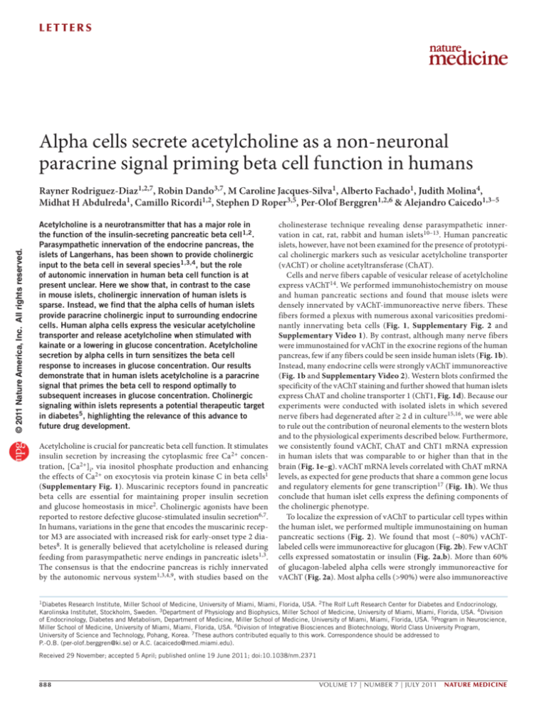
nantly innervating beta cells (Fig. 1, Supplementary Fig. 2 and
Supplementary Video 1). By contrast, although many nerve fibers
were immunostained for vAChT in the exocrine regions of the human
pancreas, few if any fibers could be seen inside human islets (Fig. 1b).
Instead, many endocrine cells were strongly vAChT immunoreactive
(Fig. 1b and Supplementary Video 2). Western blots confirmed the
specificity of the vAChT staining and further showed that human islets
express ChAT and choline transporter 1 (ChT1, Fig. 1d). Because our
experiments were conducted with isolated islets in which severed
nerve fibers had degenerated after ≥ 2 d in culture15,16, we were able
to rule out the contribution of neuronal elements to the western blots
and to the physiological experiments described below. Furthermore,
we consistently found vAChT, ChAT and ChT1 mRNA expression
in human islets that was comparable to or higher than that in the
brain (Fig. 1e–g). vAChT mRNA levels correlated with ChAT mRNA
levels, as expected for gene products that share a common gene locus
and regulatory elements for gene transcription17 (Fig. 1h). We thus
conclude that human islet cells express the defining components of
the cholinergic phenotype.
To localize the expression of vAChT to particular cell types within
the human islet, we performed multiple immunostaining on human
pancreatic sections (Fig. 2). We found that most (~80%) vAChTlabeled cells were immunoreactive for glucagon (Fig. 2b). Few vAChT
cells expressed somatostatin or insulin (Fig. 2a,b). More than 60%
of glucagon-labeled alpha cells were strongly immunoreactive for
vAChT (Fig. 2a). Most alpha cells (>90%) were also ­immunoreactive
1Diabetes Research Institute, Miller School of Medicine, University of Miami, Miami, Florida, USA. 2The Rolf Luft Research Center for Diabetes and Endocrinology,
Karolinska Institutet, Stockholm, Sweden. 3Department of Physiology and Biophysics, Miller School of Medicine, University of Miami, Miami, Florida, USA. 4Division
of Endocrinology, Diabetes and Metabolism, Department of Medicine, Miller School of Medicine, University of Miami, Miami, Florida, USA. 5Program in Neuroscience,
Miller School of Medicine, University of Miami, Miami, Florida, USA. 6Division of Integrative Biosciences and Biotechnology, World Class University Program,
University of Science and Technology, Pohang, Korea. 7These authors contributed equally to this work. Correspondence should be addressed to
P.-O.B. (per-olof.berggren@ki.se) or A.C. (acaicedo@med.miami.edu).
Received 29 November; accepted 5 April; published online 19 June 2011; doi:10.1038/nm.2371
888
VOLUME 17 | NUMBER 7 | JULY 2011 nature medicine
letters
nature medicine VOLUME 17 | NUMBER 7 | JULY 2011
b
c
d
HI1 HI2 HI3 HP HI4
ChAT
vAChT
MB
ChT1
e
f
30
g
h
20
vAChT mRNA
for ChAT (Fig. 2). Within the alpha cell, vAChT staining did not
overlap with glucagon staining and appeared confined to distinct
compartments (Fig. 2c). We examined the colocalization of vAChT
and glucagon immunofluorescence18 and found a Pearson’s correlation coefficient significantly smaller than that of C peptide and insulin colocalization and closer to that of the clearly segregated nuclear
DAPI and glucagon staining (Fig. 2d). These findings concur with
studies showing that in neuroendocrine cells, vAChT localizes preferentially to synaptic-like microvesicles and is excluded from hormone
granules19,20. Furthermore, human alpha cells have been reported to
possess secretory vesicles of different sizes21. To determine whether
acetylcholine and glucagon are stored in different secretory granules,
however, would require electron microscopy studies.
Our immunohistochemical results suggested that in human alpha
cells acetylcholine is packaged in secretory vesicles for exocytotic
release. We therefore examined human islets for acetylcholine secretion using cellular biosensors, namely Chinese hamster ovary (CHO)
cells expressing the muscarinic receptor M3 (Fig. 3). We monitored
acetylcholine secretion from human islets in real time by recording
[Ca2+]i responses from biosensors loaded with the [Ca2+]i indicator
Fura-2 and placed in apposition to isolated human islets (Fig. 3a).
The biosensors showed large responses to KCl-mediated depolarization (25 mM) of human islets (Fig. 3d), indicating that acetylcholine
release was induced from excitable islet cells and ruling out a contribution from exocrine tissue. Stimulation with kainate (100 µM)
or lowering the glucose concentration from 16 mM to 3 mM, which
are both alpha cell–specific stimuli16,22,23 (Supplementary Fig. 3),
induced strong acetylcholine release, as measured by large [Ca2+]i
responses in the biosensors (Fig. 3c,d). By contrast, increases in the
glucose concentration from 3 mM to 16 mM did not elicit acetylcholine secretion (Fig. 3c,d). Biosensor responses could be blocked by
the muscarinic antagonist atropine (5 µM), and none of the stimuli
used, including KCl depolarization, induced responses in biosensors
in the absence of human islets (Fig. 3b). This confirmed that the
[Ca2+]i responses were elicited by acetylcholine released from islet
cells. We obtained similar results using an enzymatic assay to detect
acetylcholine release (Fig. 3e). Because acetylcholine was released
in response to treatments known to specifically stimulate alpha cells
and not in response to increased glucose concentration, which stimulates beta and delta cells, we conclude that human alpha cells secrete
acetylcholine.
We further used the biosensor assay to detect acetylcholine release
from mouse islets. For these experiments, we cultured mouse islets
a
mRNA expression
© 2011 Nature America, Inc. All rights reserved.
Figure 1 Endocrine cells in human pancreatic islets express cholinergic
markers. (a) Z-stack of confocal images of a mouse pancreatic section
showing an islet immunostained for vesicular acetylcholine transporter
(vAChT, green) and glucagon (red). (b) Z-stack of confocal images of a
human pancreatic section showing vAChT immunostaining in islet cells.
Merge of glucagon and vAChT immunostaining appears yellow. (c) Z-stack
of confocal images of a human pancreatic section showing lack of vAChT
staining in human islets after preincubation with control peptide. Scale
bars, 50 µm (a–c). (d) Western blotting analyses of lysates from four
separate human islet preparations (HI1–HI4) and human pancreatic
exocrine tissue (HP), with mouse brain (MB) as a positive control. Specific
bands were seen in human islet lysates for vAChT (~70 kDa; top), for
choline acetyltransferase (~63 kDa and ~68 kDa; middle) and for ChT1
(~68 kDa; bottom). A molecular marker was run in parallel (second lane).
(e–g) vAChT (e), ChAT (f) and ChT1 (g) mRNA expression in brain (B, n = 4),
human islets (I, n = 12) and human pancreas (P, n = 3). Data represent
means ± s.e.m. (h) vAChT mRNA levels were associated with ChAT mRNA
levels (r2 = 0.57; slope significantly different from 0, P < 0.01).
20
20
10
10
10
80
60
40
20
0
B I
P
0
0
0
0
B I
P
B I
P
20
40
60
80
ChAT mRNA
for 4 d after isolation to eliminate neural elements, the same time
as for human islets. Acetylcholine secretion could not be recorded
from mouse islets stimulated with 25 mM KCl or 100 µM kainate
(0 out of 26 mouse islets responded versus 17 out of 75 human islets;
P = 0.0052, Fisher’s exact test), consistent with the lack of vAChT
immunostaining in mouse endocrine cells (Fig. 1a).
What is the role of alpha cell–derived acetylcholine for islet function? Studies have shown that exposure to cholinergic agonists sensitizes beta cells to subsequent stimuli, increasing insulin secretion1,24.
Given that in human islets most beta cells are closely associated with
alpha cells25, we hypothesized that alpha cells release acetylcholine to
prime neighboring beta cells. To test this hypothesis, we first examined whether cholinergic agonists induce insulin responses in human
beta cells. At low glucose concentration (3 mM), acetylcholine and the
muscarinic agonist oxotremorine elicited concentration-dependent
insulin release from isolated human islets, indicating that activation
of muscarinic receptors can induce insulin secretion at basal glucose
concentrations (Fig. 4a,b).
To infer the role of acetylcholine as a paracrine signal we manipulated
endogenous levels of acetylcholine. Applying the acetylcholinesterase
inhibitor physostigmine (30 µM) at 3 mM glucose increased insulin
secretion in isolated islets cultured for 4 d (Fig. 4c). These insulin
responses to physostigmine were strongly inhibited by vesamicol, a
selective inhibitor of vAChT that blocks acetylcholine transport into
vesicles and depletes cells of releasable acetylcholine (Fig. 4d). We
also found that physostigmine-induced increases in insulin secretion were partially inhibited by the M3 antagonist J-104129 (Fig. 4e).
889
c
100
z
z
x
0.4
*
0.2
0
f
ChAT
Glucagon
Merge
is endogenously released at low glucose concentrations to stimulate
insulin secretion and that acetylcholine secretion requires vesicular
mechanisms. These results are consistent with our immunohistochemical results showing the presence of vAChT in alpha cells.
ACh release (∆340/380)
0.2
0.1
0
6G 3G
+ –16
at G
16
ro
G
–3 1 pin
G 6G e
+ –3
at G
ro
pi
ne
0
–1
3G
e
*
0.4
*
0.3
0.2
0.1
I
G
KC
–3
G
–1
1G
l
0
sa
Figure 3 Isolated human islets secrete acetylcholine (ACh) in response to alpha cell–specific stimuli. (a) Photomicrograph of an ACh
biosensor (colorized green) apposed to an isolated human islet to monitor ACh secretion evoked by stimulation of islet cells.
Responses in the biosensor were recorded by loading biosensors with Fura-2 and imaging cytoplasmic [Ca2+]. Scale bar, 50 µm.
(b) ∆[Ca2+]i (∆340/380) responses in biosensors, in the absence of human islets, to direct application of ACh (10 µM), kainate
(100 µM), KCl (25 mM), changes in glucose concentration (from 3 mM to 16 mM (high glucose) or from 16 mM to 3 mM
(low glucose)) or ACh (10 µM) in the presence of the muscarinic antagonist atropine (5 µM). (c) Trace of the 340/380 Fura-2 ratio
in an ACh biosensor positioned against the islet as in a shows stimulus-induced secretion of ACh from endocrine cells in a human islet.
Horizontal lines denote application of kainate (Kai, 100 µM), a transient increase in glucose concentration from 3 mM to 16 mM (16G)
or application of atropine (red, 5 µM). (d) Summary of data from experiments conducted as those shown in c. Bars show means ± s.e.m.
for ACh biosensor signals (∆340/380) in response to stimulation of islets with KCl (25 mM) depolarization (n = 8 experiments), kainate
(100 µM, n = 11), increases in glucose concentration (from 3 mM to 16 mM, 3G–16G) or decreases in glucose concentration (from
16 mM to 3 mM, 16G–3G, n = 4). Biosensor responses were blocked by atropine (5 µM). (e) ACh release in response to increasing the
glucose concentration from 3 mM to 11 mM (3G–11G), lowering the glucose concentration from 11 mM to 3 mM (11G–3G) or to
depolarization with KCl (25 mM), as determined with a fluorescent enzymatic assay (see Online Methods; n = 6 islet preparations;
ANOVA followed by multiple comparison, *P < 0.05). Data represent means ± s.e.m.
Ba
0
11
ACh release (∆340/380)
0.3
3G
Time (s)
3,000
0.2
ACh (µM)
2,000
0.4
at Ka
ro i
pi
ne
1,000
16G
0.6
+
0
Kai
I+
Kai 16G
0
0.5
KC
0.5
1.0
Ka
i
Atropine
1.5
0.8
at KC
ro I
pi
ne
340/380
2.0
ACh release (∆340/380)
d 1.5
1.0
h
at
e
ig
h KC
Lo glu I
w cos
gl e
uc
os
AC
e
h
+ AC
at
ro h
pi
ne
0.6
vAChT
e
1
in
Glucagon
go
n
su
lin
So
m
a
ca
y
In
lu
y
2.5
Ka
0
C peptide
0
c
H
PI
Insulin
Cells (%)
40
2
890
255
DAPI
20
b
∆340/380
PI
60
Biosensor
Islet
d0
x
80
Somatostatin
vAChT
Pearson’s correlation coefficient
b
This antagonist by itself reduced insulin secretion at 3 mM glucose,
and this effect was negligible at 11 mM glucose (data not shown).
Physostigmine-induced increases in insulin secretion were reduced at
11 mM glucose (Fig. 4f). These experiments indicate that acetylcholine
a
Insulin
vAChT
Glucagon
vAChT
Glucagon
a
G
Figure 2 Human alpha cells express vAChT and
ChAT. (a) Confocal images of human pancreatic
sections showing vAChT immunostaining (green)
co-stained with glucagon immunostaining
(red, left), with insulin immunostaining (red,
middle) or with somatostatin immunostaining
(red, right). Colocalization appears yellow.
(b) Quantification of the percentage of
vAChT immunostained cells also labeled for
glucagon, insulin or somatostatin (n = 3 human
pancreata). Percentages do not add exactly
to 100% because analyses were performed
on different sections. (c) Glucagon (red) and
vAChT immunostaining (green) in alpha cells
at high magnification. Shown are three optical
planes through an alpha cell. (d) Scatter plots
of pixel intensities (PI) of glucagon and DAPI
immunofluorescence in alpha cells (left, top),
insulin and C peptide immunofluorescence in
beta cells (left, middle) and glucagon and
vAChT immunofluorescence in alpha cells
(left, bottom). Bar graph (right) shows the
thresholded Pearson’s correlation coefficient
values for glucagon-DAPI (left column)
insulin–C peptide (middle column) and
glucagon-vAChT colocalization (right column;
n = 12 cells, analysis of variance (ANOVA)
followed by multiple comparison, *P < 0.05).
(e) ChAT immunostaining (green, left) in
glucagon-labeled alpha cells (red, middle).
Colocalization appears yellow (merge, right).
(f) High magnification confocal image of an
alpha cell stained for glucagon (red) and ChAT
(green). Scale bars, 50 µm (a,e) and 5 µm (c,f).
Data represent means ± s.e.m.
AC
© 2011 Nature America, Inc. All rights reserved.
letters
VOLUME 17 | NUMBER 7 | JULY 2011 nature medicine
nature medicine VOLUME 17 | NUMBER 7 | JULY 2011
60
30
0.01
0
10 µM
1
0.1
3 mM
11 mM
100
+ atropine
∆Insulin (mU per liter)
Insulin (mU per liter)
b
Oxotremorine
Nicotine
ACh
90
50
0
30
20
10
40
0.01
Time (min)
d
∆Insulin (mU per liter)
150
100
50
0
40
*
20
10
20
Time (min)
100
11G
0
11G
30
50
50
*
11G 11G KCI
60
90
Time (min)
0
–
+
J-104129
h
200
0
*
100
0
–
+
Vesamicol
Physostigmine/J-104129
300
120
10
f
100
0
0
g
e
60
∆Insulin (mU per liter)
c
1
0.1
10
[ACh] (µM)
∆Insulin (mU per liter)
0
Control response (%)
In vivo, the secretion of insulin and glucagon fluctuates constantly
with periods of approximately 10 min26,27. We hypothesized that these
fluctuations allow alpha cells to increase acetylcholine secretion and
influence beta cells. We reproduced these hormonal fluctuations
in vitro by subjecting isolated human islets to an experimental protocol
in which we stimulated beta cells and alpha cells intermittently while
modulating cholinergic signaling (Fig. 4g,h and Supplementary Fig. 3).
When acetylcholine degradation was inhibited with physostigmine
(30 µM), insulin release increased during repeated exposure to high
glucose (11 mM) (Fig. 4g,h). Blocking muscarinic receptors with the
general antagonist atropine (10 µM) produced variable results, most
likely because multiple receptors on different cells were activated (data
not shown). By contrast, adding the M3 receptor-specific antagonist
J-104129 (50 nM) consistently reduced insulin responses (Fig. 4g,h).
These results show that endogenously released acetylcholine contributed
to the enhanced beta cell response by activating M3 receptors. Thus,
in the absence of any influence from the autonomic nervous system,
endogenously released acetylcholine in human islets is able to sensitize
the beta cell to subsequent increases in glucose concentration.
On the basis of our results, we propose that acetylcholine is a
paracrine signal secreted by alpha cells in human islets. Our findings
showing that alpha cells express vAChT and that alpha-specific stimuli
induce acetylcholine secretion indicate that acetylcholine is stored
in alpha cells for exocytotic release. In our model, alpha cells release
acetylcholine when activated by lowering glucose concentration to
prime the beta cell response to a subsequent increase in glucose concentration. Although additional paracrine effects of acetylcholine on
other cells within the human islet (for example, delta cells) remain to
be investigated, our results suggest that acetylcholine serves as a feedforward signal to keep the beta cell responsive to future challenges,
thus limiting plasma glucose fluctuations. Moreover, the intracellular
signaling pathways activated by acetylcholine may promote long-term
survival of beta cells28, thus further suggesting that alpha cell–derived
acetylcholine may act as a trophic factor.
This paracrine interaction is only possible because of the unique
cytoarchitecture of the human islet, where most beta cells are closely
associated with alpha cells25,29,30. With beta cells comprising 64%
a
Insulin (mU per liter)
Figure 4 Endogenously released ACh amplifies glucose-induced insulin
secretion in human islets. (a) Insulin release from human islets elicited
by ACh, the muscarinic agonist oxotremorine and nicotine. Horizontal
lines denote stimulus application. Representative traces of n = 3 islet
preparations. (b) Summary of data from experiments similar to those
shown in a but conducted in the presence of low (3 mM) and high
(11 mM) glucose (n = 3 preparations). (c) Insulin secretion elicited by the
acetylcholinesterase inhibitor physostigmine (30 µM) at 3 mM glucose
(n = 5 human islet preparations). (d–f) Physostigmine-induced increases
in insulin secretion (∆Insulin) in the absence (−) or presence (+) of the
vAChT blocker vesamicol (10 µM, d), the M3 receptor antagonist
J-104129 (50 nM, e) or when the experiment was performed at different
glucose concentrations (3 mM, 3G; 11 mM, 11G, f; Student’s t test,
P < 0.05). (g) Insulin secretion induced by repeatedly raising glucose
from 3 mM to 11 mM in the presence of physostigmine (30 µM) or
in the presence of J-104129 (50 nM); representative traces of four
experiments. A control experiment with untreated islets was run in
parallel (gray symbols). Horizontal lines at bottom denote drug
application. 11G indicates 15 min of elevated glucose (11 mM). Islets
were stimulated four times with glucose followed by 25 mM KCl.
(h) Summary of data from experiments such as those shown in c.
Responses are expressed as percentage of the respective insulin response
of control islets (100%, gray dashed line; n = 4 preparations for J-104129
treatment; n = 5 preparations for physostigmine treatment). One-sample
t tests were used to compare the actual mean to a theoretical mean of
100% (control; *P < 0.05). Data represent means ± s.e.m.
Insulin secretion
(scaled to first response)
© 2011 Nature America, Inc. All rights reserved.
letters
150
100
3G 11G
J-104129
Physostigmine
*
*
*
*
*
50
0
2nd
3rd
Response
4th
and alpha cells most of the remaining volume in the human islet 31,
there is a high probability for a beta cell to be close to an alpha cell.
Indeed, most beta cells (70–80%) face alpha cells 25,30 and maintain
a strong association even after dispersion of the islet30. This cellular
arrangement is compatible with the notion that paracrine interactions
occur via the interstitial space between endocrine cells, although the
vascular route may also be used32. Thus, in human islets, alpha cells
seem optimally placed to influence beta cells.
Although we cannot rule out a contribution of parasympathetic nervous input, our results suggest that cholinergic innervation of human
islets may be sparse. This is consistent with studies showing that the
influence of neural input on insulin secretion occurring before the actual
absorption of nutrients (cephalic phase) has a relatively minor role in
humans33,34. Along these lines, vagotomized patients have normal postprandial serum insulin levels35, and individuals with type 1 diabetes
who have undergone pancreas transplantation (and thus have denervated islets) remain euglycemic without therapy36–38. Furthermore, it is
possible that the reported parasympathetic influence on islet function in
human beings may be mediated by peptidergic axons12. The human islet
may thus be self reliant in terms of cholinergic input. That acetylcholine
is a paracrine signal, and not only a neural signal as in rodents, further
implies that ­cholinergic signaling in human islets is activated under
circumstances that ­cannot be modeled with rodent studies, highlighting
the importance of species divergence in the pancreatic islet. Because
cholinergic ­ signaling pathways have been proposed as intervention
points to promote beta cell function and survival5, our study has major
­implications for ­therapies in diabetes mellitus.
Methods
Methods and any associated references are available in the online
version of the paper at http://www.nature.com/naturemedicine/.
891
letters
Note: Supplementary information is available on the Nature Medicine website.
© 2011 Nature America, Inc. All rights reserved.
Acknowledgments
We thank A. Formoso for help with data analyses, K. Johnson for histological
work and M. Correa-Medina for assistance with RT-PCR. This work was funded
by the Diabetes Research Institute Foundation, US National Institutes of Health
grants R56DK084321 (A.C.), R01DK084321 (A.C.), R01DC000374 (S.D.R.),
R01DC007630 (S.D.R.), F32DK083226 (M.H.A.) and 5U01DK070460-07 (C.R.),
the Juvenile Diabetes Research Foundation, the Swedish Research Council, the
Novo Nordisk Foundation, the Swedish Diabetes Association, the Family ErlingPersson Foundation and the World Class University program through the National
Research Foundation of Korea funded by the Ministry of Education, Science and
Technology (R31-2008-000-10105-0). Human islets were made available through
the Islet Cell Resource basic science islet distribution program, Islet Cell Resource
Centers Consortium, Division of Clinical Research, National Center for Research
Resources, US National Institutes of Health.
AUTHOR CONTRIBUTIONS
R.R.-D., M.C.J.-S., A.F. and J.M. performed hormone assay experiments and
ELISAs; R.D. performed experiments with biosensor cells to detect acetylcholine
secretion; R.R.-D. and M.C.J.-S. conducted Amplex assays to measure
acetylcholine secretion; R.R.-D. and M.H.A. collected, analyzed and quantified
immunohistochemical data, and R.R.-D. performed RT-PCR and western blotting.
R.R.-D., R.D., M.H.A., C.R., S.D.R., P.-O.B. and A.C. designed the study, analyzed
data and wrote the paper. R.R.-D. and R.D. contributed equally to the study. All
authors discussed the results and commented on the manuscript.
COMPETING FINANCIAL INTERESTS
The authors declare no competing financial interests.
Published online at http://www.nature.com/naturemedicine/.
Reprints and permissions information is available online at http://www.nature.com/
reprints/index.html.
1. Gilon, P. & Henquin, J. Mechanisms and physiological significance of the cholinergic
control of pancreatic beta-cell function. Endocr. Rev. 22, 565–604 (2001).
2. Gautam, D. et al. A critical role for beta cell M3 muscarinic acetylcholine receptors
in regulating insulin release and blood glucose homeostasis in vivo. Cell Metab. 3,
449–461 (2006).
3. Ahrén, B. Autonomic regulation of islet hormone secretion—implications for health
and disease. Diabetologia 43, 393–410 (2000).
4. Conn, P.M., Goodman, H.M. & Kostyo, J.L. The Endocrine System: The Endocrine
Pancreas and Regulation of Metabolism. 153 (Oxford University Press, New York,
1998).
5. Gautam, D. et al. Role of the M3 muscarinic acetylcholine receptor in beta-cell function
and glucose homeostasis. Diabetes Obes. Metab. 9 (Suppl 2), 158–169 (2007).
6. Guenifi, A., Simonsson, E., Karlsson, S., Ahrén, B. & Abdel-Halim, S. Carbachol
restores insulin release in diabetic GK rat islets by mechanisms largely involving
hydrolysis of diacylglycerol and direct interaction with the exocytotic machinery.
Pancreas 22, 164–171 (2001).
7. Doliba, N.M. et al. Restitution of defective glucose-stimulated insulin release of
sulfonylurea type 1 receptor knockout mice by acetylcholine. Am. J. Physiol.
Endocrinol. Metab. 286, E834–E843 (2004).
8. Guo, Y. et al. CHRM3 gene variation is associated with decreased acute insulin
secretion and increased risk for early-onset type 2 diabetes in Pima Indians.
Diabetes 55, 3625–3629 (2006).
9. Woods, S.C. & Porte, D.J. Neural control of the endocrine pancreas. Physiol. Rev.
54, 596–619 (1974).
10.Coupland, R.E. The innervation of pan creas of the rat, cat and rabbit as revealed
by the cholinesterase technique. J. Anat. 92, 143–149 (1958).
11.Amenta, F., Cavallotti, C., de Rossi, M., Tonelli, F. & Vatrella, F. The cholinergic
innervation of human pancreatic islets. Acta Histochem. 73, 273–278 (1983).
892
12.Ahrén, B., Taborsky, G.J. & Porte, D.J. Neuropeptidergic versus cholinergic and
adrenergic regulation of islet hormone secretion. Diabetologia 29, 827–836 (1986).
13.Brunicardi, F.C., Shavelle, D. & Andersen, D. Neural regulation of the endocrine
pancreas. Int. J. Pancreatol. 18, 177–195 (1995).
14.Weihe, E., Tao-Cheng, J., Schäfer, M., Erickson, J. & Eiden, L. Visualization of the
vesicular acetylcholine transporter in cholinergic nerve terminals and its targeting
to a specific population of small synaptic vesicles. Proc. Natl. Acad. Sci. USA 93,
3547–3552 (1996).
15.Karlsson, S., Myrsén, U., Nieuwenhuizen, A., Sundler, F. & Ahrén, B. Presynaptic
sympathetic mechanism in the insulinostatic effect of epinephrine in mouse
pancreatic islets. Am. J. Physiol. 272, R1371–R1378 (1997).
16.Cabrera, O. et al. Glutamate is a positive autocrine signal for glucagon release.
Cell Metab. 7, 545–554 (2008).
17.Eiden, L.E. The cholinergic gene locus. J. Neurochem. 70, 2227–2240 (1998).
18.Barlow, A.L., Macleod, A., Noppen, S., Sanderson, J. & Guérin, C.J. Colocalization
analysis in fluorescence micrographs: verification of a more accurate calculation of
pearson’s correlation coefficient. Microsc. Microanal. 16, 710–724 (2010).
19.Liu, Y. & Edwards, R. Differential localization of vesicular acetylcholine and
monoamine transporters in PC12 cells but not CHO cells. J. Cell Biol. 139, 907–916
(1997).
20.Krantz, D.E. et al. A phosphorylation site regulates sorting of the vesicular
acetylcholine transporter to dense core vesicles. J. Cell Biol. 149, 379–396 (2000).
21.Greider, M.H., Bencosme, S. & Lechago, J. The human pancreatic islet cells and
their tumors. I. The normal pancreatic islets. Lab. Invest. 22, 344–354 (1970).
22.Cabrera, O. et al. Automated, high-throughput assays for evaluation of human
pancreatic islet function. Cell Transplant. 16, 1039–1048 (2008).
23.Jacques-Silva, M.C. et al. ATP-gated P2X3 receptors constitute a positive autocrine signal
for insulin release in the human pancreatic beta cell. Proc. Natl. Acad. Sci. USA 107,
6465–6470 (2010).
24.Zawalich, W.S., Zawalich, K. & Rasmussen, H. Cholinergic agonists prime the
beta-cell to glucose stimulation. Endocrinology 125, 2400–2406 (1989).
25.Cabrera, O. et al. The unique cytoarchitecture of human pancreatic islets has
implications for islet cell function. Proc. Natl. Acad. Sci. USA 103, 2334–2339
(2006).
26.Stagner, J.I., Samols, E. & Weir, G.C. Sustained oscillations of insulin, glucagon,
and somatostatin from the isolated canine pancreas during exposure to a constant
glucose concentration. J. Clin. Invest. 65, 939–942 (1980).
27.Lang, D.A., Matthews, D.R., Peto, J. & Turner, R.C. Cyclic oscillations of basal
plasma glucose and insulin concentrations in human beings. N. Engl. J. Med. 301,
1023–1027 (1979).
28.Wessler, I. & Kirkpatrick, C. Acetylcholine beyond neurons: the non-neuronal
cholinergic system in humans. Br. J. Pharmacol. 154, 1558–1571 (2008).
29.Brissova, M. et al. Assessment of human pancreatic islet architecture and
composition by laser scanning confocal microscopy. J. Histochem. Cytochem. 53,
1087–1097 (2005).
30.Bosco, D. et al. Unique arrangement of alpha- and beta-cells in human islets of
Langerhans. Diabetes 59, 1202–1210 (2010).
31.Pisania, A. et al. Quantitative analysis of cell composition and purity of human
pancreatic islet preparations. Lab. Invest. 90, 1661–1675 (2010).
32.Stagner, J.I. & Samols, E. The vascular order of islet cellular perfusion in the
human pancreas. Diabetes 41, 93–97 (1992).
33.Taylor, I.L. & Feldman, M. Effect of cephalic-vagal stimulation on insulin, gastric
inhibitory polypeptide, and pancreatic polypeptide release in humans. J. Clin.
Endocrinol. Metab. 55, 1114–1117 (1982).
34.Teff, K.L., Mattes, R., Engelman, K. & Mattern, J. Cephalic-phase insulin in obese
and normal-weight men: relation to postprandial insulin. Metabolism 42, 1600–1608
(1993).
35.Becker, H.D., Börger, H. & Schafmayer, A. Effect of vagotomy on gastrointestinal
hormones. World J. Surg. 3, 615–622 (1979).
36.Pozza, G. et al. Metabolic control of type I (insulin dependent) diabetes after
pancreas transplantation. Br. Med. J. (Clin. Res. Ed.) 291, 510–513 (1985).
37.Diem, P. et al. Glucagon, catecholamine and pancreatic polypeptide secretion in
type I diabetic recipients of pancreas allografts. J. Clin. Invest. 86, 2008–2013
(1990).
38.Blackman, J.D. et al. Insulin secretory profiles and C-peptide clearance kinetics at
6 months and 2 years after kidney-pancreas transplantation. Diabetes 41,
1346–1354 (1992).
VOLUME 17 | NUMBER 7 | JULY 2011 nature medicine
ONLINE METHODS
Pancreatic islets. We obtained human pancreatic islets from the Human Islet
Cell Processing Facility at the Diabetes Research Institute, University of Miami
Miller School of Medicine, or from the Islet Cell Resource basic science islet
distribution program, Islet Cell Resource Centers Consortium, Division of
Clinical Research, National Center for Research Resources, National Institutes
of Health. Mouse islets were obtained from C57BL/6 mice killed by exsanguination under general anesthesia. Mouse islet isolation was performed
using collagenase digestion followed by purification on density gradients. All
experimental protocols using mice were approved by the University of Miami
Animal Care and Use Committee.
Determination of acetylcholine secretion with biosensor cells. We adapted
real time measurements of acetylcholine secretion from a previously published study39. We used Fura-2–loaded CHO cells stably expressing muscarinic
M3 receptors40.
© 2011 Nature America, Inc. All rights reserved.
Determination of acetylcholine secretion with Amplex assay. We measured acetylcholine secretion with the Amplex red Acetylcholine Assay Kit
(Invitrogen).
Insulin and glucagon secretion. We measured insulin and glucagon secretion
as previously described16,22,23. We purchased kainate, oxotremorine, physo­
stigmine hemisulfate, vesamicol hydrochloride and J-104129 fumarate from
Tocris Bioscience and atropine sulfate and nicotine from Sigma.
Immunohistochemistry. We performed immunostaining as previously
described16,23,25 in sections of >20 human pancreata. Antibodies used
included rabbit antibody to vAChT (Synaptic Systems, 139103; control peptide 139-1P), rabbit antibody to vAChT (Sigma, V5387), rabbit antibody to
ChT1 (Chemicon, AB5966), mouse antibody to somatostatin (Chemicon,
MAB354), mouse antibody to glucagon (Sigma, G2654), guinea pig antibody to insulin (Dako, A0564) and rabbit antibody to C peptide (GeneTex,
GTX14181). In control experiments, we incubated primary antibodies with
the corresponding control peptide at a ratio of 50 µg antigenic peptide to
1 µg antibody at 22 °C for 5 h.
doi:10.1038/nm.2371
For ChAT immunostaining we used rabbit antibody to ChAT (Chemicon,
AB143), goat antibody to ChAT (Chemicon, AB144P), or rabbit antibody to
ChAT (Pierce, OSC00008W). Only antibody AB144P gave reliable results, but it
required signal amplification (ABC method followed by tyramide signal amplification). The strong signal obtained after amplification may explain why the
proportion of ChAT-stained alpha cells (90%) was higher than that of vAChTstained alpha cells (60%).
Colocalization studies. We quantified the degree of association of glucagon and vAChT staining within alpha cells with the colocalization macros of
Volocity software (PerkinElmer) as previously described18.
Western blotting. We carried out immunoblot analysis by standard methods
with the antibodies used for immunohistochemistry (1 in 1,000).
RT-PCR. We extracted RNA from total human brain (Applied Biosystems/
Ambion), total human pancreas (Applied Biosystems/Ambion), or human
islets using the RiboPure Kit (Applied Biosystems/Ambion), and we prepared
cDNA using the High-Capacity cDNA reverse transcription kit (Applied
Biosystems). We ran PCR reactions in duplicate using Taqman gene expression assays (Applied Biosystems) in a StepOnePlus Real-Time PCR System
(Applied Biosystems). We performed relative quantification (RQ) of gene
expression based on the equation RQ = 2− ∆Ct ×10,000, where ∆Ct is the difference between the Ct value (number of cycles at which amplification for a
gene reaches a threshold) of the target gene and the Ct value of the ubiquitous
housekeeping gene GAPDH.
Statistical analyses. For statistical comparisons we used Student’s
t test, ANOVA followed by multiple comparisons (Bonferroni) or Fisher’s
exact test.
39.Huang, Y.J. et al. Mouse taste buds use serotonin as a neurotransmitter. J. Neurosci.
25, 843–847 (2005).
40.Buck, M.A. & Fraser, C. Muscarinic acetylcholine receptor subtypes which selectively
couple to phospholipase C: pharmacological and biochemical properties. Biochem.
Biophys. Res. Commun. 173, 666–672 (1990).
nature medicine


