treatment strategies in liver abscess our experience
advertisement
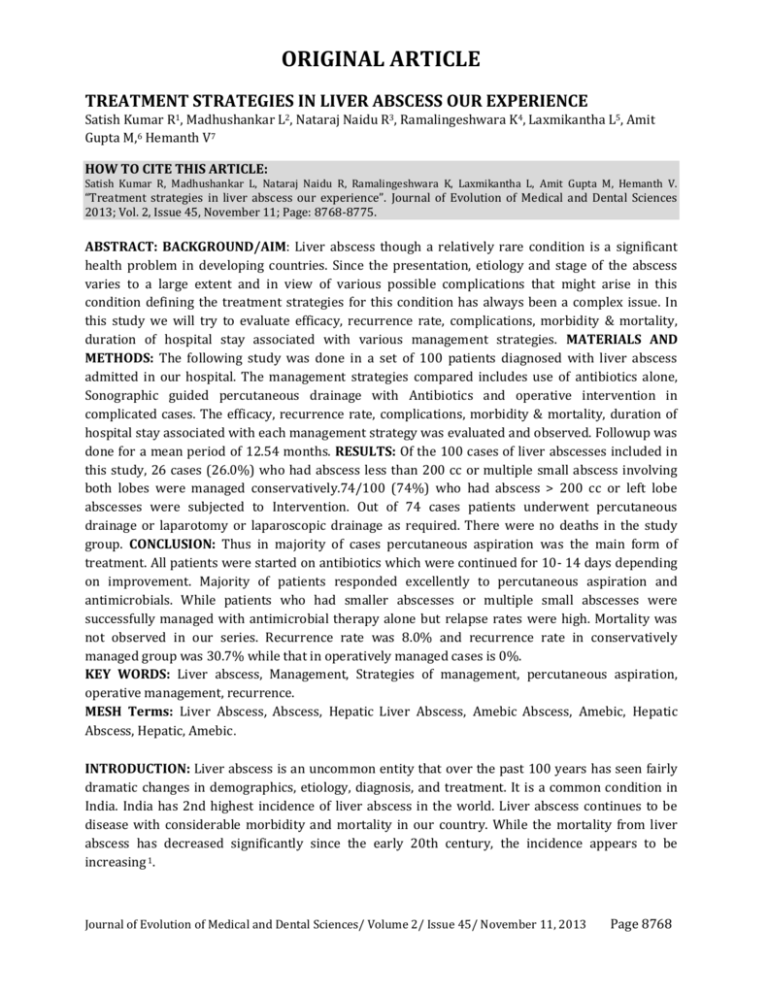
ORIGINAL ARTICLE TREATMENT STRATEGIES IN LIVER ABSCESS OUR EXPERIENCE Satish Kumar R1, Madhushankar L2, Nataraj Naidu R3, Ramalingeshwara K4, Laxmikantha L5, Amit Gupta M,6 Hemanth V7 HOW TO CITE THIS ARTICLE: Satish Kumar R, Madhushankar L, Nataraj Naidu R, Ramalingeshwara K, Laxmikantha L, Amit Gupta M, Hemanth V. “Treatment strategies in liver abscess our experience”. Journal of Evolution of Medical and Dental Sciences 2013; Vol. 2, Issue 45, November 11; Page: 8768-8775. ABSTRACT: BACKGROUND/AIM: Liver abscess though a relatively rare condition is a significant health problem in developing countries. Since the presentation, etiology and stage of the abscess varies to a large extent and in view of various possible complications that might arise in this condition defining the treatment strategies for this condition has always been a complex issue. In this study we will try to evaluate efficacy, recurrence rate, complications, morbidity & mortality, duration of hospital stay associated with various management strategies. MATERIALS AND METHODS: The following study was done in a set of 100 patients diagnosed with liver abscess admitted in our hospital. The management strategies compared includes use of antibiotics alone, Sonographic guided percutaneous drainage with Antibiotics and operative intervention in complicated cases. The efficacy, recurrence rate, complications, morbidity & mortality, duration of hospital stay associated with each management strategy was evaluated and observed. Followup was done for a mean period of 12.54 months. RESULTS: Of the 100 cases of liver abscesses included in this study, 26 cases (26.0%) who had abscess less than 200 cc or multiple small abscess involving both lobes were managed conservatively.74/100 (74%) who had abscess > 200 cc or left lobe abscesses were subjected to Intervention. Out of 74 cases patients underwent percutaneous drainage or laparotomy or laparoscopic drainage as required. There were no deaths in the study group. CONCLUSION: Thus in majority of cases percutaneous aspiration was the main form of treatment. All patients were started on antibiotics which were continued for 10- 14 days depending on improvement. Majority of patients responded excellently to percutaneous aspiration and antimicrobials. While patients who had smaller abscesses or multiple small abscesses were successfully managed with antimicrobial therapy alone but relapse rates were high. Mortality was not observed in our series. Recurrence rate was 8.0% and recurrence rate in conservatively managed group was 30.7% while that in operatively managed cases is 0%. KEY WORDS: Liver abscess, Management, Strategies of management, percutaneous aspiration, operative management, recurrence. MESH Terms: Liver Abscess, Abscess, Hepatic Liver Abscess, Amebic Abscess, Amebic, Hepatic Abscess, Hepatic, Amebic. INTRODUCTION: Liver abscess is an uncommon entity that over the past 100 years has seen fairly dramatic changes in demographics, etiology, diagnosis, and treatment. It is a common condition in India. India has 2nd highest incidence of liver abscess in the world. Liver abscess continues to be disease with considerable morbidity and mortality in our country. While the mortality from liver abscess has decreased significantly since the early 20th century, the incidence appears to be increasing 1. Journal of Evolution of Medical and Dental Sciences/ Volume 2/ Issue 45/ November 11, 2013 Page 8768 ORIGINAL ARTICLE Traditionally classified into pyogenic and amoebic liver abscess, latter being common in the developing countries. Though the advent of modern diagnostic techniques like ultrasound and computed tomography to locate and drain the abscesses has reduced the mortality to 2-12% there is still high morbidity due to the complications of liver abscess especially the amoebic liver abscess 2, 3. Management of liver abscess is multifaceted and results are also varied. Since the various presentations of liver abscess need to be managed accordingly standard management strategies cannot be rigid and the treating clinician should change strategies accordingly. Our own experience prompted a retrospective review of cases admitted in a single tertiary center in Bengaluru, India. The present study aims at studying the various treatment strategies adopted at our institute and the outcome in different clinical scenarios and different treatment strategies we will try to evaluate efficacy, recurrence rate, complications, morbidity & mortality, duration of hospital stay associated with various management strategies. MATERIALS AND METHODS: The present study was carried out at Kempegowda Institute of Medical Sciences and Research Centre, Bangalore. The study subjects included 100 patients admitted with liver abscess diagnosed by ultrasonography or CT scan between January 2011 to August 2012. The study design and protocol was approved by the hospital Research and Ethics Committee. The inclusion criteria for the study were diagnosis of liver abscess single or multiple on ultrasonography , irrespective of their age/gender/ background /socio economic status. Exclusion criteria included age less than 18 yrs, clinical signs of peritonitis on presentation(suggestive of ruptured abscess), patients of poor general condition with disorientation not fit for giving informed consent, recurrent abscess and traumatic liver abscess. Detailed history of patient was entered in proforma. Complete haemogram , LFT, Prothrombin time, Serology for amoebic antigen will be sent immediately on presentation. Preliminary Ultrasound of Abdomen and Pelvis was done on the same day of presentation. Patient was put on conservative line of management. Patient was followed up daily clinically and LFT & USG Abdomen was repeated on the 3rd day if patient symptomatically not relieved. Repeat Ultrasound / CT /MRI Abdomen & pelvis will be done immediately if patient condition does not improve/worsens or after 3-4 days as a routine. If the patient develops any of the complications like ruptured liver abscess into any of the serosal cavity, patient was immediately taken up for surgery (Laparotomy drainage or Laparoscopic drainage). Pus was sent for gram’s stain and culture and sensitivity. Anaerobic cultures were not done as the facility was not available in our hospital. Blood cultures were not routinely performed in all cases. Patient was informed about any surgical procedure and consent taken. The following management strategies were followed4: 1. Antibiotics alone ( in uncomplicated Abscess measuring <200cc) 2. Sonographic guided percutaneous drainage (Percutaneous Aspiration or Percutaneous Pigtail Catheter Drainage) + Antibiotics Coverage ( in un ruptured Abscess measuring >200cc) 3. In ruptured liver abscess a. Open Surgical drainage b.Laparoscopic Surgical drainage(Extraperitoneal & Transperitoneal) Journal of Evolution of Medical and Dental Sciences/ Volume 2/ Issue 45/ November 11, 2013 Page 8769 ORIGINAL ARTICLE Patients were followed up for a period of minimum six months: 1) Monthly for first 3 months 2) Then once after 6 months, for recurrent attacks or development of complications and to monitor the efficacy of the treatment given. Cure was defined as improvement clinically with subsidence of fever, and local signs, symptoms, decrease in WBC count and follow-up ultrasonography showed reduction in size < 3 cm in diameter and no evidence of relapses. Statistical Methods: Descriptive statistical analysis has been carried out in the present study. Results on continuous measurements are presented on Mean SD (Min-Max) and results on categorical measurements are presented in Number (%). Significance is assessed at 5 % level of significance. Chi-square/ Fisher Exact test has been used to find the significance of study parameters on categorical scale between two or more groups. 95% Confidence Interval has been computed to find the significant features. Confidence Interval with lower limit more than 50% is associated with statistical significance. Statistical software: The Statistical software namely SAS 9.2, SPSS 15.0, Stata 10.1, MedCalc 9.0.1, Systat 12.0 and R environment ver.2.11.1 were used for the analysis of the data and Microsoft word and Excel have been used to generate graphs, tables etc. RESULTS: Out of the 100 cases of liver abscesses 26 cases (26.0%) that had abscess: less than 200 cc or multiple small abscess involving both lobes were managed conservatively. 74/100 (74%) who had abscess > 200 cc or left lobe abscesses were subjected to Intervention. Out of 74 cases 49 cases underwent percutaneous aspiration under antibiotic coverage with a 95% CI (39.42 – 58.65) which is significant. 4 cases underwent Pigtail catheter drainage under USG guided as abscess cavity was big and not completely liquefied (In our study size of abscess cavity was >10cms). 12 Cases underwent Laparotomy procedure and 9 cases underwent laparoscopic drainage for ruptured liver abscess cases (21.0%) and 3 patients required ICD insertion. One case underwent thoracoscopy and diagnostic laparoscopy. Number of patients % 95%CI (n=100) Antibiotic coverage only (Conservative) 26 26.0 18.40-35.37 ASP (Percutaneous Aspiration under 49 49.0 39.42-58.65 Antibiotic Coverage) Laparoscopy Drainage 09 9.0 4.81-16.23 Laparotomy & Procedure 12 12.0 7.00-19.81 Pig Tail Catheter 4 4.0 1.57-9.84 Thoracoscopy + Diagnostic laparoscopy 1 1.0 0.2-5.45 ICD Insertion 3 3.0 1.03-8.45 Table 1: Different strategies of management followed and the number of patients undergoing each modality of treatment. Treatment Journal of Evolution of Medical and Dental Sciences/ Volume 2/ Issue 45/ November 11, 2013 Page 8770 ORIGINAL ARTICLE Out of the 49 patients who underwent percutaneous aspiration, 71.4% of patients (95% CI (57.59-82.15) required percutaneous aspiration only once for resolution of the abscess which is statistically significant. In 35/49 (69%) of the cases single aspiration was adequate. While 10/49 (19%) required 2 aspirations were required and 4/49 (9%) required 3 aspirations. Number of patients % 95%CI (n=49) Single aspiration 35 71.4 57.59-82.15 Two aspirations 10 20.4 11.48-33.64 Three aspirations 04 8.2 3.23-19.19 Table 2: Rate of repeat aspirations in patients undergoing percutaneous aspiration of abscess. Number of aspirations done The various complications developed by the patients were analyzed. Intra abdominal rupture with peritonitis was seen in 21/100 (21.0%) of cases. Features of shock in 4 cases (4%), Cholangitis was seen in 3% of cases. Pleural rupture was seen in 3/100 (3.0%) of cases. The patients were followed up for a mean period of 12.64 months which was significant (with Mean+S.D = 12.64+/-6.81). In the follow up period there were 8 (8%) cases of recurrence and 92 patients were free of any symptoms. It was also noted that all the recurrences occurred in patients treated conservatively without any aspiration of the abscess. And those patients who underwent ultrasound guided aspiration/pigtail Insertion /operative intervention did not develop any recurrence of the liver abscess. Journal of Evolution of Medical and Dental Sciences/ Volume 2/ Issue 45/ November 11, 2013 Page 8771 ORIGINAL ARTICLE Management Incidence of Recurrence Yes No Cons (liver abscess < 200cc) (n=26) 8(30.8%) 18(69.2%) PCA (liver abscess > 200 cc) (n=53) 0 53(100.0%) Ruptured liver abscess (n=21) 0 21(100.0%) Table 3: Correlation of incidence of recurrence with management DISCUSSION: Controversies in the management of liver abscess still exist. Surgical drainage of liver abscess has been an accepted therapy for decades. The diagnosis and treatment of liver abscess has changed due to advances in imaging techniques. Out of the 100 cases in this study, 26 patients who had multiple small abscess and solitary abscess with volume < 200 cc or < 5cms size were treated conservatively. 74/100 (74%) who had abscess > 200 cc or left lobe abscesses were subjected to Intervention as compared to Hyo Min Yo et al Study5 where 100.0% patients underwent intervention Out of 74 cases 49% cases underwent Percutaneous aspiration under antibiotic coverage as compared to Hyo Min Yo et al Study5 where 79.0% patients underwent Percutaneous Aspiration. 4 cases underwent Pigtail catheter drainage under USG guided as abscess cavity was big and not completely liquefied (In our study size of abscess cavity was >10cms). Laparotomy as initial line of treatment was performed in 12/74 ruptured liver abscess cases (16.21%) and Laparoscopy drainage in 9/74 (12.16%) ruptured liver abscess as compared to Hyo Min Yo et al Study5 where 21.0% patients underwent surgical intervention. In our Study Intercostal drainage was required in three cases in whom the abscess had ruptured into pleural cavity, Patients survived. Conservative management: While patients who had smaller abscesses or multiple small abscesses were successfully managed with antimicrobial therapy alone but relapse rates were high. The relapse rates in conservatively treated group were 30.76% (95% C.I-15.9-51.9) which was statistically significant. Thus incidence of recurrence is significantly associated with conservative management with p<0.001.Thus though it is a decent strategy for management of liver abscess in view of high recurrence rate this strategy should be limited to cases of multiple small abscess or abscess deep in location where aspiration is not possible as percutaneous aspirations had nearly 100% cure rates in our studies. Journal of Evolution of Medical and Dental Sciences/ Volume 2/ Issue 45/ November 11, 2013 Page 8772 ORIGINAL ARTICLE Per Cutaneous aspiration/Pig tail Catheter insertion: In majority of cases percutaneous aspiration was the main form of treatment6. All patients were started on antibiotics which were continued for 10- 14 days depending on improvement. Majority of patients responded excellently to percutaneous aspiration and antimicrobials. This was a very successful strategy as there was 100% cure rate and no complications in patients undergoing these procedures7. However need for repeat aspiration was the main problem in this method. Thick viscous pus was the main reason for repeat aspirations. Average volume of the abscesses was larger in patients who required repeat aspirations in our study. Hence in presence of thick viscous pus/partially liquefied abscess and large abscess cavity it is better to put a pigtail catheter in situ for few days until serial USG reveals complete drainage of the cavity8. Operative Intervention: Though operative drainage gives 100% cure rates in our study this modality of treatment should be reserved for those cases which develop complications like intraabdominal/pleural/pericardial rupture with peritonitis/ cholangitis etc9, 10. The overall Complications (21% )(95% C.I-13.75-30.67) like Intra-abdominal rupture with peritonitis (21.0%), pleural rupture (4.0%), Pericardial rupture (0.0%) were much less as compared to Study by Hyo Min Yoo et al (59%) which is statistically significant, which implies that the management strategies followed in this study were superior to those that were followed in Hyo Min Yoo et al as the recurrence rate and mortality rate were less compared to the earlier study. Study Complications Recurrence Mortality Hyo Min Yoo et al 59.0% 9% 11% Present Series 21.0% 8% 0% Table4: Comparison of Hyo Min Yoo et al with present study. REFERENCES: 1. Meddings L, Myers RP, Hubbard J, et al: A population-based study of pyogenic liver abscesses in the United States: Incidence, mortality, and temporal trends. Am J Gastroenterol 105:117, 2010. 2. Ribaudo JM, Ochsner A. Intrahepatic abscesses: Amoebic and pyogenic. Am J Surg 1973; 125:570-574. 3. Rahimian J, Wilson T, Oram V, Holzman RS. Pyogenic liver abscess: recent trends in etiology and mortality. Clin Infect Dis 2004; 39:1654-1659. 4. Ochsner A. Debakey M. Amoebic Hepatitis and hepatic abscess (an analysis of 181 cases with review of literature). Surgery 1943; 13: 612-49 5. Yonsei Medical Journal Vol. 34, No. 4, 1993 – The Changing Patterns of Liver Abscess During the Past 20 Years – A study of 482 cases – Hyo Min Yoo et al. 6. Hootan AM. The treatment of abscess of the liver by aspiration and injection of quinine. British Med.J. 1908; 24: 1251-52. 7. Wong KP. Percutaneous drainage of pyogenic liver abscess. World Journal of Surgery 1990; 14: 492-497. Journal of Evolution of Medical and Dental Sciences/ Volume 2/ Issue 45/ November 11, 2013 Page 8773 ORIGINAL ARTICLE 8. McDermott VGM. Questions and Answers : What is the role of percutaneous drainage for treatment of Amebic abscess of liver. American Journal of Roentgenology 1995; 165: 10051006. 9. Bertel CK, Van Heuden JA. Treatment of pyogenic hepatic abscess, surgical vs. percutaneous drainage. Archives of Surgery 1986; 121: 554-558. 10. Shimada H, Ohta S. Diagnostic and therapeutic strategies of pyogenic liver abscess. 1993. Fig 1: Showing Pigtail Catheter Introduced over Guide Wire in Liver Abscess Cavity. Fig 3: Showing Open Drainage of Liver Abscess Cavity with Ragged Edges Fig 2: USG Picture Showing Pigtail Catheter in Abscess Cavity Fig 4: Showing Laparoscopy Drainage in Case of Ruptured Liver Abscess (Right Lobe) Fig 5: Showing Percutaneous Aspiration (Creamy White Pus) USG guided (pyogenic Liver Abscess) Journal of Evolution of Medical and Dental Sciences/ Volume 2/ Issue 45/ November 11, 2013 Page 8774 ORIGINAL ARTICLE AUTHORS: 1. Satish Kumar R. 2. Madhushankar L. 3. Nataraj Naidu R. 4. Ramalingeshwara K. 5. Laxmikantha L. 6. Amit Gupta M. 7. Hemanth V. PARTICULARS OF CONTRIBUTORS: 1. Professor, Department of General Surgery, Kempegowda Institute of Medical Sciences and Research Centre, Bangalore. 2. Assistant Professor, Department of General Surgery, Kempegowda Institute of Medical Sciences and Research Centre, Bangalore. 3. Senior Resident, Department of General Surgery, Kempegowda Institute of Medical Sciences and Research Centre, Bangalore. 4. Senior Resident, Department of General Surgery, Kempegowda Institute of Medical Sciences and Research Centre, Bangalore. 5. 6. 7. Resident, Department of General Kempegowda Institute of Medical and Research Centre, Bangalore. Resident, Department of General Kempegowda Institute of Medical and Research Centre, Bangalore. Resident, Department of General Kempegowda Institute of Medical and Research Centre, Bangalore. Surgery, Sciences Surgery, Sciences Surgery, Sciences NAME ADDRESS EMAIL ID OF THE CORRESPONDING AUTHOR: Dr. Satish Kumar R., Professor of Surgery, Kempegowda Institute of Medical Sciences and Research Centre, K.R. Road, V.V. Puram, Bangalore, Karnataka, PIN – 560004. Email – hemumbbs@gmail.com Date of Submission: 28/10/2013. Date of Peer Review: 29/10/2013. Date of Acceptance: 02/11/2013. Date of Publishing: 06/11/2013 Journal of Evolution of Medical and Dental Sciences/ Volume 2/ Issue 45/ November 11, 2013 Page 8775
