Protocol for preliminary Shape Change experiment
advertisement
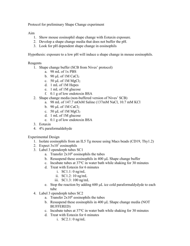
Protocol for preliminary Shape Change experiment Aim 1. Show mouse eosinophil shape change with Eotaxin exposure. 2. Develop a shape change media that does not buffer the pH. 3. Look for pH dependent shape change in eosinophils Hypothesis: exposure to a low pH will induce a shape change in mouse eosinophils. Reagents 1. Shape change buffer (SCB from Nives’ protocol) a. 98 mL of 1x PBS b. 90 L of 1M CaCl2 c. 50 L of 1M MgCl2 d. 1 mL of 1M Hepes e. 1 mL of 1M glucose f. 0.1 g of low endotoxin BSA 2. Shape change media (non-buffered version of Nives’ SCB) a. 98 mL of 147.7 mOsM Saline (137mM NaCl, 10.7 mM KCl b. 90 L of 1M CaCl2 c. 50 L of 1M MgCl2 d. 1 mL of 1M glucose e. 0.1 g of low endotoxin BSA 3. Eotaxin 4. 4% paraformaldehyde Experimental Design 1. Isolate eosinophils from an IL5 Tg mouse using Macs beads (CD19, Thy1.2) 2. Expect 3x10⁷ eosinophils 3. Label 3 ependorph tubes SC1 a. Transfer 2x10⁶ eosinophils the tubes b. Resuspend these eosinophils in 400 L Shape change buffer c. Incubate tubes at 37oC in water bath while shaking for 30 minutes d. Treat with Eotaxin for 6 minutes i. SC1.1: 0 ng/mL ii. SC1.2: 10 ng/mL iii. SC1.3: 100 ng/mL e. Stop the reaction by adding 600 L ice cold paraformaldydyde to each tube 4. Label 3 ependorph tubes SC2 a. Transfer 2x10⁶ eosinophils the tubes b. Resuspend these eosinophils in 400 L Shape change media (NOT BUFFERED) c. Incubate tubes at 37oC in water bath while shaking for 30 minutes d. Treat with Eotaxin for 6 minutes i. SC2.1: 0 ng/mL ii. SC2.2: 10 ng/mL iii. SC2.3: 100 ng/mL e. Stop the reaction by adding 600 L ice cold paraformaldydyde to each tube 5. Label 3 ependorph tubes SC3 a. Transfer 2x10⁶ eosinophils the tubes b. Resuspend these eosinophils in 400 L Shape change media (NOT BUFFERED) c. Incubate tubes at 37oC in water bath while shaking for 30 minutes d. Treat with DMEM for 6 minutes i. SC3.1: pH= 7.5 ii. SC3.2: pH: 7.0 iii. SC3.3: pH: 6.0 iv. SC4.3: pH 6.5 e. Stop the reaction by adding 600 L ice cold paraformaldydyde to each tube 6. Facs stain for left over eosinophils (5x10⁵ – 106 cells per stain) a. CCR3 or Siglec F b. Ly6G c. 1:1000 CXCR2 d. 1:10,000 CXCR2 e. 1:100,000 CXCR2 7. Facs calibur set up: a. FSC E-1 log b. SSC 258 log c. FL2 544 log d. Hopefully mouse eosinophils will have autofluorescents in FL2, look at only the FSC of those cells.
