Tutorial Skin Cancer
advertisement
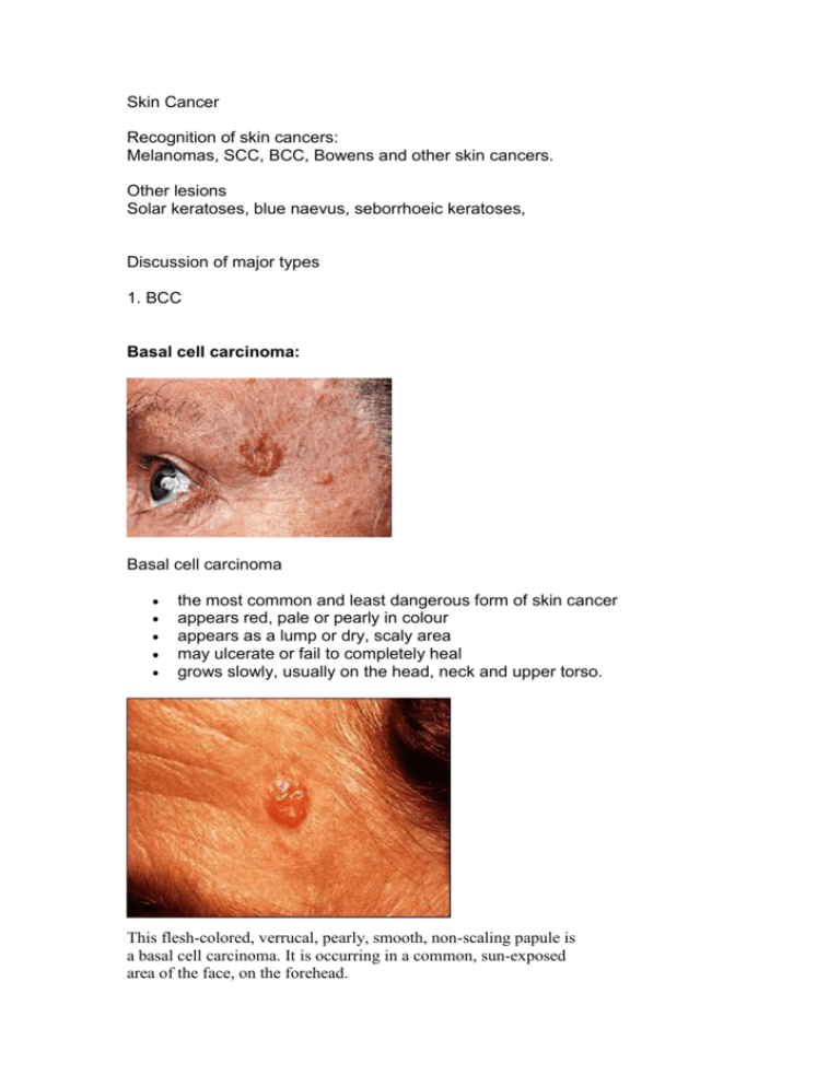
Skin Cancer Recognition of skin cancers: Melanomas, SCC, BCC, Bowens and other skin cancers. Other lesions Solar keratoses, blue naevus, seborrhoeic keratoses, Discussion of major types 1. BCC Basal cell carcinoma: Basal cell carcinoma the most common and least dangerous form of skin cancer appears red, pale or pearly in colour appears as a lump or dry, scaly area may ulcerate or fail to completely heal grows slowly, usually on the head, neck and upper torso. This flesh-colored, verrucal, pearly, smooth, non-scaling papule is a basal cell carcinoma. It is occurring in a common, sun-exposed area of the face, on the forehead. This skin cancer, a basal cell carcinoma, is 5 to 6 centimeters across, red (erythematous), with well defined (demarcated) borders and sprinkled brown pigment along the margins. This cancer is located on the person's back. The typical basal cell skin cancer appears as a small, pearly, dome-shaped nodule with small visible blood vessels (telangiectasias). This skin cancer appears as a 2 to 3 centimeter skin spot. The tissue has become destroyed (forming an atrophic plaque). There is a brownish color because of increased skin pigment (hyperpigmentation) and a slightly elevated, rolled, pearl-colored margin. This growth is located along the hair line. 2 SCC Squamous cell carcinoma: Squamous cell carcinoma not as dangerous as melanoma appears as a thickened, red, scaly spot that may bleed easily, crust or ulcerate appears on skin most often exposed to the sun grows over some months more likely to occur in people over fifty years. Metastasis common 3. Melanoma Melanoma is cancer of the skin's melanocytes (pigment cells) and the most deadly form of skin cancer. If untreated, it can spread to other parts of the body. There are two main types of melanoma. Melanoma a) Common (or superficial spreading) melanoma: appears as a new spot or an existing spot that changes colour, size or shape has an uneven, smudgy outline and is an irregular mix of colours may appear on skin not normally exposed to the sun. b) Nodular melanoma is not as common. It: grows quickly looks different from common melanomas - is raised from the start and even in colour may be red or pink; some are brown or black is firm to the touch and dome-shaped will begin to bleed and crust, after a while. Nodular melanoma c) acral lentiginous melanoma also known as subungual melanoma. It is seen on the palms, soles and under the nails. This is the most common form of melanoma in Asians and Blacks. The average age at diagnosis is between sixty and seventy years. It also occurs in Caucasians and in young people. This type of melanoma occurs on non hair baring surfaces of the body which may or may not be exposed to sunlight. Typical symptoms include: longitudinal tan, black, or brown streak on a finger or toe nail (melanonychia striata) pigmentation of proximal nail fold areas of dark pigmentation on palms of hands or soles of feet Any new area of pigmentation or an existing one that shows change should be checked by a dermatologist. If caught early this type of melanoma has a similar cure rate as the other types of superficial spreading melanoma. d) lentigo maligna (melanoma) Lentigo maligna is a melanoma in situ: it consists of malignant cells but does not show invasive growth. It can remain in this non- invasive form for years. It is normally found in the elderly (peak incidence in the 9th decade), on skin areas with high levels of sun exposure (for example, face and forearms). When it develops into melanoma, the resulting lesion is called lentigo maligna melanoma. The transition to melanoma is marked by the appearance of a bumpy surface (vertical growth, invasion). Therapy of lentigo maligna consists of surgical excision (best) or radiotherapy (only in special cases). Lentigo maligna melanoma Any of the above types may produce melanin (and be dark in colour) or not (and be amelanotic - not dark). Similarly any subtype may show desmoplasia (dense fibrous reaction with neurotropism) which is a marker of aggressive behaviour and a tendency to local recurrence. Popular method for remembering the signs and symptoms of melanoma is the mnemonic "ABCDE": Asymmetrical skin lesion. Border of the lesion is irregular. Color: melanomas usually have multiple colors. Diameter: moles greater than 5 mm are more likely to be melanomas than smaller moles. Evolution: The evolution (ie change) of a mole or lesion may be a hint that the lesion is becoming malignant --or-- Elevation: The mole is raised or elevated above the skin. Early melanoma Level 3 Melanoma Level 4 Melanoma Breslow's depth is most accurately measured by evaluating the entire tumor via an excisional biopsy. Determination from specimens obtained using other biopsy techniques, such as a wedge or punch biopsy, are less accurate. Breslow's depth should never be calculated from a shave biopsy; these specimens contain only a portion of the tumor and can easily lead to an underestimation of its thickness. Clark's levels is a related staging system, often used in conjunction with Breslow's depth, which depends on multiple anatomical landmarks in the skin. In fact, Clark's level was the primary factor in earlier AJCC staging schemae for melanoma. However, with further study, it has been shown that Clark's level has a lower predictive value, is less reproducible, and is more operatordependent as compared with Breslow's depth. Thus, in the current (2002) AJCC staging system, Clark's level has prognostic significance only in patients with very thin (Breslow depth <1 mm) melanomas. Solar keratoses or sunspots: a warning sign you are prone to sun damage and skin cancer appear as red, flattish, scaling dry skin that may sting if scratched appear on areas of skin most often exposed to the sun, like hands and face are most common in people over forty years Solar keratosis Seborrhoeic keratoses: Seborrhoeic keratoses spots with a clear edge; they look like they sit on top of the skin most people have at least one or two of these spots by the age of sixty vary in colour from pale brown to orange or black vary in size from a few millimetres to 2 cm. Cutaneous Markers of internal malignancy See http://www.dermnetnz.org/systemic/malignancy.html Atypical Naevus Atypical naevi can be considered funny looking moles. They may be larger than average (5-15mm) They may be an unusual shape or have a notched or blurred border. The colour is variable and may be pink, brown or black. The surface may be bumpy or smooth. They have characteristic dermoscopic features. They may have atypical or dysplastic features on biopsy. Blue Naevus Halo Mole

