outline3903
advertisement
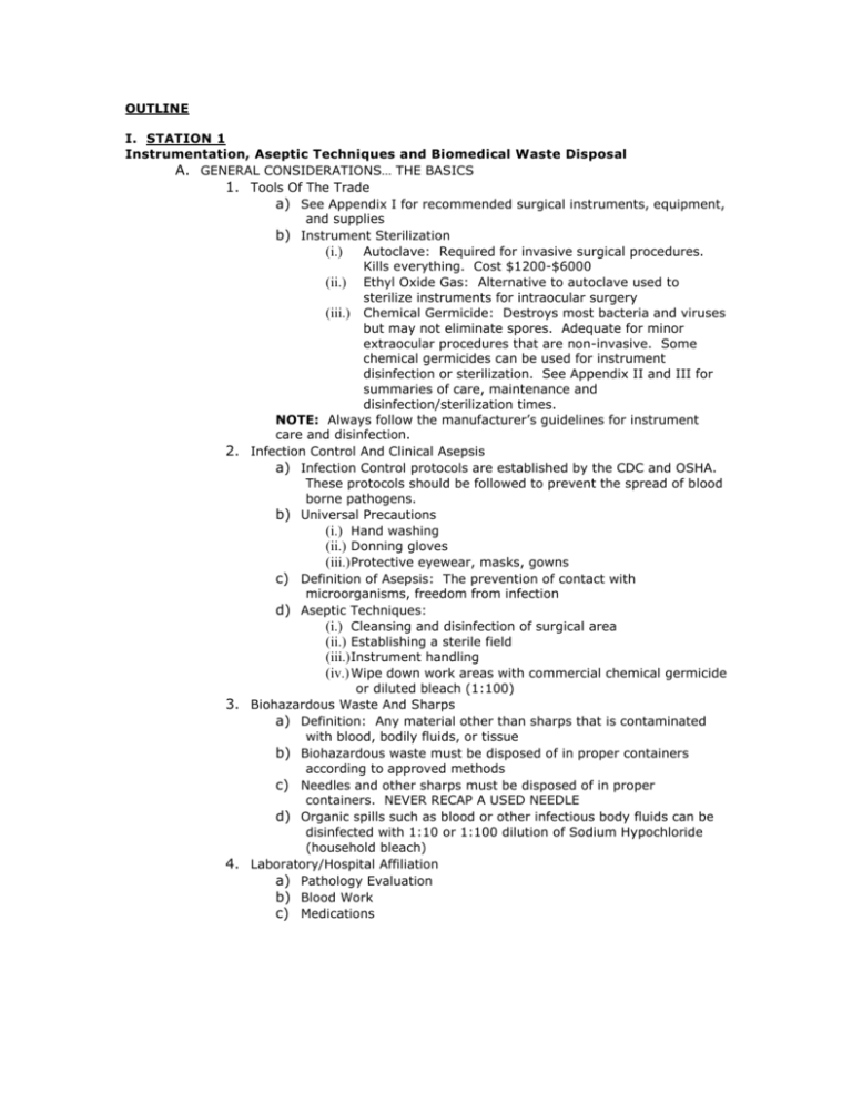
OUTLINE I. STATION 1 Instrumentation, Aseptic Techniques and Biomedical Waste Disposal A. GENERAL CONSIDERATIONS… THE BASICS 1. Tools Of The Trade a) See Appendix I for recommended surgical instruments, equipment, and supplies b) Instrument Sterilization (i.) Autoclave: Required for invasive surgical procedures. Kills everything. Cost $1200-$6000 (ii.) Ethyl Oxide Gas: Alternative to autoclave used to sterilize instruments for intraocular surgery (iii.) Chemical Germicide: Destroys most bacteria and viruses but may not eliminate spores. Adequate for minor extraocular procedures that are non-invasive. Some chemical germicides can be used for instrument disinfection or sterilization. See Appendix II and III for summaries of care, maintenance and disinfection/sterilization times. NOTE: Always follow the manufacturer’s guidelines for instrument care and disinfection. 2. Infection Control And Clinical Asepsis a) Infection Control protocols are established by the CDC and OSHA. These protocols should be followed to prevent the spread of blood borne pathogens. b) Universal Precautions (i.) Hand washing (ii.) Donning gloves (iii.) Protective eyewear, masks, gowns c) Definition of Asepsis: The prevention of contact with microorganisms, freedom from infection d) Aseptic Techniques: (i.) Cleansing and disinfection of surgical area (ii.) Establishing a sterile field (iii.) Instrument handling (iv.) Wipe down work areas with commercial chemical germicide or diluted bleach (1:100) 3. Biohazardous Waste And Sharps a) Definition: Any material other than sharps that is contaminated with blood, bodily fluids, or tissue b) Biohazardous waste must be disposed of in proper containers according to approved methods c) Needles and other sharps must be disposed of in proper containers. NEVER RECAP A USED NEEDLE d) Organic spills such as blood or other infectious body fluids can be disinfected with 1:10 or 1:100 dilution of Sodium Hypochloride (household bleach) 4. Laboratory/Hospital Affiliation a) Pathology Evaluation b) Blood Work c) Medications 5. Medicolegal Considerations a) Informed Consent (Appendix IV) b) Standard of Care c) Documentation II. STATION 2 Basic Injection Techniques A. ROUTES OF PARENTERAL ADMINISTRATION 1. Intradermal a) Between epidermis and the dermal layers b) Least used route c) Less blood supply, therefore, longer absorption time d) Used for diagnostic tests, such as, tuberculin skin test and allergy tests e) Body’s reaction to drug is easily visible f) 26-27 gauge, ¼ to ½ inch needle 2. Subcutaneous a) Loose connective tissue underlying dermis b) Blood supply not as rich as muscle c) Absorption time about 30 minutes d) Used for epinephrine, insulin, tetanus toxoid, vaccine, narcotics, vitamin B12 e) 25-27 gauge, ½ to 7/8 inch needle 3. Intramuscular a) For larger volume (3ml) and quicker absorption b) Less irritation from drug due to less sensory fibers c) Absorption time 10-30 minutes d) 19-23 gauge, 1 to 1 ½ inch needle 4. Intravenous a) Highest risk to patient b) Impossible to reverse effects of drug once administered c) Mix drug within large volume of IV fluids d) Piggyback drug through existing IV line e) Inject a bolus of drug B. BASIC INJECTION PROCEDURES 1. Procedural Safety Precautions a) Informed consent b) Blood borne pathogens c) Gloves d) Hand washing e) Handling sharp instruments f) Do not recap or remove contaminated needles 2. Intradermal, Subcutaneous, or Intramuscular Injection a) Glove b) Select injection site and wipe with alcohol swab c) Select needle size d) Recheck volume of medication e) Remove needle cap f) Inject needle g) Aspirate to avoid intravascular injection h) Inject medication slowly i) Withdraw needle quickly and cover puncture with antiseptic swab j) Dispose of needle and syringe 3. Intravenous Bolus Injection a) Prepare IV tray b) Glove c) Inject infusion line into vein (bevel up); do not inject if injection site appears puffy or edematous Attach syringe Inject medication Remove needle and apply gauze Ask patient how they feel Discard supplies Inspect injection site and apply bandage Discard gloves C. PERIOCULAR INJECTIONS 1. Subconjunctival a) Advantages (i.) Between anterior conjunctiva and Tenon’s capsule (ii.) Provides high local concentration of drug with less systemic side effects (iii.) Allows for high tissue concentration of drugs which have poor intraocular penetration (iv.) Good delivery of drug for unreliable patients (v.) Eliminate need for adjunctive topical drug therapy b) Technique (i.) Apply topical anesthetic (use soaked cotton-tipped applicator over injection site) (ii.) May choose to grasp and tent up conjunctiva with forceps (iii.) Use 25 gauge, 3/8 inch needle (iv.) Face bevel towards sclera (v.) Insert needle at desired location (usually inferior) (vi.) Withdraw plunger to guard against intravascular penetration (vii.) Inject contents of syringe and remove needle (viii.) Apply gentle pressure through closed eyelids (ix.) Discard waste c) Complications (i.) Perforation of the glove (ii.) Chemosis (iii.) Subconjunctival hemorrhage (iv.) Pain (v.) Retained drug deposits 2. Subtenons a) Advantages (i.) Same as with subconjunctival injection (ii.) Beneficial for posterior segment drug therapy b) Disadvantages (i.) Slightly greater risk of perforation than with subconjunctival injection (ii.) Slower absorption c) Technique (i.) Inject between Tenons capsule and sclera (ii.) Use 25 gauge, 5/8 inch needle (iii.) Follow same procedure as with subconjunctival injection d) Complications (i.) Perforation of the globe (ii.) Chemosis (iii.) Subconjunctival hemorrhage d) e) f) g) h) i) j) (iv.) Pain (v.) Retained drug deposits 3. Retrobulbar (and Peribulbar) a) Advantages (i.) Main use is for globe anesthesia (ii.) Blocks CN’s III, IV, VI, and ciliary nerves b) Technique for retrobulbar (and Peribulbar) injection (i.) Use 23-25 gauge, 1 3/8 inch needle with blunt tip (ii.) Face bevel towards globe (iii.) Insert needle through lower eyelid (iv.) Penetrate muscle cone (v.) Aspirate before injection (vi.) Inject contents (1.5 to 6.0 ml) in the vicinity of orbital apex (vii.) Remove needle (viii.) Apply pressure to globe (ix.) Discard waste c) Complications (i.) Ecchymosis of eyelid (ii.) Extraocular muscle palsies (iii.) Upper eyelid ptosis (iv.) Perforation of the globe (v.) Central retinal artery occlusion (vi.) Optic atrophy (vii.) Retrobulbar hemorrhage (viii.) Proptosis and exposure keratopathy (ix.) Elevated intraocular pressure (x.) Excitation of CNS (anesthesia) (xi.) Depression of cardiovascular system (anesthesia) 4. Intracameral a) Advantages (i.) Injection directly into anterior chamber (ii.) Produces rapid high concentration of drug (iii.) Uses very minute volume of drug b) Technique (i.) Use cotton-tipped applicator dipped in 2-4% cocaine to anesthetize the base of the medial rectus muscle (for 1 minute) (ii.) Retract eyelid with eyelid speculum (iii.) Use forceps to fixate globe by holding base of medial rectus muscles (iv.) Use 30 gauge needle on a tuberculin syringe (v.) Pass needle through cornea into anterior chamber (vi.) Enter anterior chamber temporally at the limbus w/ bevel pointed up (vii.) Withdraw fluid (usually 0.1-0.2 cc) until anterior chamber shallows (viii.) Withdraw needle and place one drop topical antibiotic in eye c) Disadvantages (i.) Carries high risk for drug toxicity 5. Intravitreal a) Injection into vitreous cavity b) Main use is for endophthalmitis and air-fluid gas exchange III. STATION 3 Minor Surgical Procedures A. THE 5 P’s 1. Preop a) Asymmetry b) Borders/bleeding c) Color/care d) Duration/diameter e) Evaluate 2. Prep a) Set-up tray b) Informed consent c) Wash hands and glove d) Clean surgical site e) Administer anesthetic 3. Procedure a) Anesthetic test b) Reassure patient c) Perform procedure d) Control bleeding e) Wound closure 4. Postop a) Apply antibiotic/steroid/combination to surgical site b) Write Rx for antibiotic and pain control medication if necessary c) Schedule follow-up PO 5. Paperwork a) Complete medical record documentation b) Complete pathology report and arrange for pick-up if necessary B. EYELID PROCEDURES 1. Epilation 2. Cyst Evacuation 3. Papilloma Removal 4. Chalazion: Incision and Curettage 5. Xanthelasma Excision 6. Entropion Repair C. LACRIMAL PROCEDURES 1. Dilation and Irrigation 2. Punctal Plug Insertaion 3. Punctal Cautery D. CORNEAL PROCEDURES 1. Foreign Body Removal 2. Rust Ring Removal 3. Debridement 4. Anterior Stromal Puncture E. CONJUNCTIVAL PROCEDURES 1. Foreign Body Removal 2. Cyst Drainage * APPENDIX I-IV WILL BE GIVEN OUT AT APPROPRIATE STATIONS
