Tertiary Species - Reptiles and Amphibians
advertisement
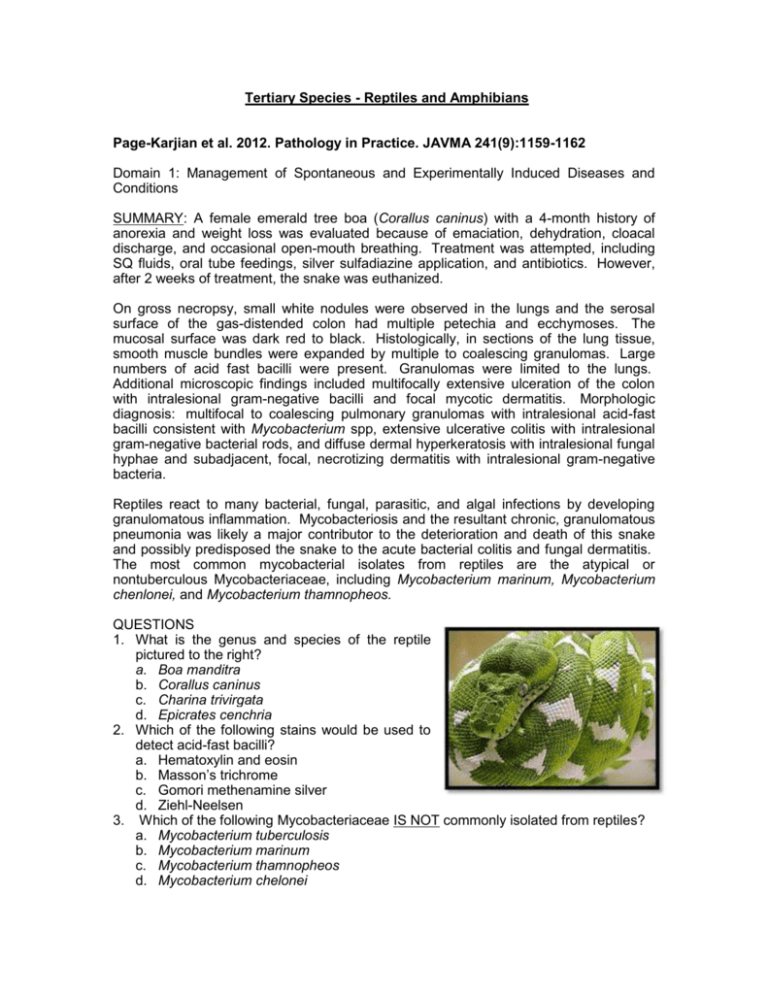
Tertiary Species - Reptiles and Amphibians Page-Karjian et al. 2012. Pathology in Practice. JAVMA 241(9):1159-1162 Domain 1: Management of Spontaneous and Experimentally Induced Diseases and Conditions SUMMARY: A female emerald tree boa (Corallus caninus) with a 4-month history of anorexia and weight loss was evaluated because of emaciation, dehydration, cloacal discharge, and occasional open-mouth breathing. Treatment was attempted, including SQ fluids, oral tube feedings, silver sulfadiazine application, and antibiotics. However, after 2 weeks of treatment, the snake was euthanized. On gross necropsy, small white nodules were observed in the lungs and the serosal surface of the gas-distended colon had multiple petechia and ecchymoses. The mucosal surface was dark red to black. Histologically, in sections of the lung tissue, smooth muscle bundles were expanded by multiple to coalescing granulomas. Large numbers of acid fast bacilli were present. Granulomas were limited to the lungs. Additional microscopic findings included multifocally extensive ulceration of the colon with intralesional gram-negative bacilli and focal mycotic dermatitis. Morphologic diagnosis: multifocal to coalescing pulmonary granulomas with intralesional acid-fast bacilli consistent with Mycobacterium spp, extensive ulcerative colitis with intralesional gram-negative bacterial rods, and diffuse dermal hyperkeratosis with intralesional fungal hyphae and subadjacent, focal, necrotizing dermatitis with intralesional gram-negative bacteria. Reptiles react to many bacterial, fungal, parasitic, and algal infections by developing granulomatous inflammation. Mycobacteriosis and the resultant chronic, granulomatous pneumonia was likely a major contributor to the deterioration and death of this snake and possibly predisposed the snake to the acute bacterial colitis and fungal dermatitis. The most common mycobacterial isolates from reptiles are the atypical or nontuberculous Mycobacteriaceae, including Mycobacterium marinum, Mycobacterium chenlonei, and Mycobacterium thamnopheos. QUESTIONS 1. What is the genus and species of the reptile pictured to the right? a. Boa manditra b. Corallus caninus c. Charina trivirgata d. Epicrates cenchria 2. Which of the following stains would be used to detect acid-fast bacilli? a. Hematoxylin and eosin b. Masson’s trichrome c. Gomori methenamine silver d. Ziehl-Neelsen 3. Which of the following Mycobacteriaceae IS NOT commonly isolated from reptiles? a. Mycobacterium tuberculosis b. Mycobacterium marinum c. Mycobacterium thamnopheos d. Mycobacterium chelonei ANSWERS 1. a. Boa manditra (Madagascar tree boa) b. Corallus caninus (Emerald tree boa) c. Charina trivirgata (Rosy boa) d. Epicrates cenchria (Rainbow boa) 2. d. Ziehl-Neelsen 3. a. Mycobacterium tuberculosis Gumber et al. 2012. Pathology in Practice. JAVMA 241(3):327-330 [Boa Constrictor] SUMMARY: An 11-year-old female Rainbow boa constrictor (197 cm in length and weighing 4 kg [8.8 lb]) was evaluated because of a 1.5-month history of constipation. On physical examination, the boa constrictor was weak and lethargic and had a large swelling in the body wall. Ultrasonographic examination revealed a mass that was compressing the colon and invading approximately 80% of the coelomic cavity and a small amount of effusion cranial and caudal. Postmortem examination revealed a pale gray, firm, multilobulated mass (12 cm in diameter) in the body wall. On cut surface, the mass encompassed the deep dermis, ribs, and vertebrae and had multifocal areas of fat necrosis. The coelom contained approximately 100 mL of clear fluid. Feces were dry and firm, consistent with the history of constipation. The body wall mass was a large, unencapsulated, infiltrative, moderately cellular subcutaneous neoplasm composed of spindle cells and arranged in bundles. There were 2 to 3 mitoses/10 hpf. Multifocal areas of fat necrosis, myodegeneration, and myonecrosis were observed. The renal architecture at the cranial poles of both kidneys was effaced by broad streams of neoplastic spindle cells resembling the body wall mass. Similarly, aggregates of adipocytes in fat bodies were interlaced with numerous bundles smooth muscle actin. The prevalence of neoplastic disease in captive reptiles is increasing as a result of increased life expectancy associated with improved husbandry and management. The commonly reported neoplasms included mesenchymal, epithelial, and lymphoid or hematopoietic neoplasms. Among ophidian species, neoplasms are most commonly reported for aged colubrids, followed by crotalids, vipers, and boids. The differential diagnoses (determined on the basis of clinical history of constipation and abdominal swelling) were subcutaneous granulomas caused by various infectious organisms (fungi, mycobacteria, or parasites), bacterial abscess, egg binding, gastrointestinal tract obstruction, and a neoplastic process. The histopathologic features of the body wall mass were consistent with leiomyosarcoma in the body wall with extension into the kidneys and fat bodies in a Rainbow boa constrictor. QUESTIONS 1. Which is the most commonly reported species among ophidians for neoplasm? a. Crotalids b. Colubrids c. Vipers d. Boids 2. The prevalence of neoplastic disease in captive reptiles is increasing as a result of : a. Nutrition b. Genetics c. Increased life expectancy d. All above are false 3. The differential diagnosis for a clinical history of constipation and abdominal swelling includes: a. Bacterial abscess b. Neoplastic process c. Egg binding d. a, b, c ANSWERS 1. b 2. c 3. d Mans and Sladky. 2012. Endoscopically guided removal of cloacal calculi in three African spurred tortoises (Geochelone sulcata). JAVMA 240(7):869-875 SUMMARY: This is a case report detailing the removal of cloacal calculi from 3 African spurred tortoises. Each one presented with anorexia and constipation. The calculi were diagnosed on the basis of radiography and cloacoscopy. Serology revealed hyperuricemia in 2 of the cases. No bloodwork was reported for the third case. The cloaca is the distal terminus of the gastrointestinal, reproductive and urinary systems in tortoises. Cloacal calculi are caused by the migration of cystic calculi (bladder stones) from the urinary bladder to the pelvic canal where they can get wedged in the cloacal. The options for removal of these calculi include endoscopic intervention, plastron osteotomy and surgical approach through the prefemoral fossa. Endoscopy is the least invasive procedure which allows for a shorter anesthetic exposure and quicker recovery. In each of these 3 cases, the tortoises were sedated with ketamine, medetomidine, midazolam and morphine. The procedures lasted from 30 – 60 minutes. One tortoise required a second procedure. Another tortoise had a second calculus visible in the bladder which was not accessed. The sedation was reversed with atipamezole and flumazenil which allowed the animals to return to their homes within a few hours of the procedure. The difference between this procedure and those previously described that the visual field was maintained by lavaging with water in between drilling (with a dental drill) into the calculi with a cutting burr. The procedure was successful in all 3 cases. QUESTIONS 1. What is the genus and species of the African spurred tortoises? 2. What is the reversal agent for medetomidine? For midazolam? 3. What organ systems are affected with an obstructive cloacal calculi? 4. What are the options for removal of cloacal calculi in tortoises? 5. Name potential causes of the formation of cystic calculi in tortoises. ANSWERS 1. Geochelone sulcata 2. Atipamezole; Flumazenil 3. Gastrointestinal, reproductive and urinary systems 4. Plastron osteotomy, cloacoscopy and surgical approach through the prefemoral fossa 5. Vitamin deficiencies, calcium deficiency, excess protein and oxalates in the diet, bacterial infections of the urinary bladder and suture remnants have all been suggested, but the underlying cause is not known. Burcham et al. 2011. Pathology in Practice. JAVMA 239(10):1305-1310 [Anole] Domain 1: Management of Spontaneous and Experimentally Induced Diseases and Conditions Tasks T2. Control spontaneous or unintended disease or condition T3. Diagnose disease or condition as appropriate T4. Treat disease or condition as appropriate SUMMARY History: Two adult brown anoles (Anolis sagrei), also known as Bahama anoles, were evaluated because of multiple skin lesions. These lizards were from a pet store, where 3 groups of 10 to 15 lizards each were similarly affected Clinical Findings: Physical Exam: Skin lesions in the 2 evaluated anoles were similar in appearance but varied in size and location. Lesions were round to ovoid, gray to black plaques that ranged from1mm in diameter to an area of 7X3mm. Foci were usually well demarcated from adjacent, more normal-appearing skin and contained white, powdery material on their surfaces. Smaller lesions were completely black and lacked powdery material. Histopathology: Diagnosis: Skin plus subcutaneous tissue plus muscle plus blood vessels, necrosis plus mild granulomatous inflammation associated with septate rarely branching mycotic hyphae, mycotic dermatitis plus cellulitis plus myositis. Comments: Based on histopathology, the major cause of disease in these anoles is a species of nonpigmented fungus (hyalohyphomycosis). The microscopic characteristics of the fungus (including arthroconidiating hyphae at the skin surface), the extensive nature of the lesion (i.e., cellulitis and myositis), and the clinical history of multiple infected lizards strongly suggest that the etiologic agent of the skin lesions is the Chrysosporium anamorph of Nannizziopsis vriesii (CANV). CANV has been implicated as the cause of dermatomycosis and deep mycosis in multiple lizard species, as well as snake species and salt water crocodiles. In bearded dragons, the disease is known as yellow fungus disease. CANV is considered a highly contagious, primary pathogen of reptiles. Although resident cutaneous fungi are common in reptiles, the CANV appears to be a rare inhabitant of healthy reptile skin. Control and treatment o Treatment plan should include isolation of affected animals, when feasible. Reduction of overcrowding or other environmental stressors may mitigate future outbreaks. Adjustment of the environmental temperature to the upper end of the species’ preferred temperature range may be beneficial. o Cutaneous lesions caused by the CANV can be treated surgically, with excision or debridement of affected tissue and subsequent topical application of antifungal agents. o Concurrent systemic treatment with antifungal agents is warranted. Itraconazole is the drug of choice for systemic antifungal treatment in reptiles; fluconazole has little efficacy against the CANV and should be avoided. Treatment regimens should continue until lesions regress, barring development of medication-related adverse effects. QUESTIONS: 1. The following agent is the cause of skin and deep mycosis in many reptile species: a. Coccidioides immitis b. Candida albicans c. Chrysosporium anamorph of Nannizziopsis vriesii (CANV) d. Histoplasma capsulatum 2. True or False: Yellow fungus disease in bearded dragons is caused by Chrysosporium anamorph of Nannizziopsis vriesii (CANV) 3. True or False: Fluconazole is very effective for treatment of CANV infection in reptiles. 4. What is the drug of choice for treatment of CANV infection in reptiles? ANSWERS: 1. c. Chrysosporium anamorph of Nannizziopsis vriesii (CANV) 2. True 3. False 4. Itraconazole is the drug of choice for systemic antifungal treatment in reptiles Folland et al. 2011. Diagnosis and management of lymphoma in a green iguana (Iguana iguana). JAVMA 239(7):985-991 Domain 1: Spontaneous and Experimentally Induced Diseases and Conditions SUMMARY: A 2-year-old female green iguana was examined for anorexia, swelling, and pain on palpation of the cranial cervical area. The iguana was considered to be 50% underweight. Marked soft tissue swelling with corresponding cystic swellings in the pharynx was noted. The WBC count was markedly elevated. Surgical exploration identified lymphoma with secondary infection, as confirmed by histological and microbial testing. The tumor was initially treated with a single 10-Gy fraction of radiation. A canine chemotherapy protocol was modified for use in the iguana, and a vascular access port was placed in the ventral abdominal vein. The iguana appeared to be in remission at 1,008 days after initiation of treatment. This is probably the first reported use of radiation with doxorubicin, vincristine, cyclophospamide and prednisone to successfully manage lymphoma in a reptile. QUESTIONS: 1. How was the lymphoma found? a. Exploratory surgery with histological testing b. Fine needle aspirate c. Physical exam d. Biopsy 2. T or F: The use of radiation and chemotherapy to manage tumors is welldocumented in reptiles. 3. How was the chemotherapy treatment administered to the iguana? a. Intramuscularly b. Intracolonically c. Sedation for intravenous treatment d. Vascular access port ANSWERS: 1. a 2. F 3. d Hughes et al. 2011. What Is Your Diagnosis? JAVMA 239(5):573-574 [Box Turtle] SUMMARY: An adult female free-ranging box turtle, found along the roadside, presented with an open carapacial wound. The wound was cleansed with chlorhexidine gluconate and bandaged. Ceftiofur, meloxicam, and fluids were administered subcutaneously. The turtle was also noted to have hind limb deficits. Whole body radiographs revealed four intact mineralized eggs and several fracture lines along the carapace with ventral displacement of some fracture fragments. Computed tomography was used to assess impingement of the carapace fragments on the vertebral column and spinal cord. One of the displaced fracture fragments was compromising the spinal canal. The CT also showed settling of hyperdense contents in the dependent part of the egg in the scan field. Conservative treatment was pursued consisting of wound lavage, bandage changes, and administration of ceftiofur and meloxicam. Fracture fragments remained stable. Laser treatment of the wound was used to prevent excessive inflammation as well as increase formation of new collagen. Three of the eggs noted on radiographs were laid but never hatched and the final egg was never laid. Gradual improvement of hind limb function was observed. QUESTIONS: 1. Which of the following is an adequate probe placement location for conducting a caudal coelomic cavity ultrasound on a turtle? a. Over the carapace b. Over the plastron c. In the dependent prefemoral region d. In the dependent prescapular region 2. T/F. Normal turtle eggs are characterized on ultrasonographic examination by a hypoechoic or anechoic layer of albumin surrounding the central hyperechoic yolk. ANSWERS: 1. c 2. True Cardona et al. 2011. Incomplete ovariosalpingectomy and subsequent malignant granulose cell tumor in a female green iguana (Iguana iguana). JAVMA 239(2):237242 Domain 1: Management of Spontaneous and Experimentally Induced Diseases and Conditions, Task 3: Diagnose disease or condition as appropriate SUMMARY: Case Report 9 year old, spayed female iguana CS: enlarged coelom and progressive weight loss Hx: one episode of egg binding followed by bilateral ovariosalpingectomy (2 yrs prior) PE: generalized muscle wasting, large mass in mid-coelomic region on the left side Diagnostics: U/S with FNAs, CBC, serum biochemistry U/S: body cavity effusion, irregular mass with multiple hypoechoic regions in the midcoelomic region (unable to determine affected organ) CBC: heterophilia, monocytosis, lymphopenia, and basophilia Chem: hypocholesterolemia, hypoproteinemia, and hypercalcemia FNA (body fluid): transudate (no infectious agent) FNA (mass): suggestive of malignant epithelial neoplasia Main R/O: Ovarian tumor Surgical exploration – large left ovary, normal right ovary, and mass in the fat body. All were removed and iguana recovered well from surgery Histo: Granulosa cell tumor (GCT) with metastasis to fat body Follow-up: U/S recheck 4 months after surgery documented multiple irregular nodules t/o coelomic cavity. Chemotherapy (carboplatin) was attempted. Nodules decreased in size at first but then continued to grow and the iguana died 11 months after initial presentation Histo: Masses had 3 cell populations (neoplastic cells similar to original mass, large well-differentiated mast cells, and cords of atrophic adipocytes); mast cell infiltrates were also present in other parts of the body not associated with neoplastic cells Discussion Dystocia is common in reptiles (10% reported incidence) and neutering is a common treatment or prophylaxis If oophorectomy is incomplete in reptiles, even small remnants can regrow and folliculogenesis will occur; if oviducts were also removed, normal oviposition cannot occur and problems will develop Use of hemostatic clips and microsurgical instruments is recommended to ensure complete excision of ovarian tissue Systemic mastocytosis may be due to hyperestrogenism from GCT (unable to document increased estrogen in this case), an inflammatory response, or development of a concurrent neoplasia First report of a granulosa cell tumor in reptiles that has antemortem cytological, histological, and ultrastructural documentation QUESTIONS: 1. Describe how the ovarian tissue in the iguana differs from that of the cat or dog. 2. Name the 3 broad categories of primary ovarian tumors in mammals. 3. Name 3 other species that GCTs are found in. 4. Give the name of the structure that is considered to be the histological hallmark of GCTs. ANSWERS: 1. Iguana ovarian tissue is diffuse and intimately associated with the vena cava and adrenal gland. This makes oophorectomy technically challenging in this species. 2. These categories are on the basis of embryological cell of origin of the predominating neoplastic cell: a. Epithelial tumors (adenocarcinoma and adenoma) b. Germ cell tumors (dysgerminoma and teratoma) c. Sex cord tumors (GCT, thecoma, granulosa-theca cell, and luteoma) – usually associated with excessive sex hormone production and related CS (irregular estrus, aggression, masculine behavior, nymphomania, and infertility) 3. Humans (5% of all ovarian tumors), horses (85% of all reproductive tract tumors; 2.5% of all tumors), and dogs (0.5-1.2% of all tumors) 4. a. Call-Exner bodies. b. Not observed in every case of a GCT. c. Composed of a round, eosinophilic center of proteinaceous material rimmed by an acinar-like aggregate of tumors cells d. Occasionally seen along with capillaries that appear as linear structures containing erythrocytes lined by endothelial cells with elongated nuclei Baker et al. 2011. Evaluation of the analgesic effects of oral and subcutaneous tramadol administration in red-eared slider turtles. JAVMA 238(2):220-227 Domain 2: Management of Pain and Distress SUMMARY: The need exists for identifying an efficacious analgesic in reptiles that does not result in significant respiratory depression. Tramadol, a noncontrolled, commonly used analgesic in small animal practice, results in analgesia in mammals by activating m-opioid receptors and by inhibiting serotonin and norepinephrine reuptake in the CNS. The drug binds with the m-opiod receptors with 6000 times less affinity than morphine, having the potential for producing fewer related adverse effects. The purpose of this study was to determine the dose- and time-dependent changes in analgesia and respiration caused by tramadol administration in red-eared slider turtles. Analgesia experiments were conducted by applying infrared thermal stimuli to the plantar surface of the turtle hind limbs and measuring hind limb thermal withdrawal latencies at baseline and 7 time points after administration. The first group received either oral tramadol dissolved in 0.1 ml water or the same volume of water (control), were tested and then allowed to rest 2 weeks. The group then received either 0.1 ml saline SC or tramadol SC at 10 and 25 mg/kg dissolved in saline. Respiratory experiments measured ventilation (ml/min/kg) in conscious, freely swimming turtles using a pneumotachometer. Turtles were either given water PO or tramadol PO at 5, 10, and 25 mg/kg in water after acclimation and 2 hours of baseline breathing in the respiratory tank. For short-term trials, turtles were returned to the tank for another 12 continuous hours and for separate long-term trials, they were not returned to the tank immediately, but instead at 24, 48 and 72 hours following drug administration. In this study, tramadol resulted in analgesia with mild respiratory depression in red-eared slider turtles. 5 to 10 mg/kg of tramadol PO appeared to be the ideal dose range for providing pain relief without respiratory depression. Tramadol administered at 25 mg/kg resulted in flaccid limbs and neck as well as respiratory depression within the first 12 hours. Oral administration was more effective for analgesia as SC administration resulted in lower withdrawal latencies, slower onset, and decreased duration of action. QUESTIONS: 1. T/F. Tramadol’s metabolite O-desmethyl-tramadol (M1) has 200 times as great an affinity for m-opioid receptors than tramadol. 2. Which dose is associated with the best analgesia and least respiratory depression: a. 1 mg/kg PO b. 15 mg/kg PO c. 15 mg/kg SQ d. 25 mg/kg SQ 3. Which of the following is not a limitation of using tramadol SC in turtles: a. An injectable tramadol formulation is not commercially available b. SC tramadol is absorbed directly into systemic circulation, bypassing the liver c. SC injection results in higher withdrawal latencies d. SC tramadol results in slower onset and decreased duration of action as compared to PO ANSWERS: 1. T 2. B. 15 mg/kg PO 3. C. tramadol SQ results in LOWER withdrawal latencies Ruder et al. 2010. Pathology in Practice. JAVMA 237(7):783-787 [Turtle] SUMMARY: An adult female eastern box turtle (Terrapene carolina carolina) was presented moribund to the University of Georgia after being found along a pond in Kentucky in the vicinity of 7 other dead turtles. Clinical signs included severe depression, dehydration and lethargy, mucopurulent, bilateral nasal discharge and respiratory distress. The animal was euthanized and submitted for necropsy. Gross findings included a firm, swollen right side of the face with numerous multifocal, raised, tan-white plaques along the mouth, hard palate, esophagus, coelomic cavity and on the serosal surfaces of organs. Plaques were prominent on the mesentery, liver, spleen and stomach. Morphologic diagnosis was severe, chronic, focally extensive, ulcerative and necrotizing pharyngitis and esophagitis with multicentric heterophilic vasculitis, thrombosis and fibrinoid change. Differential diagnoses for this case included ranavirus infection, herpesvirus infection and septicemia with or without mycoplasmosis. Ranavirus was isolated from the spleen, kidneys, liver and lungs. Samples were cultured on Terrapene carolina heart cells. The ranavirus was further identified as Frog virus-3 or FV-3 by gene sequencing and PCR. There was no concurrent mycoplasma or herpesvirus infection. Clinical signs of ranaviral infections in chelonians include dyspnea, depression, palpebral edema, oculonasal discharge and death. Gross findings generally include conjunctivitis and yellow-tan caseous plaques on the tongue, palate, pharynx and esophagus. Microscopic lesions include necroulcerative glossitis, esophagitis and pharyngitis, as well as splenitis and multicentric vasculitis with fibrinoid change. Basophilic intracytoplasmic inclusion bodies are sometimes present in infected tissues. Infected animals should be euthanized. There is increasing evidence of the ability of ranaviruses to infect reptiles. QUESTIONS: 1. Which of the following statements is false regarding ranavirus? a. It is an iridovirus b. Ranavirus can cause mass mortality in reptiles, amphibians and fish. c. Eosinophilic intranuclear inclusion bodies are sometimes present in infected tissues. d. Clinical signs of a ranaviral infection include depression, dyspnea and oculonasal discharge. 2. True or False: Medical treatment, rehabilitation and/or release of free ranging chelonians and amphibians with suspected or confirmed ranaviral infection should not be attempted. ANSWERS: 1. c 2. True Domain 1 – Management of Spontaneous and Experimentally-induced Disease Conditions; T3 – Diagnose disease or condition as appropriate SUMMARY: History: Wild eastern box turtle (Terrapene carolina carolina) found moribund near the edge of a 1-acre pond. 7 additional box turtles were found dead nearby. Physical Exam: Severe depression, dehydration, lethargic and poorly responsive to external stimuli. Mild bilateral mucopurulent nasal discharge, slight respiratory difficulty, right palpebral swelling, face right side moderately swollen and firm. Euthanasia performed due to grave prognosis. Necropsy: Skin overlying the swollen face was yellow-tan and partially sloughed. The hard palate was covered by a light-tan plaque with a rough undulating surface, and underlying ulcer. Similar lesion in the proximal esophagus. Multiple 1 to 3 mm diameter nodules present in coelomic cavity, serosal surfaces, liver, spleen and stomach. Many muscles of the head were pale, dry and firm. Histopathology: Widespread, multifocal infiltration of blood vessel walls by moderate to marked numbers of heterophils and fewer lymphocytes, sometimes associated with surrounding tissue coagulative necrosis and more heterophilic inflammation. Morphologic Diagnosis 1) Severe, chronic, focally extensive, ulcerative and necrotizing pharyngitis and esophagitis with multicentric heterophilic vasculitis, thrombosis and fibrinoid change 2) Severe, chronic, multifocal to coalescing, necrotizing, fibrinoheterophilic myositis and osteomyelitis of the head 3) Mild, chronic, multifocal, heterophilic meningitis with vasculitis 4) Severe, subacute, diffuse, fibrinoheterophilic, necrotizing splenitis 5) Moderate, multifocal, subacute, heterophilic, necrotizing multinodular pneumonia with vasculitis and thrombosis. QUESTIONS 1. Differential diagnosis for this case includes: a. Ranavirus infection, with or without concurrent mycoplasmosis b. Herpesvirus infection, with or without concurrent mycoplasmosis c. Systemic bacterial infection, with or without concurrent mycoplasmosis d. a and b e. a, b and c 2. Clinical signs of ranavirus infection in chelonians include: 3. True or false – ranavirus can cause intracytoplasmic inclusion bodies 4. What groups of animals are susceptible to ranavirus infection? a. Amphibians b. Fish c. Chelonians d. a and c e. All of the above 5. True or false – medical treatment, rehabilitation and release of free-ranging chelonians and amphibians with suspected or confirmed ranavirus infection is an acceptable practice ANSWERS: 1. e (in this case, the diagnosis was ranavirus infection – Frog Virus-3. All bacterial and mycoplasma cultures were negative) 2. Depression, dyspnea, palpebral edema, oculonasal discharge, death 3. True; however, the basophilic cytoplasmic inclusion bodies are often are not present. 4. E 5. False – Such animals may still harbor and shed the virus and are a risk for populations of free-ranging amphibians and chelonians Stern. 2009. Pathology in Practice. JAVMA 235(6):669-671 Task 1- Prevent, Diagnose, Control, and Treat Disease Tertiary Species – Reptiles SUMMARY: A juvenile green iguana was evaluated because of anorexia and progressive weakness of 1 month’s duration. Abnormal findings included an obtunded attitude, body condition score of 0.5 (on a scale of 5), marked soft swelling of the mandible and maxilla, bilateral swelling and thickening of the appendicular skeleton. Because the prognosis for recovery was poor, the iguana was euthanized. Gross findings at necropsy included poor nutritional condition, absence of intracoelomic fat stores. The mandible, maxilla, humerus, femur, radius, ulna, tibia, and fibula were soft and thickened by white to tan fibrous connective tissue; this tissue also encroached the nasal cavity. The parathyroid glands could not be identified. Histologically, the normal architecture of numerous bones was disrupted by resorption of bone and deposition of fibrocartilage and bone marrow was hypocellular and infiltrated by connective tissue. The morphologic diagnosis was marked, chronic, diffuse resorption of bone with marked fibrocartilage deposition (consistent with fibrous osteodystrophy). Fibrous osteodystrophy as a result of nutritional secondary hyperparathyroidism is common in iguanas and often develops as a result of an inadequate diet or husbandry mismanagement. Factors most commonly associated with this disease include a prolonged dietary deficiency of vitamin D3 or calcium, an imbalance of dietary calcium to phosphorous, or inadequate exposure to UV radiation in diurnal animals. Because of the iguana’s small body size, the parathyroid glands could not be identified during necropsy. In green iguanas, there are 2 pairs of parathyroid glands. The anterior parathyroid glands are located along the medial surface of the mandibular ramus. The posterior glands are located at the bifurcation of the internal and external carotid arteries. After initial treatment of serious problems such as fractures and tetany, affected reptiles require oral treatment with a calcium supplement, parenteral administration of vitamin D3, and IM administration of calcitonin. Improper diet and inadequate lighting should also be corrected. The prognosis for reptiles with nutritional secondary hyperparathyroidism varies from good to poor. The prognosis for reptiles with fibrous osteodystrophy, pathologic fractures, and paralysis is poor. QUESTIONS: 1. Fibrous osteodystrophy in iguanas often develops because of? 2. What are 2 factors commonly associated with fibrous osteodystrophy? 3. How many parathyroid glands do green iguanas have? 4. What is the prognosis for reptiles with fibrous osteodystrophy? ANSWERS: 1. Fibrous osteodystrophy as a result of nutritional secondary hyperparathyroidism often develops as a result of an inadequate diet or husbandry mismanagement. 2. Factors commonly associated with fibrous osteodystrophy include prolonged dietary deficiency of vitamin D3 or calcium, an imbalance of dietary calcium to phosphorus, or inadequate exposure to UV radiation in diurnal animals. 3. Green iguanas have 2 pairs of parathyroid glands. 4. The prognosis for reptiles with fibrous osteodystrophy is poor. Frye et al. 2009. Pathology in Practice. JAVMA 235(5):511-513 Domain 1, T3: Diagnose disease and condition Tertiary Species – Reptile SUMMARY: This case describes primary osteoma cutis diagnosed in a wild-caught adult female European pond turtle (Emys orbicularis). This is the formation of bone within the skin in the absence of a pre-existing lesion and is the first known report in reptiles. The turtle was observed with a dorsal cervical mass that party affected her ability to retract her head and neck into the shell and was captured for mass excision. The mass was bilaterally symmetrical, originating from the epidermis, and had a median raphe-like groove. Histological examination revealed highly sclerotic mature lamellar bone covered with keratinized squamous epithelium. This is a benign lesion and the turtle recovered well after excision, regained full function of her neck and there was no evidence of recurrence three years after surgery. QUESTIONS: 1. What is the clinical significance of the lesion in this turtle? 2. What is a similar bony lesion that has been reported in reptiles? ANSWERS: 1. She was unable to fully retract her head and neck into the shell, an important passive defense postural behavior in the wild. 2. Osteomata – but these lesion are exostoses, origination from the integument is unusual Raiti. 2009. What’s your diagnosis? JAVMA 234(7):877-878 Species: Tertiary (Red-eared slider turtle =Trachemys scripta elegans) Domain: 1; Task3: Diagnose disease or condition as appropriate SUMMARY: A 3–year-old female red-eared slider turtle (Trachemys scripta elegans) weighing 240 g (0.53 lb) was evaluated because of recurring episodes of cloacal prolapse. Prolapse usually recurred several days after each suture removal. The turtle has been maintained in an-outdoor pond with other turtle for 1 year prior to being moved indoors. Within 1 month after being transferred to an indoor enclosure, the turtle developed a cloacal prolapse. Treatment consisted of multiple reductions and placement of purse-string sutures that were kept in place for 1-week durations. Bacteria identified from cloacal culture were Aeromonas hydrophila, Pasteurella sp., and Acinetobacter sp. On physical examination, the turtle was active and alert. Body scoring system was judged to be 2 (underfed). Plasma biochemical and hematologic abnormalities were as follows: hypoalbuminemia, low total protein concentration, hypocalcemia, hypophosphatemia, hypoglycemia, low Hematocrit and leucopenia. Whole-body radiography was performed. Oral administration of 2.3 ml contrast agent (an ionic iodinated) was administered via gastric tube. Dorsoventral radiographic views were obtained immediately and at 15 minutes, 2 hours, 22 hours and 144 hours after administration. At 144 hours, the contrast agent is still within the transverse colon and has not reached the descending colon (a foreign body is not identified). A suspected blockage of the descending colon causes an obstructive ileus. An exploratory celiotomy was recommended; however the owner requested euthanasia. On necropsy, the descending colon appeared atrophied. Ulcerative colitis with sclerosis, chronic coelomitis with adhesions, and coelomic granulomas were identified on histological examination. Findings were considered secondary to colonic perforation from an ingested foreign body with subsequent stricture formation. Iodinated contrast agents have a substantially faster transit time than barium suspensions. Red-eared slider turtles normally have a total transit time of 24 to 72 hours. Iodinated contrast agents reach the distal colon within 24 hours after administration and are particularly useful for evaluating gastrointestinal tract motility when obstructive lesions (perforation of intestines) are suspected. QUESTIONS: 1. Treatment of cloacal prolapse consisted of multiple reductions and placement of purse-string sutures that were kept in place for 1-week durations. True or False. 2. Bacteria identified from cloacal culture were: a. Aeromonas hydrophila b. Pasteurella sp c. Acinetobacter sp. d. All of the above 3. Plasma biochemical and hematologic abnormalities were as follows: a. Hypoalbuminemia b. Low total protein concentration c. Hypocalcemia, hypophosphatemia and hypoglycemia d. Low Hematocrit and leukopenia e. All of the above 4. Oral administration of 2.3 ml contrast agent of ………………. was administered via gastric tube 5. At 144 hours, the ionic iodinated contrast agent is still within the transverse colon and has not reached the descending colon True of False 6. Barium sulfate contrast agents are particularly useful for evaluating gastrointestinal tract motility when obstructive lesions (perforation of intestines) are suspected. True or False ANSWERS: 1. True 2. D 3. E 4. An ionic iodinated 5. True 6. False Ziolo and Bertelsen. 2009. Effects of propofol administered via the supravertebral sinus in red-eared sliders. JAVMA 234(3):390-393. Domain: 2 Species: red-eared slider; tertiary species SUMMARY: In this study, propofol was administered to 10 healthy red-eared sliders at a dose of 10 mg/kg and 20 mg/kg. Propofol was administered via the supraventebral sinus using a 23g needle. With the head retracted, the sinus was entered by injection in the midline above the head, a few millimeters caudal to the point where the skin meets the ventral aspect of the carapace. Mean induction time was 1.7 ± 2.4 minutes and 0.9 ± 1.4 minutes for 10 and 20 mg/kg respectively. Total anesthetic duration was 63.2 ± 23.8 minutes and 90.5 ± 32.3 minutes, respectively. Significant differences were also found in the duration of the plateau phase and the time to recovery of skeletal muscle tone. Loss of deep pain sensation and loss of palpebral reflexes were seen in a significantly greater number of turtles at the 20 mg/kg dose. Corneal and tap reflexes were maintained in most turtles at both doses. This study concluded that propofol administered via the supraventebral sinus in red-eared sliders is safe, rapidly acting, and reliable. Although complete induction was achieved in all turtles, longer anesthetic duration and better skeletal muscle relaxation were seen at the higher dose. The authors recommend additional analgesia with the lower dose based on findings that only 2 of 10 turtles lost deep pain sensation. QUESTIONS: 1. The use of inhalants in reptile anesthesia is impractical because of a. The high potential for toxicity b. Long induction times caused by apneic episodes c. The potential for hyperthermia d. All of the above 2. T/F Propofol at 10 mg/kg provided sufficient analgesia in most of the turtles 3. Which is FALSE concerning propofol administered at 20 mg/kg? a. Led to a loss of deep pain sensation in most turtles b. Had a longer anesthetic duration than propofol at 10 mg/kg c. Led to a loss of corneal reflexes in most turtles d. Provided better skeletal muscle relaxation than propofol at 10 mg/kg ANSWERS: 1. b 2. False 3. c Adkesson et al. 2007. Vacuum-assisted closure for treatment of a deep shell abscess and osteomyelitis in a tortoise. JAVMA 231(8):1249-1254. Task 1 Species: Other Reptile - Tertiary SUMMARY: This case report follows the initial evaluation, diagnostics and treatment course of a female Aldabra tortoise with focal necrosis of the carapace. The 49 year old tortoise presented with a focal carapacial lesion. Her CBC revealed a mild leukocytosis due to heterophilia and monocytosis and her chemistry panel was within normal limits. After extensive debridement, a 14.5 cm x 11.5 cm wound involving shell necrosis, deep abscess formation and osteomyelitits was revealed. The area was contaminated with bacterial (Staphylococcus aureus, Pseudomonas spp. and Klebsiella spp) and fungal (Paecilomyces spp and Candida tropicalis) pathogens. Initial management included extensive debridement, wet-to-dry bandaging, systemic antibiotics (amikacin), topical chlorhexadine lavage and application of 1% silver sulfadiazine cream. After almost a month of this management scheme, the deep infection appeared resolved and granulation was developing across the defect. Vacuum-assisted wound closure using silver-impregnated bandaging material was utilized to speed wound healing. The VAC applied continuous negative pressure of 125 mm Hg to the wound. While the tortoise generally only displayed mild discomfort during bandage changes and scrubbing of the wound, not during the application of negative pressure, ketoprofen and buprenorphine were administered for analgesia during the wound healing process. Initially the foam bandage used for the VAC was changed daily, and then bandage changes were reduced to every other day and finally every four days. The wound was considered healed in 55 days when a layer of epidermal tissue with progressing keratinization was present with smooth underlying ossification. Keratin continued to develop and was considered of normal thickness by 122 days post initiation of therapy. In order to use vacuum-assisted wound closure, a porous foam pad is inserted into/over a wound and covered with an air-occlusive, plastic, adherent dressing. The bandage is connected via suction tubing to a vacuum pump that provides controlled negative. It the tortoise’s case, the vacuum apparatus was mounted above the animal’s enclosure with plenty of suction tubing length provided allowing the tortoise to roam around the enclosure while connected to the machine. VAC has been successfully used to manage wounds in humans, dogs, horse and a tiger. It has alarm functions to protect against air leaks in the system that may lead to deterioration and desiccation of the wound. It can be applied over any tissue or material including hardware, bone, grafts, dermis, fascia, tendon, fat and muscle. This case suggests that VAC with silver-impregnated bandaging materials may have advantages over traditional methods of treating chelonian shell lesions. VAC may lead to faster wound healing, improved cosmetic appearance of the healed wound, superior control of microbial contamination and lower overall treatment costs. QUESTIONS: 1. T/F Maintaining negative pressure across a wound positively influences wound healing in multiple ways. 2. A VAC system can be applied to which of the following tissues: a. Fat b. Bone c. Tendon d. Dermis e. All the above 3. Bandage changes during vacuum-assisted wound closure in humans are recommended every: a. 6 - 12 hrs b. 12 - 24 hrs c. 24 - 72 hrs d. 5 days e. 10 days ANSWERS: 1. True 2. e. all the above 3. c. 24 - 72 hrs Sladky et al. 2007. Analgesic efficacy and respiratory effects of butorphanol and morphine in turtles. JAVMA 230(9):1356-1362. Task 2: Prevent, Alleviate, and Minimize Pain and Distress SUMMARY: This study assessed the effects of morphine and butorphanol as analgesic agents in red-eared slider turtles. Authors planned to determine whether either analgesic agent induced antinociception with minimal respiratory depression in conscious turtles. Authors used a thermal hind limb withdrawal latency test to assess the extent and time course of any drug associated respiratory depression. Heat was applied to the surface of one limb. An increase in temperature caused the turtle to withdraw the limb and time to withdrawal was measured before and at intervals after subcutaneous administration of saline, butorphanol at high and low doses, or morphine at high and low doses. Ventilation was assessed in turtles before and after subcutaneous administration of those agents. Authors determined that thermal withdrawal latencies did not differ after injection of saline or high or low dose of butorphanol. Morphine administered at high and low doses significantly increased hind limb withdrawal latencies (by 8 hours). Authors found that ventilation rates were depressed by both agents; however much more significantly depressed by morphine. Both agents depressed ventilation by decreasing breathing frequency. Authors concluded that morphine appeared to be a better choice than butorphanol for antinociception in turtles; however, this agent caused longer lasting respiratory depression. QUESTIONS: 1. What is the scientific name for red-eared slider turtle? 2. Which is the most common analgesic agent administered to reptiles? a. Carprofen b. Butorphanol c. Morphine d. Ketaprofen e. Meloxicam 3. Which of the analgesic agents listed above in Q2 was determined to work better for red-eared slider turtles? 4. What is a limiting factor with opium drug use for analgesia? ANSWERS: 1. Trachemys scripta 2. b 3. c 4. Respiratory depression Royal et al. 2007. Internal fixation of a femur fracture in an American bullfrog. JAVMA 230(8):1201-1204. Task 1 Species: Amphibian - Tertiary SUMMARY: An adult male American bullfrog (Rana catesbeiana) presented with a closed midshaft comminuted fracture of the left femur following vehicular trauma. No other injuries or abnormalities were noted and surgical fixation was attempted 24 hours after the initial injury. Anesthesia was induced with tricaine methanesulfonate dissolved in deionized water (3 g/L). The patient was placed on an anesthetic soaked pad for surgery and a surgical plane of anesthesia was maintained by periodically drenching the bullfrog with the induction solution. Anesthetic depth was monitored by assessment of voluntary movement. The surgical site was lightly scrubbed with a 1:5 dilution of chlorhexadine solution and saline. Using a lateral approach, transcortical K wires (4 proximal & 3 distal) were placed above and below the fracture. The K wires were cut so that less than 3 mm of length extended outside of the bone. A positive profile pin was cut to the length of the femur diaphysis and placed parallel to the femur and perpendicular to the protruding K wires. Simple interrupted encircling sutures (4-0 polyglyconate) were used to appose the femur and pin. Polymethylmethacrylate (PMMA) was molded around the pin and protruding K wires to secure the apparatus. The skin was closed over the implants. Postoperatively, butorphanol was used for analgesia and enrofloxacin was used for antibiotic coverage. The enrofloxacin was injected into crickets and one injected cricket was given ever five days for four treatments to minimize stress and handling. The animal was using the affected limb the day following surgery. Fracture healing was monitored through periodic whole body radiography. The fracture was completely healed within one year following surgery and the animal was released back into the wild. This is the first report of internal fixation of an amphibian fracture. One report of external fixation exists and external coaptation devices (splints, Robert Jones bandages) are generally contraindicated due the aquatic environment of amphibians and porous nature of their skin. Amputation should be considered with open or contaminated fractures. When attempting fracture repair in amphibians several factors must be taken into consideration: 1) they exhibit prolonged fracture healing, 2) they live in a moist environment and 3) their unique behavior and associated anatomical changes may complicate fracture repair surgery, stabilization and fracture healing. QUESTIONS: 1. The scientific name of the American Bullfrog is: a. Rana pipiens b. Bufo sp. c. Xenopus laevis d. Rana catesbeiana 2. T/F Fracture healing in amphibians is different from fracture healing in reptiles and mammals. 3. Fracture healing in amphibians may be influenced by environmental temperature. Fracture repair rates are ___________ (increased/decreased) at higher temperatures. ANSWERS: 1. d. Rana catesbeiana 2. True 3. Increased Read. 2004. Evaluation of the use of anesthesia and analgesia in reptiles. JAVMA 224(4):547-552. SUMMARY: Members of the ARAV (American Association of Reptile and Amphibian Veterinarians) were surveyed on anesthetic and analgesic techniques used in reptiles in their respective practices; approximately 1/3 of the members responded. Reptiles are increasing in popularity as pets (and a small percentage are also used research – reviewer’s note) and use of anesthesia/analgesia for these species becoming more sophisticated than in previous times. Knowledge anesthesia/analgesia lags behind in these species when compared to the body information available for mammals. in is of of The majority of respondents used inhalant anesthetics. The most commonly used anesthetics included isoflurane, ketamine, butorphanol and local anesthetics. Patient monitoring and support varied widely with the most common methods being intubation, pulse oximetry, ultrasonic Doppler, fluid support and a dedicated anesthetist. Problems commonly encountered included respiratory depression, apnea, difficulty monitoring, prolonged recovery, intra- or postoperative hypothermia and difficulty maintaining anesthetic depth. Almost all respondents felt that reptiles are capable of feeling pain, however less than half of the respondents used analgesics routinely. QUESTIONS: 1. Which inhalant anesthetic is most commonly used? 2. What are some problems encountered with the use of ketamine in reptiles? 3. Why might the non-use of analgesics in reptiles be problematic? ANSWERS: 1. Isoflurane. Because it results in more rapid induction and recovery with better control of depth of anesthesia. Becoming more attractive is sevoflurane, as it results in even faster induction and recovery. Benefits of inhalant anesthesia are similar to use in mammals in that there is better control over anesthetic depth, utilizes oxygen as a carrier gas for physiologic support and, if intubated, can be ventilated. 2. High doses needed for induction that results in often prolonged (sometimes extremely prolonged) recovery, respiratory depression, apnea and unpredictable depth of anesthesia. Use of medetomidine with lower doses of ketmine or replacement with propofol is becoming more common. 3. Most practitioners feel that reptiles can feel pain and the basic anatomic, physiologic and biochemical components of pain perception exist in reptiles. There is no defensible rationale for non-use of pain relieving procedures in reptiles undergoing procedures that might cause more than momentary pain or distress. The basic problem is that there is little scientific evidence for either the assessment of pain in reptiles or appropriate use/dose of analgesics in reptiles. Until such time as this occurs, the assumption that procedures that may cause pain in humans should be extrapolated to reptiles. Reviewer’s note: reptiles are: Two good sources of information on anesthesia and analgesia in Medicine and Surgery of Tortoises and Turtles; S. McArthur, R. Wilkinson & J. Meyer, 2004 and Vet Clinics of North America, Exotic Animal Practice, January 2001 (Analgesia and Anesthsia); D. Heard Ed. Association of Reptilian and Amphibian Veterinarians (ARAV). 1998. Association of Reptilian and Amphibian Veterinarians guidelines for reducing risk of transmission of Salmonella spp from reptiles to humans. JAVMA 213(1):51-52. SUMMARY: A position statement, guidelines for reducing risk zoonoses of Salmonella from reptiles and a client education handout developed by ARAV is included in this article. Salmonella spp. carriage is highly prevalent in reptiles and may be shed intermittently in the feces of these species; consequently it is not possible to determine whether any individual living reptile is free of Salmonella. All reptiles should be presumed to be carrying Salmonella in their intestinal tract. Bacterial culture of fecal specimens is discouraged, since shedding may be intermittent and a negative culture would be meaningless. If asked to assist health officials in determining source of salmonellosis in a person, a bacterial culture of combined fecal and cloacal specimens from reptiles in contact with infected person is recommended. It is not recommended to treat healthy reptiles with antimicrobials in an attempt to eliminate Salmonella from the intestinal tract. It is thought that this would increase the likelihood of emergence of antimicrobial-resistant Salmonella strains of even greater threat to humans. Client education (also good for caretakers) consists of recommendations for hand washing after handling reptiles, their feces, cages, and equipment. Do not allow reptiles access to areas where food is prepared or eaten and do not eat, drink or smoke while handling reptiles, cages, or equipment. Do not use sinks, counters, or shared use tubs to bathe reptiles. Purchase of dedicated basins or tubs, and disposal of wash water by flushing down the toilet is recommended. Children less than 5 years of age should avoid contact with reptiles, as should immunocompromised people. QUESTIONS: 1. True/False. It is possible to rear reptiles free of Salmonella spp. 2. The best way to diagnose Salmonella carriage in reptiles is by? a. Fecal culture b. Combined fecal and cloacal culture c. It is impossible to definitively diagnose Salmonella carriage in a live reptile ANSWERS: 1. False 2. c [However, if asked to attempt carriage, b is the best sample and it would be best to do this upon multiple occasions) Austin et al. 1998. Reptile-associated salmonellosis. JAVMA 212(6):866-867. SUMMARY: Salmonellosis is the second most common cause of bacterial diarrhea in people in the U.S. There are 2300 serotypes of Salmonella which have been classified into 6 subspecies (I-VI). Salmonella may be transmitted through consumption of contaminated undercooked meat, contact with uncooked food items, person-to-person, or contact with infected animals. Contact with turtles, iguanas, corn snakes, savannah monitor lizards, komodo dragons, bearded dragon lizards and boa constrictors has been associated with disease, but it is likely that all reptiles carry Salmonella. Reptiles become infected through direct contact with other infected reptiles or through transovarial transmission. Many do not manifest signs of disease and shed the organism intermittently during times of stress. Transmission to people occurs via the fecal/oral route. Indirect transmission is possible and has been documented in cases when infants have disease but have had no direct contact with a reptile. Clinical signs in people include diarrhea, fever, vomiting and abdominal cramps. The incubation period is 6-73 hours and symptoms can persisted from 24 hours to 12 days. Treatment of reptiles has generally been unsuccessful. The government has created regulations regarding the sale of certain reptiles. In 1975, the FDA prohibited the sale or distribution of turtles less than 4 inches in diameter. This has resulted in a decrease of 100,000 cases of Salmonellosis in children ages 1-9. The CDC recommends that persons at increased risk for disease avoid contact with reptiles and that reptiles should not be kept in child care facilities. They further recommend that veterinarians and pet store owners provide information about salmonellosis transmission. QUESTIONS 1. How is Salmonella transmitted to people? 2. What are the symptoms of salmonellosis in reptiles? 3. What regulations governing the sale of reptiles have resulted in fewer Salmonella cases in children? 4. Which agency has created precautions for human/reptile interactions in an effort to control Salmonella infections? ANSWERS: 1. Undercooked meat, contaminated food, person-to-person, contact with infected animals. 2. Most are asymptomatic. 3. FDA prohibits the sale of turtles less than 4 inches in diameter. 4. CDC
