418_2011_884_MOESM1_ESM - Springer Static Content Server
advertisement
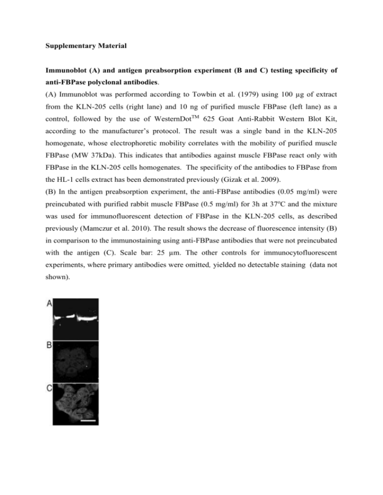
Supplementary Material Immunoblot (A) and antigen preabsorption experiment (B and C) testing specificity of anti-FBPase polyclonal antibodies. (A) Immunoblot was performed according to Towbin et al. (1979) using 100 µg of extract from the KLN-205 cells (right lane) and 10 ng of purified muscle FBPase (left lane) as a control, followed by the use of WesternDotTM 625 Goat Anti-Rabbit Western Blot Kit, according to the manufacturer’s protocol. The result was a single band in the KLN-205 homogenate, whose electrophoretic mobility correlates with the mobility of purified muscle FBPase (MW 37kDa). This indicates that antibodies against muscle FBPase react only with FBPase in the KLN-205 cells homogenates. The specificity of the antibodies to FBPase from the HL-1 cells extract has been demonstrated previously (Gizak et al. 2009). (B) In the antigen preabsorption experiment, the anti-FBPase antibodies (0.05 mg/ml) were preincubated with purified rabbit muscle FBPase (0.5 mg/ml) for 3h at 37oC and the mixture was used for immunofluorescent detection of FBPase in the KLN-205 cells, as described previously (Mamczur et al. 2010). The result shows the decrease of fluorescence intensity (B) in comparison to the immunostaining using anti-FBPase antibodies that were not preincubated with the antigen (C). Scale bar: 25 µm. The other controls for immunocytofluorescent experiments, where primary antibodies were omitted, yielded no detectable staining (data not shown). SDS-PAGE of nuclear proteins that interacted with Sepharose 4B-FBP2. Nuclear proteins were extracted with low and, subsequently, with high salt buffer from the KLN-205 cells (see Materials and Methods section), subjected to Sepharose 4B-FBP2 chromatography and electrophoretically separated. In contrast to the low salt homogenization, the high salt buffer enabled extraction of several DNA-interacting low molecular weight proteins (e.g., histones). Electrophoregram shows proteins interacting with Sepharose 4BFBP2 and extracted with a low salt buffer (lane B) and by an additional treatment with high salt buffer (lane C). Molecular mass of marker proteins (PageRulerTM Prestained Protein Ladder) are indicated in lane A. Preparation of the KLN-205 cell extracts for enzyme activity measurement The cells were trypsinized, counted, washed twice with PBS (phosphate-buffered saline: 137 mM NaCl, 2.7 mM KCl, 8 mM Na2HPO4, 1.5 mM KH2PO4; pH 7.5, RT) and homogenized in homogenization buffer (50 mM Tris, 250 mM KCl, 1 mM phenylmethylsulfonyl fluoride, 1% Triton X-100, 1 mM EDTA, 1 mM EGTA, 0.014 mg/ml leupeptin; pH 7.4, 4oC). The homogenate was centrifuged (20 min, 20,000g, 4oC) and the supernatant was assayed for the enzyme activity and protein concentration. Fluorescence in situ hybridization (FISH) Studying subcellular localization of mRNA for FBPase, we found that the mRNA was located also in nucleoli of the KLN-205. Such nucleolar localization was unexpected and thus, to exclude the possibility that the probe associated nonspecifically with nucleolar RNA’s, we tested subcellular localization of mRNA for FBP2 using two additional 5’-end FITC-labeled oligonucleotides which were complementary to other fragments of mRNA for FBP2. oligo-1: (FITC)-5’-TGTCACATTCACGCTCCCCGAAATCCCATACAGGTTGGCC oligo-2: (FITC)-5’-CTCACTGCCATCCTCGGGGAATTTCTTTTTCTGTACATAC oligo-3: (FITC)-5’-GGGTACATGAAGATTCCTCCATAGACCAAGGTGCGATGCA Localization of the oligos within FBP2 mRNA sequence: 162 5’- 201 oligo-1 678 717 oligo-2 975 1011 oligo-3 -3’ The hybridization experiment revealed practically the same pattern of staining, which strongly suggests that mRNA for FBP2 localizes in nucleoli. We also tested the effect of various hybridization conditions: fixation (methanol or paraformaldehyde), temperature, olinucleotides concentration, digestion with proteinase K and treatment with mixture of triethanolamine/acetic acid etc. In most cases, practically only cytoplasmic and nucleolar localization was observed. Hybridization in higher temperature significantly increased autofluoroscence of the fixed cells.
