COTM0307 - California Tumor Tissue Registry
advertisement

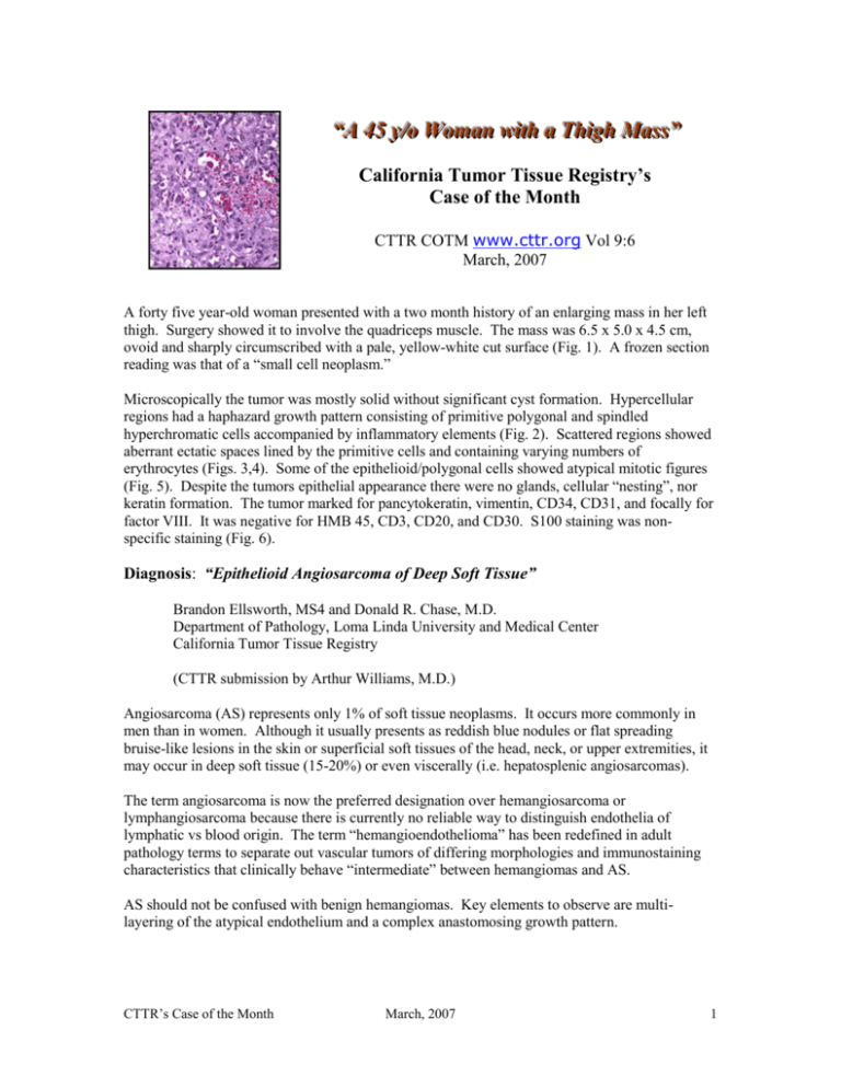
““A A 4455 yy//oo W Woom maann w wiitthh aa TThhiigghh M Maassss”” California Tumor Tissue Registry’s Case of the Month CTTR COTM www.cttr.org Vol 9:6 March, 2007 A forty five year-old woman presented with a two month history of an enlarging mass in her left thigh. Surgery showed it to involve the quadriceps muscle. The mass was 6.5 x 5.0 x 4.5 cm, ovoid and sharply circumscribed with a pale, yellow-white cut surface (Fig. 1). A frozen section reading was that of a “small cell neoplasm.” Microscopically the tumor was mostly solid without significant cyst formation. Hypercellular regions had a haphazard growth pattern consisting of primitive polygonal and spindled hyperchromatic cells accompanied by inflammatory elements (Fig. 2). Scattered regions showed aberrant ectatic spaces lined by the primitive cells and containing varying numbers of erythrocytes (Figs. 3,4). Some of the epithelioid/polygonal cells showed atypical mitotic figures (Fig. 5). Despite the tumors epithelial appearance there were no glands, cellular “nesting”, nor keratin formation. The tumor marked for pancytokeratin, vimentin, CD34, CD31, and focally for factor VIII. It was negative for HMB 45, CD3, CD20, and CD30. S100 staining was nonspecific staining (Fig. 6). Diagnosis: “Epithelioid Angiosarcoma of Deep Soft Tissue” Brandon Ellsworth, MS4 and Donald R. Chase, M.D. Department of Pathology, Loma Linda University and Medical Center California Tumor Tissue Registry (CTTR submission by Arthur Williams, M.D.) Angiosarcoma (AS) represents only 1% of soft tissue neoplasms. It occurs more commonly in men than in women. Although it usually presents as reddish blue nodules or flat spreading bruise-like lesions in the skin or superficial soft tissues of the head, neck, or upper extremities, it may occur in deep soft tissue (15-20%) or even viscerally (i.e. hepatosplenic angiosarcomas). The term angiosarcoma is now the preferred designation over hemangiosarcoma or lymphangiosarcoma because there is currently no reliable way to distinguish endothelia of lymphatic vs blood origin. The term “hemangioendothelioma” has been redefined in adult pathology terms to separate out vascular tumors of differing morphologies and immunostaining characteristics that clinically behave “intermediate” between hemangiomas and AS. AS should not be confused with benign hemangiomas. Key elements to observe are multilayering of the atypical endothelium and a complex anastomosing growth pattern. CTTR’s Case of the Month March, 2007 1 AS usually falls into one of five categories: 1. cutaneous (nos) 2. cutaneous associated with lymphedema 3. radiation-associated without lymphedema 4. mammary 5. deep. The most common variety is the cutaneous form presenting as bruise-like discoloration(s) on the skin of the head or neck. Despite a relatively bland microscopic appearance, these tumors may metastasize to cervical lymph nodes, lung, liver, or spleen. Angiosarcomas associated with lymphedema are classically seen in post-radical mastectomy patients years after the operation. This association was first described by Stewart and Treves in 1948. It is unknown what causes the malignant transformation. Although diffuse lymphangiomatosis is present, these tumors are histologically identical to the head and neck tumors. Metastasis is usually to the pleura, lung, or chest wall. Although the radiation-associated type is found mainly in women who have had breast-sparing surgery and radiation with or without lymph node dissection, it may occur in any body site which has been previously irradiated. By definition, however, the patients do not have lymphedema. Mammary AS is very rare. They are deeply located, rapidly growing masses causing diffuse enlargement of the breast with blue-red discoloration of the skin. Deep AS such as hepatosplenic angiosarcomas have also been reported due to exposure to a chemical agent such as arsenic, vinyl chloride, or thorotrast as well as in other tissues with foreign material, defunctionalized AV fistulas, or port wine stains. Histologically, angiosarcomas display varying degrees of vascular differentiation. Welldifferentiated specimens have irregularly infiltrative or dissecting vascular channels. Endothelial cells can be plump and show pleomorphism, nuclear atypia, high mitotic activity, multi-layering, and tufts or papillae. Poorly differentiated versions usually assume a more spindled or rarely, an epithelioid appearance with smaller vascular channels. The subtle nuclear features, delicate dissecting growth pattern, and tendency for discontinuous tumor growth render frozen section microscopy useless in determining resection margins. A morphologic variant of AS, characterized within the past 10 years, is epithelioid angiosarcoma (EAS). These tumors may occur at any age and have an even age distribution. Usual sites are the deep tissues of the extremities and abdominal cavity. EAS usually presents as a large, hemorrhagic mass easily confused with a chronic hematoma. Very large tumors, particularly in the very young, can demonstrate hematosequestration and arteriovenous shunting resulting in cardiac failure. Microscopically, EAS is composed of plump cells which resemble epithelial cells. The cells are relatively large, show vesicular nuclei with a prominent nucleolus, abundant mitotic figures but usually lack bizarre pleomorphism. The cytoplasm contains abundant intermediate filaments and usually marks for vimentin, cytokeratin as well as for vascular CTTR’s Case of the Month March, 2007 2 markers. EH, on the other hand, tends to have a sclerotic pattern with a very low mitotic rate and usually fails to mark for epithelial markers. Immunohistochemistry studies are the best method to differentiate AS from metastatic carcinoma, melanoma, and epithelioid malignant peripheral nerve sheath tumor. The most sensitive vascular immunohistochemical marker is CD 31, although markers such as CD34, cytokeratin and HF VIII-AG are also usually positive. When evaluating an epithelial-appearing tumor in deep tissues when a primary site is not known, a CD31 stain should be included in the immunostain battery to rule out vascular origin. Angiosarcomas are very aggressive tumors with high metastatic rates and only a 10-15% 5-year survival. The preferred regions of metastasis are skin, soft tissues, lung, lymph nodes, liver, and bone. The only reasonable prognostic factor is tumor size less than 5cm. Suggested reading: Mills, Stacey E, M. D., Sternberg’s Diagnostic Surgical Pathology, 4th ed. 2 vols. Philadelphia: Lippincott Williams & Wilkins, 2004. Kempson, Richard L, M.D. et al. Atlas of Tumor Pathology: Tumors of the Soft Tissues. Washington D.C: Armed Forces Institute of Pathology, 1998. Franz M., M.D. Enzinger, Sharon W., M.D. Weiss. Soft Tissue Tumors, 3rd ed. C.V. Mosby, 1995. Tsang WY, Chan JK, Fletcher CD. Recently characterized vascular tumours of skin and soft tissues. Histopath 19(6):489-501; 1991. Goodlad JR, Fletcher CD. Recent developments in soft tissue tumors. Histopath 27 (2):103-20, 1995. Seo IS, Min KW. Post-irradiation epithelioid angiosarcoma of the breast: a case report with immunohistochemical and electron microscopic study. Ultrastruct Pathol 27(3):197-203; May2003. Farina MC et al. Epithelioid angiosarcoma of the breast involving the skin: a highly aggressive neoplasm readily mistaken for mammary carcinoma. J Cutan pathol 30(2):152-6; 2003. Fletcher CD et al. Epithelioid angiosarcoma of deep soft tissue: a distinctive tumor readily mistaken for an epithelial neoplasm. Am J Surg Pathol 15(10):915-24; 1991. Hallel-Halevy D et al. Stewart-Treves syndrome in a patient with elephantiasis. J Am Acad Dermatol 41(2 Pt 2):349-50; 1999. CTTR’s Case of the Month March, 2007 3


