NCL: Nachweis der PR-Produkte durch Agarose
advertisement
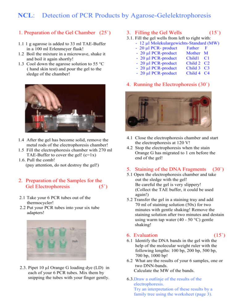
NCL: Detection of PCR Products by Agarose-Gelelektrophoresis 1. Preparation of the Gel Chamber (25`) 3. 3. Filling the Gel Wells (15`) 1.1 1 g agarose is added to 33 ml TAE-Buffer in a 100 ml Erlenmeyer flask! 1.2 Boil the mixture in a microwave, shake it and boil it again shortly! 1.3 Cool down the agarose solution to 55 °C ( hand skin test) and pour the gel to the sledge of the chamber! 3.1 3.1. Fill the gel wells from left to right with: - 12 µl Molekulargewichts-Standard (MW) - 20 µl PCR- product Father F - 20 µl PCR-product Mother M - 20 µl PCR-product Child1 C1 - 20 µl PCR-product Child 2 C2 - 20 µl PCR-product Child 3 C3 - 20 µl PCR-product Child 4 C4 4. 4. Running the Electrophoresis (30`) 1.4 After the gel has become solid, remove the metal rods of the electrophoresis chamber! 1.5 Fill the electrophoresis chamber with 270 ml TAE-Buffer to cover the gel! (c=1x) 1.6. Pull the comb! (pay attention, do not destroy the gel!) 4.1 Close the electrophoresis chamber and start the electrophoresis at 120 V! 4.2 Stop the electrophoresis when the stain Orange G has migrated to 1 cm before the end of the gel! 5. 5. Staining of the DNA Fragments 2. Preparation of the Samples for the Gel Electrophoresis (5`) 2.1 Take your 6 PCR tubes out of the thermocycler! 2.2 Put your PCR tubes into your six tube adapters! 5. in 5.1 Open the electrophoresis chamber and take out the sledge with the gel! Be careful the gel is very slippery! (Collect the TAE buffer, it could be used again!) 5.2 Transfer the gel in a staining tray and add 70 ml of staining solution (50x) for two minutes with gentle shaking! Remove the staining solution after two minutes and destain using warm tap water (40 - 50 °C) gentle shaking! 6. 6. Evaluation 2.3. Pipet 10 µl Orange G loading dye (LD) in each of your 6 PCR tubes. Mix them by snipping the tubes with your finger gently. (30`) (15`) 6.1 Identify the DNA bands in the gel with the help of the molecular weight ruler with the following lengths: 100 bp, 200 bp, 500 bp, 700 bp, 1000 bp! 6.2 What are the results of your 6 samples, one or two DNN-bands. Calculate the MW of the bands. 6.3.Draw a outlinje of the results of the electrophoresis. Try an interpretation of these results by a family tree using the worksheet (page 3).


