Flow Cytometry Protocol
advertisement
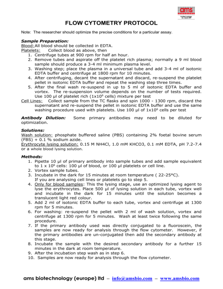
FLOW CYTOMETRY PROTOCOL Note: The researcher should optimize the precise conditions for a particular assay. Sample Preparation: Blood:All blood should be collected in EDTA. Platelets: Collect blood as above, then 1. Centrifuge tubes at 900 rpm for half an hour. 2. Remove tubes and aspirate off the platelet rich plasma; normally a 9 ml blood sample should produce a 3-4 ml minimum plasma level. 3. Washing step; place the plasma in a universal tube and add 3-4 ml of isotonic EDTA buffer and centrifuge at 1800 rpm for 10 minutes. 4. After centrifuging, decant the supernatant and discard, re-suspend the platelet pellet in isotonic EDTA buffer and repeat the washing step three times. 5. After the final wash re-suspend in up to 5 ml of isotonic EDTA buffer and vortex. The re-suspension volume depends on the number of tests required. Use 100 µl of platelet rich (1x10⁶ cells) mixture per test Cell Lines: Collect sample from the TC flasks and spin 1000 - 1300 rpm, discard the supernatant and re-suspend the pellet in isotonic EDTA buffer and use the same washing procedure used with platelets. Use 100 µl of 1x10⁶ cells per test Antibody Dilution: optimization. Some primary antibodies may need to be diluted for Solutions: Wash solution: phosphate buffered saline (PBS) containing 2% foetal bovine serum (FBS) + 0.1 % sodium azide. Erythrocyte lysing solution: 0.15 M NH4Cl, 1.0 mM KHCO3, 0.1 mM EDTA, pH 7.2-7.4 or a whole blood lysing solution. Methods: 1. Pipette 10 µl of primary antibody into sample tubes and add sample equivalent to 1 x 10⁶ cells: 100 µl of blood, or 100 µl platelets or cell line. 2. Vortex sample tubes. 3. Incubate in the dark for 15 minutes at room temperature ( 22-25°C). If you are analysing cell lines or platelets go to step 5. 4. Only for blood samples: This the lysing stage, use an optimized lysing agent to lyse the erythrocytes. Place 500 µl of lysing solution in each tube, vortex well and incubate in the dark for 15 minutes until the solution becomes a translucent light red colour. 5. Add 2 ml of isotonic EDTA buffer to each tube, vortex and centrifuge at 1300 rpm for 5 minutes. 6. For washing: re-suspend the pellet with 2 ml of wash solution, vortex and centrifuge at 1300 rpm for 5 minutes. Wash at least twice following the same procedure. 7. If the primary antibody used was directly conjugated to a fluoroscein, the samples are now ready for analysis through the flow cytometer. However, if the primary antibodies are un-conjugated then add the secondary antibody at this stage. 8. Incubate the sample with the desired secondary antibody for a further 15 minutes in the dark at room temperature. 9. After the incubation step wash as in step 6. 10. Samples are now ready for analysis through the flow cytometer. ams biotechnology (europe) ltd – info@amsbio.com – www.amsbio.com Please note 1. We recommend that each laboratory determine an optimum working titre for use of reagents in its particular application. 2. To block Fc receptor binding of antibody to human cells, and to reduce non-specific binding in general, pre-incubate the cells with normal serum or purified IgG from the same species as the specific antibody. For example, when using a mouse anti-human monoclonal antibody, block with mouse serum or purified mouse IgG. This can be done by incubating the cells for 10 minutes using 10 µg of purified IgG per one million cells. After 10 minutes, add the antibodies being used for immunofluorescent staining directly to the mixture of cells and blocking IgG. Incubate for the appropriate amount of time as per the method above. 3. All protocols are optimized for a Coulter Epics XL Flow Cytometer instrument. ams biotechnology (europe) ltd – info@amsbio.com – www.amsbio.com
