Lecture notes
advertisement
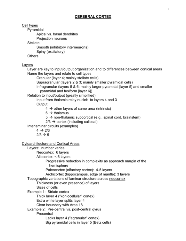
1 CEREBRAL CORTEX Cell types Pyramidal Apical vs. basal dendrites Projection neurons Stellate Smooth (inhibitory interneurons) Spiny (excitatory) Others Layers Layer are key to input/output organization and to differences between cortical areas Name the layers and relate to cell types Granular (layer 4; mainly stellate cells) Supragranular (layers 2 & 3; mainly smaller pyramidal cells) Infragranular (layers 5 & 6; mainly larger pyramidal [layer 5] and smaller pyramidal and fusiform [layer 6]) Relation to input/output (greatly simplified) Input from thalamic relay nuclei: to layers 4 and 3 Output 4 other layers of same area (intrinsic) 6 thalamus 5 non-thalamic subcortical (e.g., spinal cord, brainstem) 2/3 cortex (including callosal) Interlaminar circuits (examples) 4 2/3 2/3 5 Cytoarchitecture and Cortical Areas Layers: number varies Neocortex: 6 layers Allocortex: < 6 layers Progressive reduction in complexity as approach margin of the hemisphere Paleocortex (olfactory cortex): 4-5 layers Archicortex (hippocampus, edge of mantle): 3 layers Topographic variations of laminar structure across neocortex Thickness (or even presence) of layers Sizes of cells Example 1: Striate cortex Thick layer 4 ("koniocellular" cortex) Extra white layer splits layer 4 Clear boundary with Area 18 Example 2: Pre-central vs. post-central gyrus Precentral Lacks layer 4 ("agranular" cortex) Big pyramidal cells in layer 5 (Betz cells) 2 Postcentral Thick layer 4 Biggest pyramids in are in layer 3 instead of layer 5 Cytoarchitectonic maps -- Brodmann Good agreement on boundaries near primary areas among various cytoarchitectonic maps and with modern maps based on other methods Less agreement in association cortex Modern criteria for mapping areas Physiological Topography Maps of monkey cortex (Van Essen) Difficulties in remote areas Big receptive fields Scrambled topographies Receptive-field properties Anatomical Histochemical Myelin architecture (e.g., middle temporal visual area or MT) Cytochemistry (eg. blobs-to-stripes transition at V1/V2 border in primate visual cortex) Connectivity Corticocortical connections Thalamic afferent connectivity Efferent connectivity Summary on mapping areas Convergent evidence is best when trying to define borders Association areas may not have crisp boundaries Cortico-cortical connections as related to areas "Feedforward" (conventional, long-recognized pathway) Primary secondary association limbic/frontal Laminar signature: terminates in layer 4 of target area Old (simplistic) idea about “meaning” of forward projections: Mediates transformation from pure sensation to object recognition to integration with other senses and emotion, memory, behavior But parallel network more realistic "Feedback" (a part of the networking architecture) Laminar signature: terminates outside layer 4 of target area (both supragranular and infragranular) Possible function? Alteration of or integration with sensory processing based on higher-level perceptual, cognitive processes Hierarchies of cortical areas based on analysis of "feedforward" and "feedback" patterns of connectivity Two other classes of corticocortical connections Intrinsic (integration within a modality over space) 3 Commissural Homotopic links In primary cortex, regions of maps away from the midline tend to lack commissural connections One role of callosum may be to form a "seam" between two hemi-representations Comparative (i.e., inter-species) differences in cortical organization Location of "continental divide" between sensory and motor (central sulcus in humans) Frontal cortex expansion in human vs. cat/rat Functional correlate: differences in sophistication of planning, contingent behavior. Expansion of "association" cortex in human Correlate: complexity of cognition. e.g. Language function involves auditory, motor, symbolic, memory and visual elements Columns Definition Vertically arrayed modules Pia to white matter Shared physiology within a column Physiological properties change abruptly at boundary between adjacent columns First hint: radial fibers and cell bands Mountcastle (father of the column concept; work done in somatosensory cortex) Submodalities segregated in columnar zones (rapidly vs. slowly adapting cutaneous) Hubel and Wiesel Ocular dominance Orientation Problem: some parameters vary continuously with cortical distance. How discrete is discrete enough to indicate columnar organization? Retinotopic location - clearly not columnar Orientation preference - is it a continuous or discrete variable? No anatomical marker of discreteness Ocular dominance - even this property, except in layer 4, has a continuous, not a discrete representation But some evidence of anatomical modules Ocular dominance stripes in layer 4 Blobs in striate cortex Whisker barrels Corticocortical connections (intrinsic and long-range) 4 5
