Cells - need help with revision notes?
advertisement
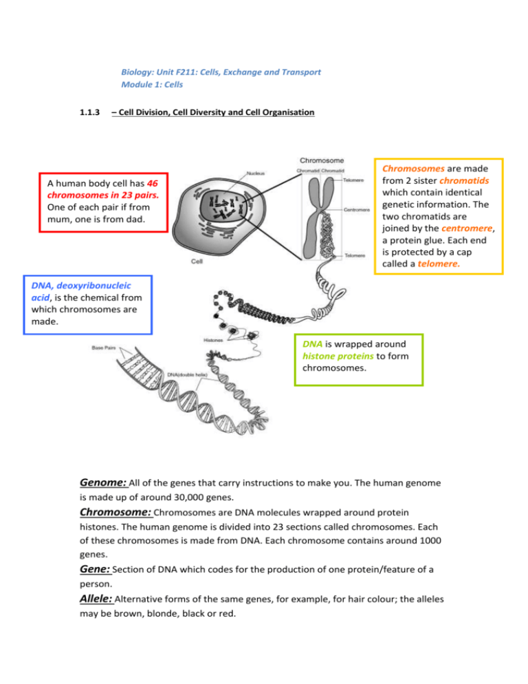
Biology: Unit F211: Cells, Exchange and Transport Module 1: Cells 1.1.3 – Cell Division, Cell Diversity and Cell Organisation A human body cell has 46 chromosomes in 23 pairs. One of each pair if from mum, one is from dad. Chromosomes are made from 2 sister chromatids which contain identical genetic information. The two chromatids are joined by the centromere, a protein glue. Each end is protected by a cap called a telomere. DNA, deoxyribonucleic acid, is the chemical from which chromosomes are made. DNA is wrapped around histone proteins to form chromosomes. Genome: All of the genes that carry instructions to make you. The human genome is made up of around 30,000 genes. Chromosome: Chromosomes are DNA molecules wrapped around protein histones. The human genome is divided into 23 sections called chromosomes. Each of these chromosomes is made from DNA. Each chromosome contains around 1000 genes. Gene: Section of DNA which codes for the production of one protein/feature of a person. Allele: Alternative forms of the same genes, for example, for hair colour; the alleles may be brown, blonde, black or red. Homologous Chromosomes are chromosomes in a diploid cell which have the same genes at the same loci. Members of an homologous pair of chromosomes pair up during mitosis. They may have different alleles. The Human Genome is made from around 30,000 genes. The 23 chromosome pairs are numbered 1-22, with the sex chromosomes being the 23rd pair. Chromosome pairs 1-22 are the autosomes. The Chromosomes of pair 23 are the sex chromosomes. Males have XY chromosomes and females have XX. This is a diploid karyotype – there are 2 copies of every chromosome. Normal human body cells are diploid. One of each pair comes from mum, one from dad. This is a haploid karyotype – there is just one copy of every chromosome. Human gametes, egg and sperm cells are haploid. Mitosis is the process of nuclear division where 2 genetically identical nuclei are formed from one parent cell nucleus. Due to the accuracy of mitosis, every cell in the human body is an exact genetic copy of the first cell that formed you; when the sperm fertilised the egg. Mitosis works to maintain genetic stability, keeping the genome constant. This is vital to health, because any changes which occur are mutations which could increase risk of developing tumours. Mitosis has 3 functions; 1. Growth of the organism, producing more cells 2. Repair of tissues and organs after injury 3. Asexual reproduction, increasing the population by division of cells Mitosis is very tightly controlled: no cell can undergo mitosis without receiving a number of signals to check it is safe to divide and desired by the rest of the body. If the signals regulating mitosis breakdown, mitosis goes on uncontrolled; this is cancer and the result is a tumour. Hormones or signal molecules must bind to the glycoprotein receptors on the cell surface in order to release the ‘brakes’ on the cell and allow it to undergo mitosis. When a cell has passed all of the relevant checkpoints and received all the necessary signals, the cell is ready to undergo mitotic cell division. The Cell Cycle describes the events that take place as one parent cell divides to produce two new daughter cells which then grow to full size. The cell cycle includes interphase, the phase between divisions (split into G1, S and G2), and mitotic phase, which is further divided into prophase, metaphase, anaphase, telophase and cytokinesis. It is controlled by enzymes. A cell spends 95% of its time in interphase. The cell goes about its normal functions as well as preparing itself for mitosis. In G1 (biosynthesis), organelles are replicated and new proteins are made. Mitotic phase involves prophase, metaphase, anaphase, telophase and cytokinesis. This is the nuclear division creating two genetically identical daughter cells. S phase is DNA synthesis, where chromosomes are replicated. G2 is growth, from a small cell into a big cell, by making more membrane and taking up more water. In every tissue, there is a small specialised subset of cells which can divide – these are called stem cells. After mitosis, they must specialise, by switching on particular genes. The process of becoming a particular type of cell is called differentiation. Cells can differentiate by changing the number of a particular organelle, changing the shape of the cell, or changing the contents of the cell. Cells, tissues and organs are specialised to perform their specific function; A tissue is a collection of similar cells working together to perform a specific function [xylem and phloem in plants; ciliated epithelial tissue in animals] An organ is a collection of similar tissues working together to perform a specific function [leaf in plants, lungs in animals] An organ system is a collection of similar organs working together to perform a specific function [reproductive and respiratory systems] Specialist Animal Cells Red blood cells – erythrocytes o Produced by stem cells in bone marrow o They transport carbon dioxide and oxygen between lungs and body tissues Adaptations to their function o Biconcave shape: large surface area: volume ratio increases ability to carry oxygen and Carbon Dioxide o Small: travel in capillaries to get close to body cells and tissues o No nucleus: more room for haemoglobin o Lots of haemoglobin: to combine with oxygen and carbon dioxide to carry around the body White blood cells – leucocytes o Produced in stem cells in bone marrow o Circulate around blood stream identifying and helping to destroy foreign material. Adaptations to their function o Glycoprotein receptors to identify self or non self o Lysosomes in cytoplasm to destroy foreign material brought into the cell by endocytosis. Sperm cells – spermatozoan Plasma Membrane: Contains receptors which bind with the egg allowing fertilisation to take place. Flagellum: tail which helps to propel the sperm towards the egg. Acrosome: a specialised lysosome which digests egg so that fertilisation can occur. Small, long and thin: streamlined shape helps them to move easily. Mitochondria: produce Modified Cytoskeleton: made of microtubules which use ATP to move and slide over each other causing the lashing movements of the tail. ATP by aerobic respiration, providing energy for the sperm to swim. o Used to transport male genetic information o They are produced by meiosis, and so have nucleus containing 23 chromosomes. They are haploid so that when the sperm and egg gametes come together at fertilisation, the zygote formed will have the full 46 chromosomes, and will be diploid. Specialist Plant Cells Palisade Cells Palisade cells are the main photosynthetic cells of the plant. They are found on the top side of the leaf, where they can absorb light. They are packed with chloroplasts containing chlorophyll so that they are able to photosynthesise. Root Hair Cells Root hair cells are found at the tips of roots. They are designed to increase the surface area of the root to help with the maximum absorption of water and minerals. Guard Cells Guard cells are found in the lower epidermis. They have unevenly thickened cell walls; the inner walls contain spirals of cellulose. They contain chloroplasts so that they can release energy in order to take up water and minerals. When water moves into these cells and they become turgid, only the outer walls stretch, because of the cellulose thickening at the inner wall, and so the two guard cells create a pore between them. This is known as a stoma. Specialist Animal Tissue Ciliated Epithelial Tissue Cilia waft in one direction to move substances away. Ciliated epithelial cells: Lots of mitochondria and a large cytoskeleton to enable the cilia to move. Basement membrane cells have complex cytoskeleton to anchor goblet and ciliated cells. Found in the trachea and the oviducts. Goblet Cell: Lots of rough endoplasmic reticulum and large Golgi body to synthesise and secrete protein mucus. Squamous Epithelial Tissue Squamous Cell Lumen Basement membrane Nucleus Underlying tissues Squamous epithelial tissue is made up of cells which are flattened and very thin. The cells together make up a smooth, flat surface, making them ideal in environments where friction can be reduced, such as blood vessels, so that fluids can pass easily over them. They also form thin walls such as those of the alveoli in the lungs. This provides a short diffusion pathway for the exchange of gases. Specialist Plant Tissue Xylem and Phloem Phloem – have perforated end ‘sieve plates’ which allow sap (sucrose dissolved in water). Xylem – made from dead cells aligned end to end to form a continuous column. The walls are lined with extra lignin, which makes them strong, and ideal for carrying water. Cooperation - Leaves Palisade Mesophyll: parenchyma cells contain many chloroplasts for photosynthesis Vascular bundle: midrib containing xylem and phloem Upper Epidermis: Thin transparent layer which allows light to reach mesophyll. Protective, covered in waterproof cuticle to reduce water loss. Spongy Mesophyll: Large air spaces to allow carbon dioxide for photosynthesis to circulate Phloem: Transports organic solutes (sugars) made by the plant during photosynthesis. Lower Epidermis: containing stoma and guard cells. Xylem: Transports water and mineral ions Mitosis vs Meiosis Where? Why? What? How? Mitosis Meiosis Everywhere else Growth of tissue, repair, asexual reproduction Genetically identical cells - clones Diploid parent cell produces 2 diploid daughter cells which are genetically identical. Ovaries, testes Sexual reproduction Gametes – genetically variable cells Diploid parent cell produces 4 haploid non identical daughter cells. Stem cells exist in every tissue, and their role is to undergo mitosis when an organism needs to grow, asexually reproduce or repair themselves. Stem cells are undifferentiated cells with no identity; they have no specific features like other cells. They are able to undergo mitosis to produce daughter cells which are then able to differentiate into specific and specialised cells. Undifferentiated stem cell with no identity. MITOSIS Differentiated cells with an identity The two types of stem cell are: Totipotent: such as in an embryo. These are able to divide and differentiate to form any type of cell in the body. Pluripotent: such as in adults. These are able to divide and differentiate to become just a few cell types in the body. Plants are slightly different: they have meristem tissue in the growing roots and shoots. Mitosis in these cells drives the growth of the plant. The meristem tissues are sensitive to auxin plant hormone. They undergo mitosis and push the plant up and the root down, elongating the cell. Phloem Meristem – Cambium Xylem Binary fission in Bacteria In binary fission, the DNA is copied and then the cell grows larger and larger, finally splitting into two. This is a type of asexual reproduction, as the cells produced are genetically identical clones. Budding in Yeast Yeast, a single celled eukaryotic organism, with structures similar to our own, divide by a process called budding. This is another form of asexual reproduction. Yeast buds are created when a mother cell grows to a critical size at a time coinciding with DNA synthesis. There is a weakening of a small area of the cell wall and this, together with the turgor pressure of osmosis allows swelling of the plasma membrane and cytoplasm to form a bud. This process leaves a ring in the plasma membrane of the mother cell known as a bud scar, and a birth scar on the surface of the daughter cell. The number of bud scars on the surface of a yeast cell is a useful indicator of cellular age.

