Some Experimental Investigations in Micro/Nano Flows
advertisement
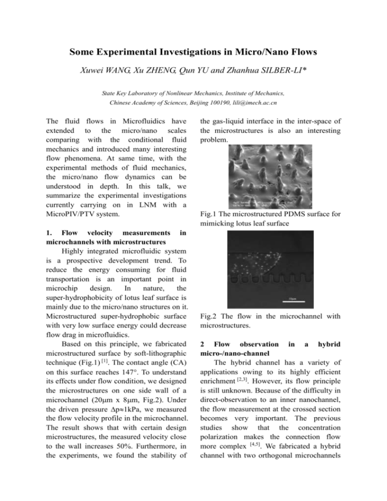
Some Experimental Investigations in Micro/Nano Flows Xuwei WANG, Xu ZHENG, Qun YU and Zhanhua SILBER-LI* State Key Laboratory of Nonlinear Mechanics, Institute of Mechanics, Chinese Academy of Sciences, Beijing 100190, lili@imech.ac.cn The fluid flows in Microfluidics have extended to the micro/nano scales comparing with the conditional fluid mechanics and introduced many interesting flow phenomena. At same time, with the experimental methods of fluid mechanics, the micro/nano flow dynamics can be understood in depth. In this talk, we summarize the experimental investigations currently carrying on in LNM with a MicroPIV/PTV system. 1. Flow velocity measurements in microchannels with microstructures Highly integrated microfluidic system is a prospective development trend. To reduce the energy consuming for fluid transportation is an important point in microchip design. In nature, the super-hydrophobicity of lotus leaf surface is mainly due to the micro/nano structures on it. Microstructured super-hydrophobic surface with very low surface energy could decrease flow drag in microfluidics. Based on this principle, we fabricated microstructured surface by soft-lithographic technique (Fig.1) [1]. The contact angle (CA) on this surface reaches 147. To understand its effects under flow condition, we designed the microstructures on one side wall of a microchannel (20m x 8m, Fig.2). Under the driven pressure p1kPa, we measured the flow velocity profile in the microchannel. The result shows that with certain design microstructures, the measured velocity close to the wall increases 50%. Furthermore, in the experiments, we found the stability of the gas-liquid interface in the inter-space of the microstructures is also an interesting problem. Fig.1 The microstructured PDMS surface for mimicking lotus leaf surface Fig.2 The flow in the microchannel with microstructures. 2 Flow observation in a hybrid micro-/nano-channel The hybrid channel has a variety of applications owing to its highly efficient enrichment [2,3]. However, its flow principle is still unknown. Because of the difficulty in direct-observation to an inner nanochannel, the flow measurement at the crossed section becomes very important. The previous studies show that the concentration polarization makes the connection flow more complex [4,5]. We fabricated a hybrid channel with two orthogonal microchannels of PDMS (50m × 100m) and a nanoporous polycarbonate nuclear track-etched membrane (PCTE, 50nm) between them. The working liquid is calcein and borax buffer. Under external electric potential 50V or 100V, the fluorescence ions (negative charge) pass through the nanoporous membrane and enrich at the cathodic side of the membrane. It shows that the rate of ionic enrichment has a non-linear relation with electric field strength and is near four times more than the classic electro-osmotic flow velocity. We also observed the propagation of the concentration depletion in the microchannel (Fig3). Fig. 3 Propagation of the concentration depletion in the microchannel Fig. 4 instance vectors of nano particle Brownian motion 3. Measurement of the Brownian motion of nano particles Brownian motion has been used to measure physical constants (e.g. Avogadro constant, Boltzmann constant) and temperature etc. Recently, It has been used for the micro-rheology study of complex fluids in cells[6]. With the development of fluorescence microscopy and nano particle technology, it is possible to use nano particles as the bio-probe for a higher accuracy measurement. Instead of tracking the particle trajectories by hand in MPT method, we programmed software for the particle detection and trajectory linking. The program posses the functions of discriminating spurious spots from real particle images, marking movement vectors of particles in time t (Fig.4). Considering the distribution of particle diameters, the average measured diffusion coefficient versus the number of particles recorded is calculated. About 1600 particle's trajectories in statistical calculation can reach an uncertainty of about 2%. The diffusion coefficients of 50nm, 200nm and 500nm particles in di-water drops are measured, 8~10nm diameter augment is observed. Acknowledgement: The authors gratefully thank the support of this work by the National Natural Science Foundation of China (10672172 and 10872203), the National Basic Research Program (2007AC744701) and the Hi-Tech Research and Development Program of China (2007AA04Z302). References: [1] X. Zheng and Z.H. Silber-Li, NTC Rev.on Adv.in Micro,Nano Molecular System.2006 [2] T.C. Kuo, D.M. Cannon, M.A. Shannon, P.W. Bohn et al., Sensors and Actuators A, 2003, 102 223-233. [3] Q. Pu, J. Yun J, H. Temkin, S. Liu, Nano Lett., 2004, 4:1099–1103 [4] Y.C. Wang, Anna L. Stevens, and J. Han, Anal. Chem., 2005, 77, 4293-4299 [5] S.J. Kim, Y.C. Wang, J.H. Lee, H. Jang, and J. Han. Pys. Rev. Lett.,2007, 99, 044501 [6] J. Liu, M.L. Gardel, K. Kroy, E. Frey, B.D. Hoffman, J.C. Crocker, A.R. Bausch and D.A. Weitz, Phys. Rev. Lett., 2006, 96, 118104
