The extraction and separation of leaf pigments by paper
advertisement
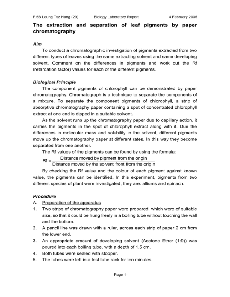
F.6B Leung Tsz Hang (29) Biology Laboratory Report 4 February 2005 The extraction and separation of leaf pigments by paper chromatography Aim To conduct a chromatographic investigation of pigments extracted from two different types of leaves using the same extracting solvent and same developing solvent. Comment on the differences in pigments and work out the Rf (retardation factor) values for each of the different pigments. Biological Principle The component pigments of chlorophyll can be demonstrated by paper chromatography. Chromatograph is a technique to separate the components of a mixture. To separate the component pigments of chlorophyll, a strip of absorptive chromatography paper containing a spot of concentrated chlorophyll extract at one end is dipped in a suitable solvent. As the solvent runs up the chromatography paper due to capillary action, it carries the pigments in the spot of chlorophyll extract along with it. Due the differences in molecular mass and solubility in the solvent, different pigments move up the chromatography paper at different rates. In this way they become separated from one another. The Rf values of the pigments can be found by using the formula: Distance moved by pigment from the origin Rf Distance moved by the solvent front from the origin By checking the Rf value and the colour of each pigment against known value, the pigments can be identified. In this experiment, pigments from two different species of plant were investigated, they are: alliums and spinach. Procedure A. Preparation of the apparatus 1. 2. 3. 4. 5. Two strips of chromatography paper were prepared, which were of suitable size, so that it could be hung freely in a boiling tube without touching the wall and the bottom. A pencil line was drawn with a ruler, across each strip of paper 2 cm from the lower end. An appropriate amount of developing solvent (Acetone Ether (1:9)) was poured into each boiling tube, with a depth of 1.5 cm. Both tubes were sealed with stopper. The tubes were left in a test tube rack for ten minutes. -Page 1- F.6B Leung Tsz Hang (29) Biology Laboratory Report 4 February 2005 B. Preparation of pigment extracts 1. 2. The leaves of the sample were washed and blot dry with tissue paper. The leaves of each were cut into small segments and were put into separate mortar. A minimum amount of extracting solvent (acetone ether 1:1) was added into each mortar. The leaf was grinded to extract the pigments 3. C. 1. Development of pigments By means of a capillary tube, the concentrated extracts were spotted onto the mid-point of the pencil line on the strip of chromatography paper. 2. Each spot was allowed to dry, and a hair dryer was employed to hasten the process. Spotting was repeated for six times until a concentrated spot is obtained. The size of the spot was not greater than 3 mm in diameter. The upper end of each strip was folded through 90°. By means of a pin, the paper was attached to the bottom of the stopper of the boiling tube. 3. 4. 5. 6. 7. 8. 9. For each paper strip, the leaf type and developing solvent used were written down on the tip of the paper in pencil. The prepared paper strip was suspended in a boiling tube such that the solvent covered the lower end of the paper but without touching the spot. The chromatogram was left to develop until the solvent front had moved up to about 1 cm from the upper end of the paper. The strips were removed and another pencil line was immediately ruled to mark the solvent front. The strips were allowed to air-dry. Each spot on the strips was outlined with pencil. The total distance moved by the solvent and the distance of each pigment spot from the origin was measured. The Rf values of each spot was calculated as follows Distance moved by pigment from the origin Rf Distance moved by the solvent front from the origin 10. Each pigment spot was identified by their colours and their relative positions on the chromatogram. -Page 2- F.6B Leung Tsz Hang (29) Biology Laboratory Report 4 February 2005 Results Pigments identified in Spinach Colour of the Distance Distance traveled Rf pigment traveled by the by pigment (cm) value solvent (cm) Yellow Green Green Yellow 13.5 Pale Yellow Name of pigment 4.3 0.319 Chlorophyll b 6.0 0.444 Chlorophyll b 9.4 0.696 Xanthophyll 13.0 0.963 Carotene Pigments identified in Allium Colour of the Distance Distance traveled Rf pigment traveled by the by pigment (cm) value solvent (cm) Green Yellow 12.0 Pale Yellow Name of pigment 6.9 0.575 Chlorophyll a 8.6 0.717 Xanthophyll 11.4 0.95 Carotene Reference: Standard Rf values for some common pigments Pigment Color Rf value Carotene Yellow 0.95 Xanthophyll Yellow-brown 0.71 Chlorophyll a Blue-green 0.65 Chlorophyll b Green 0.45 Discussion / Questions Q1 Why should you use a minimum amount of extracting solvent? It is to minimize the dilution of the concentrated extract. Q2 What should be done if the extract is found to be too viscous? To add some drops of extracting solvent Q3 What is the importance of this step? It can produce a very concentrated spot so that a clearer chromatogram can be obtained. Developed chromatogram of Allium -End of Laboratory Report-Page 3-
