A Comparison of Animal & Plants Cells
advertisement
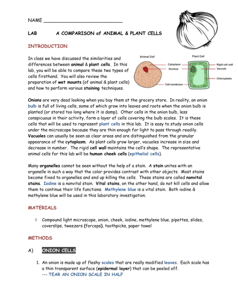
NAME ___________________________ LAB A COMPARISON of ANIMAL & PLANT CELLS INTRODUCTION In class we have discussed the similarities and differences between animal & plant cells. In this lab, you will be able to compare these two types of cells firsthand. You will also review the preparation of wet mounts (of animal & plant cells) and how to perform various staining techniques. Onions are very dead looking when you buy them at the grocery store. In reality, an onion bulb is full of living cells, some of which grow into leaves and roots when the onion bulb is planted (or stored too long where it is damp). Other cells in the onion bulb, less conspicuous in their activity, form a layer of cells covering the bulb scales. It is these cells that will be used to represent plant cells in this lab. It is easy to study onion cells under the microscope because they are thin enough for light to pass through readily. Vacuoles can usually be seen as clear areas and are distinguished from the granular appearance of the cytoplasm. As plant cells grow larger, vacuoles increase in size and decrease in number. The rigid cell wall maintains the cell’s shape. The representative animal cells for this lab will be human cheek cells (epithelial cells). Many organelles cannot be seen without the help of a stain. A stain unites with an organelle in such a way that the color provides contrast with other objects. Most stains become fixed to organelles and end up killing the cells. These stains are called nonvital stains. Iodine is a nonvital stain. Vital stains, on the other hand, do not kill cells and allow them to continue their life functions. Methylene blue is a vital stain. Both iodine & methylene blue will be used in this laboratory investigation. MATERIALS Compound light microscope, onion, cheek, iodine, methylene blue, pipettes, slides, coverslips, tweezers (forceps), toothpicks, paper towel METHODS A) ONION CELLS 1. An onion is made up of fleshy scales that are really modified leaves. Each scale has a thin transparent surface (epidermal layer) that can be peeled off. --- TEAR AN ONION SCALE IN HALF 2. Look for an edge of the epidermal layer that has been torn away from the scale. Using tweezers (or your thumb & index finger), gently peel off this epidermal layer from the scale. (The process is similar to peeling off your dead skin after recovering from sunburn.) Try not to let the thin epidermal layer wrinkle and be sure to get a piece that is small enough to fit on a microscope slide. 3. Using a drop or two of tap water, prepare a wet mount slide of your small piece of epidermal layer. 4. Under LOW power on the microscope, observe the cells. Note their shape and the presence of cell walls. Estimate the SIZE of a single cell. --- Assume LOW power FOV is 1200 µm 5. Observe the epidermal layer under HIGH power. 6. Remove the wet mount slide from the stage. Place two or three drops of iodine at one edge of the coverslip. Draw it under the coverslip by placing a piece of paper towel to the opposite edge. 7. Under LOW power, observe the epidermal layer again and note the shape & location of the stained nucleus. Also, look for the large stained vacuole. 8. SKETCH the stained onion cells under both LOW & HIGH power and LABEL the following – cell wall, cell membrane, cytoplasm, nucleus, nucleolus & vacuole. --- Also, estimate the SIZE of a single nucleus B) HUMAN CHEEK CELLS 1. With the broad end of a toothpick, gently scrape the inside of your cheek. 2. Add a drop of water to a clean microscope slide. Mix the scraping from the toothpick in with the drop of water by stirring the two together. 3. Place a coverslip on the slide. 4. Locate & observe the cheek cells under LOW power. 5. Remove the wet mount slide from the stage. Stain the cheek cells with methylene blue using the same procedure as with the onion. Use only ONE DROP of methylene blue. 6. Observe the cells under LOW & HIGH power. Estimate the SIZE of a single cell while under HIGH power. Assume HIGH power FOV is 300 µm. 7. SKETCH the stained cheek cells under both LOW & HIGH power and LABEL the following – cell membrane, cytoplasm, nucleus & nucleolus. --- Also, estimate the SIZE of a single nucleus RESULTS Onion Cells --- 2 LABELED sketches of stained cells (LOW & HIGH power) --- Estimated cell size (in µm) --- Estimated nucleus size (in µm) Cheek Cells --- 2 LABELED sketches of stained cells (LOW & HIGH power) --- Estimated cell size (in µm) --- Estimated nucleus size (in µm) NAME _________________________ THE LIVING ENVIRONMENT LAB AVERILL PARK HS LAB ANSWER SHEET A COMPARISON OF ANIMAL & PLANT CELLS RESULTS ONION CELLS (stained) LOW HIGH Estimated size of an onion cell? _________ µm Estimated size of an onion cell nucleus? _________ µm HUMAN CHEEK CELLS (stained) LOW Estimated size of a cheek cell? HIGH _________ µm Estimated size of a cheek cell nucleus? _________ µm DISCUSSION 1) What are the basic units of structure & function for all living things? -------------------------------------------------------------------------------------------------2) Name 3 features common to most animal & plant cells. --------------------------------------------------------------------------------------------------------------------------------------------------------------------------------------------------3) What cell structure is found in ALL plant cells? What is it composed of? -------------------------------------------------------------------------------------------------4) Why were NO chloroplasts found in the onion cells? -------------------------------------------------------------------------------------------------5) Excluding their color, in what way are methylene blue & iodine different stains? --------------------------------------------------------------------------------------------------------------------------------------------------------------------------------------------------6) What are the MANY differences between animal cells & plant cells? ---------------------------------------------------------------------------------------------------------------------------------------------------------------------------------------------------------------------------------------------------------------------------------------------------7) Compare the SHAPE of cheek cells & onion cells. --------------------------------------------------------------------------------------------------------------------------------------------------------------------------------------------------8) Where was the nucleus located in the cheek cells? In the onion cells? ---------------------------------------------------------------------------------------------------------------------------------------------------------------------------------------------------
