Oral Tumors Melanoma
advertisement
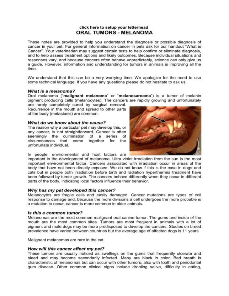
click here to setup your letterhead ORAL TUMORS - MELANOMA These notes are provided to help you understand the diagnosis or possible diagnosis of cancer in your pet. For general information on cancer in pets ask for our handout “What is Cancer”. Your veterinarian may suggest certain tests to help confirm or eliminate diagnosis, and to help assess treatment options and likely outcomes. Because individual situations and responses vary, and because cancers often behave unpredictably, science can only give us a guide. However, information and understanding for tumors in animals is improving all the time. We understand that this can be a very worrying time. We apologize for the need to use some technical language. If you have any questions please do not hesitate to ask us. What is a melanoma? Oral melanoma (“malignant melanoma” or “melanosarcoma”) is a tumor of melanin pigment producing cells (melanocytes). The cancers are rapidly growing and unfortunately are rarely completely cured by surgical removal. Recurrence in the mouth and spread to other parts of the body (metastasis) are common. What do we know about the cause? The reason why a particular pet may develop this, or any cancer, is not straightforward. Cancer is often seemingly the culmination of a series of circumstances that come together for the unfortunate individual. In people, environmental and host factors are important in the development of melanoma. Ultra violet irradiation from the sun is the most important environmental factor. Cancers associated with irradiation occur in areas of the body that have not been directly exposed. We do not know if this is the case in dogs and cats but in people both irradiation before birth and radiation hyperthermia treatment have been followed by tumor growth. The cancers behave differently when they occur in different parts of the body, indicating local factors influence their behavior. Why has my pet developed this cancer? Melanocytes are fragile cells and easily damaged. Cancer mutations are types of cell response to damage and, because the more divisions a cell undergoes the more probable is a mutation to occur, cancer is more common in older animals. Is this a common tumor? Melanomas are the most common malignant oral canine tumor. The gums and inside of the mouth are the most common sites. Tumors are most frequent in animals with a lot of pigment and male dogs may be more predisposed to develop the cancers. Studies on breed prevalence have varied between countries but the average age of affected dogs is 11 years. Malignant melanomas are rare in the cat. How will this cancer affect my pet? These tumors are usually noticed as swellings on the gums that frequently ulcerate and bleed and may become secondarily infected. Many are black in color. Bad breath is characteristic of melanomas but can occur with other tumors, also with tooth and periodontal gum disease. Other common clinical signs include drooling saliva, difficulty in eating, bleeding, displacement or loss of teeth, facial swelling, pain and swelling of the local lymph nodes (glands). How is this cancer diagnosed? Clinically, malignant oral tumors often have a fairly typical appearance. The pigment is not an infallible guide as some are not pigmented (‘amelanotic’) and other types of tumors may also contain pigmented or appear dark. X-rays (and CT scan when available) may be useful in detecting whether tumors have invaded the bones and to guide surgery. Loss of bone adjacent to the tumor usually means a poorer outlook (prognosis). Cytology, the microscopic examination of a small sample of cells, may be diagnostic in some cases. However definitive diagnosis, prediction of behavior (prognosis) and a microscopic assessment of whether the tumor has been fully removed rely on microscopic examination of tissue (histopathology). This is done at a specialized laboratory by a veterinary pathologist. The piece of tissue may be a small part of the mass (biopsy) or the whole lump but only examination of the whole lump will indicate whether the cancer has been fully removed. Histopathology also rules out other cancers. Most of these tumors invade the bone of the jaw. They need wide surgical margins usually including substantial parts of the jaw bone. This type of tissue will need decalcifying so it may take be a few weeks before the final histopathology results are available. The pathologist may also need to remove (bleach) the pigment to check malignancy with greater certainty. What types of treatment are available? Surgical removal is the standard method of treatment for all these tumors. The invasive cancers are difficult to remove completely so large pieces of the jaw bone may be taken out (hemimaxillectomy or hemimandibulectomy). The complex and extensive surgery is often done at a referral treatment center. The surgeon may want to have a CT scan performed (if available) to determine how much tissue to remove to achieve complete excision. There has been little progress with other treatments, despite much research on these tumors in people. Melanomas do not respond well to chemotherapy or radiation therapy. Immunotherapy with interferons has not improved survival in people. Currently, research is focussed on combining immunotherapy (cytokine and gene therapy) with other treatments. Vaccines are undergoing clinical trials in people. Can this cancer disappear without treatment? Curing infections and healing ulcers will help reduce superficial swelling but not cure the cancer. Very occasionally, spontaneous loss of blood supply to the cancer can make parts of it die but the dead tissue will still need surgical removal. The body’s immune system is not effective at making these tumors to regress. How can I nurse my pet? After surgery, you will probably be provided with an “Elizabethan collar” to prevent your pet from interfering with the operation site. You may be requested not to examine the surgery but inability to eat or significant swelling or bleeding should be reported to your veterinarian. Your pet may require a special diet. If you require additional advice on post-surgical care, please ask. How will I know how this cancer will behave? Histopathology will give your veterinarian the diagnosis that helps to indicate how it is likely to behave. The veterinary pathologist usually adds a prognosis that describes the probability of local recurrence or metastasis (distant spread). The completeness of excision will be assessed and other diagnoses ruled out. When will I know if the cancer is permanently cured? ‘Cured’ has to be a guarded term in dealing with any cancer. The outlook for dogs and cats with these tumors is poor. The underlying bone is invaded by over half of these tumors. Approximately 70% of tumors metastasize to regional lymph nodes (glands) and almost the same percentage to distant sites, usually the lungs. The average post surgical survival time has been only about three months with three out of four afflicted dogs dying within six months and nine out of ten within 24 months. Survival rates are however improving. Survival is unrelated to sex, site, rate of growth, histological type, amount of pigment or cancer size. Tumor stage (how far it has spread) is correlated with survival time. If there is no involvement of the local lymph nodes and no X-ray evidence of lung tumors, survival time is improved by surgery (242 days versus 65 days). Partial removal of the jaw reduces local recurrence of the tumors but does not always prevent the spread elsewhere. Are there any risks to my family or other pets? No, these are not infectious tumors and are not transmitted from pet to pet or from pets to people. This client information sheet is based on material written by Joan Rest, BVSc, PhD, MRCPath, MRCVS. © Copyright 2004 Lifelearn Inc. Used with permission under license. February 12, 2016.


