Mitophagy_20140721
advertisement
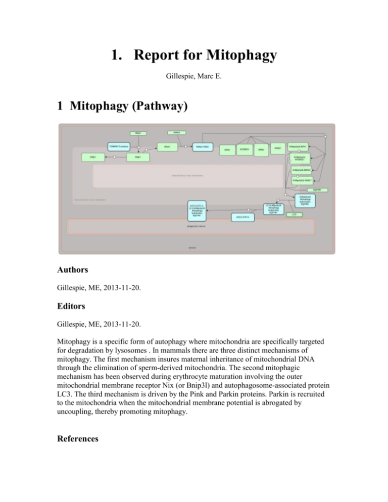
1. Report for Mitophagy Gillespie, Marc E. 1 Mitophagy (Pathway) Authors Gillespie, ME, 2013-11-20. Editors Gillespie, ME, 2013-11-20. Mitophagy is a specific form of autophagy where mitochondria are specifically targeted for degradation by lysosomes . In mammals there are three distinct mechanisms of mitophagy. The first mechanism insures maternal inheritance of mitochondrial DNA through the elimination of sperm-derived mitochondria. The second mitophagic mechanism has been observed during erythrocyte maturation involving the outer mitochondrial membrane receptor Nix (or Bnip3l) and autophagosome-associated protein LC3. The third mechanism is driven by the Pink and Parkin proteins. Parkin is recruited to the mitochondria when the mitochondrial membrane potential is abrogated by uncoupling, thereby promoting mitophagy. References Youle RJ, Narendra DP, "Mechanisms of mitophagy", Nat. Rev. Mol. Cell Biol., 12, 2011, 9-14. 1.1 Pink/Parkin Mediated Mitophagy (Pathway) Authors Gillespie, ME, 2013-11-20. Editors Gillespie, ME, 2013-11-20. This is the process of selective removal of damaged mitochondria by autophagosomes and subsequent catabolism by lysosomes. In healthy mitochondria, PINK1 is imported to the inner mitochondrial membrane, presumably through the TOM/TIM complex. The TIM complex-associated protease, mitochondrial MPP, cleaves PINK1 mitochondrial targeting sequence (MTS). PINK1 may be cleaved by the inner membrane presenilin-associated rhomboid-like protease PARL and ultimately proteolytically degraded. Loss of membrane potential in damaged mitochondria prevents the import of the PTEN-induced putative kinase 1 (PINK1) which accumulates in the defective mitochondria triggering the recruitment of the E3 ubiquitin protein ligase (Parkin). Once on the mitochondria, Parkin promotes the ubiquitination of mitochondrial substrates embedded in the OMM such as the mitofusin Mitochondrial Assembly Regulatory Factor (MARF), mitofusin 1 and 2 (Mfn1, 2) and the Voltage-Dependent Anion-Channel 1 (VDAC1). After the ubiquitination events, p62 is recruited to mitochondria, binding the Parkin-ubiquitinated substrates, linking the the ubiquitinated subbstrates to the Microtubule-associated protein Autophagy marker Light Chain 3 (LC3). Targeting the entire mitochondria for autophagic degradation. The recruitment of LC3 complexes with the autophagosome membrane and the AuTophaGy proteins 5-12 (Atg5-Atg12) complex. The mitochondria is engulfed after the isolation membrane grows to a sufficient size to engulf the mitochondria. Once the autophagocytic vesicle formation is complete, vesicle fusion with lysosomes to form the autolysosomes in which the lysosomal hydrolases (cathepsins and lipases) degrade the intra-autophagosomal content. Cathepsin also degrades LC3 on the intra-autophagosomal surface of the autophagocytic vesicle. References Youle RJ, Narendra DP, "Mechanisms of mitophagy", Nat. Rev. Mol. Cell Biol., 12, 2011, 9-14. 1.1.1 Pink1 is recruited from the cytoplasm to the mitochndria (Reaction) Authors Gillespie, ME, 2013-11-20. Editors Gillespie, ME, 2013-11-20. PINK1 is constitutively synthesized and imported into all mitochondria. In healthy mitochondria PINK1 is cleaved by voltage-sensitive proteolysis. References Narendra DP, Jin SM, Tanaka A, Suen DF, Gautier CA, Shen J, Cookson MR, Youle RJ, "PINK1 is selectively stabilized on impaired mitochondria to activate Parkin", PLoS Biol., 8, 2010, e1000298. 1.1.2 Pink1 is cleaved on healthy mitochndria (Reaction) Authors Gillespie, ME, 2013-11-20. Editors Gillespie, ME, 2013-11-20. Full-length PINK1 (63 kDa), which is in the inner mitochondrial space, is proteolytically cleaved into an 52-kDa cytosolic fragment (111 - 581) that is released back into the cytoplasm by an unknown mechanism and degraded by the proteasome. cleavage of PINK1 into an unstable cytosolic form maintains low levels of PINK1 on healthy mitochondria in order to suppress the PINK1/Parkin pathway in the absence of mitochondrial damage. At present, it is unclear which proteases mediate the cleavage of PINK1 in mammalian cells. References Jin SM, Lazarou M, Wang C, Kane LA, Narendra DP, Youle RJ, "Mitochondrial membrane potential regulates PINK1 import and proteolytic destabilization by PARL", J. Cell Biol., 191, 2010, 933-42. Narendra DP, Jin SM, Tanaka A, Suen DF, Gautier CA, Shen J, Cookson MR, Youle RJ, "PINK1 is selectively stabilized on impaired mitochondria to activate Parkin", PLoS Biol., 8, 2010, e1000298. Narendra DP, Youle RJ, "Targeting mitochondrial dysfunction: role for PINK1 and Parkin in mitochondrial quality control", Antioxid. Redox Signal., 14, 2011, 1929-38. 1.1.3 Pink1 is not cleaved on damaged mitochndria (Reaction) Authors Gillespie, ME, 2013-11-20. On damaged mitochondria that have lost their membrane potential, however, PINK1 cleavage is inhibited, leading to high PINK1 expression on the OMM of the dysfunctional mitochondria. Full-length mitochondrial PINK1 is the active form in the PINK1/Parkin pathway. References Jin SM, Lazarou M, Wang C, Kane LA, Narendra DP, Youle RJ, "Mitochondrial membrane potential regulates PINK1 import and proteolytic destabilization by PARL", J. Cell Biol., 191, 2010, 933-42. Narendra DP, Jin SM, Tanaka A, Suen DF, Gautier CA, Shen J, Cookson MR, Youle RJ, "PINK1 is selectively stabilized on impaired mitochondria to activate Parkin", PLoS Biol., 8, 2010, e1000298. Narendra DP, Youle RJ, "Targeting mitochondrial dysfunction: role for PINK1 and Parkin in mitochondrial quality control", Antioxid. Redox Signal., 14, 2011, 1929-38. 1.1.4 Pink1 recruits Parkin to the outer mitochondrial membrane. (Reaction) Authors Gillespie, ME, 2013-11-20. Editors Gillespie, ME, 2013-11-20. Parkin, an E3 ubiquitin ligase, is selectively recruited from the cytosol to damaged mitochondria to trigger their autophagy. References Narendra DP, Jin SM, Tanaka A, Suen DF, Gautier CA, Shen J, Cookson MR, Youle RJ, "PINK1 is selectively stabilized on impaired mitochondria to activate Parkin", PLoS Biol., 8, 2010, e1000298. 1.1.5 Parkin promotes the ubiquitination of mitochondrial substrates (Reaction) Authors Gillespie, ME, 2013-11-20. Editors Gillespie, ME, 2013-11-20. Parkin promotes the ubiquitination of the mitofusin mitochondrial assembly regulatory factor (MARF), mitofusin 1, mitofusin 2 and voltage-dependent anion-selective channel protein 1 (vDAC1), all of which are embedded in the OMM. References Sun F, Kanthasamy A, Anantharam V, Kanthasamy AG, "Mitochondrial accumulation of polyubiquitinated proteins and differential regulation of apoptosis by polyubiquitination sites Lys-48 and -63", J. Cell. Mol. Med., 13, 2009, 1632-43. 1.1.6 p62 binds ubiquitinated mitochondrial substrates (Reaction) Authors Gillespie, ME, 2013-11-20. Editors Gillespie, ME, 2013-11-20. After the ubiquitination events, p62 is recruited to mitochondria, binding the Parkin-ubiquitinated substrates References Tanida I, "Autophagosome formation and molecular mechanism of autophagy", Antioxid. Redox Signal., 14, 2011, 2201-14. 1.1.7 p62 links damaged mitochondria to LC3 (Reaction) Authors Gillespie, ME, 2013-11-20. Editors Gillespie, ME, 2013-11-20. p62 links to the microtubule-associated protein Autophagy marker Light Chain 3 (LC3). This begins the recruitment of the autophagic machinery to the damaged mitochondria, targeting it for autophagic degradation. 1.1.8 LC3 binds the autophagosome membrane Atg5-Atg12 complex (Reaction) Authors Gillespie, ME, 2013-11-20. Editors Gillespie, ME, 2013-11-20. The mitochondria are engulfed after elongation of the isolation membrane. Once the autophagosome is formed, its outer membrane fuses with lysosomes to form the autolysosome. The lysosomal hydrolases (cathepsins and lipases) degrade the damaged mitochondria and its associated proteins. References Tanida I, "Autophagosome formation and molecular mechanism of autophagy", Antioxid. Redox Signal., 14, 2011, 2201-14. Full List of Literature References for Pathway "Mitophagy" Jin SM, Lazarou M, Wang C, Kane LA, Narendra DP, Youle RJ, "Mitochondrial membrane potential regulates PINK1 import and proteolytic destabilization by PARL", J. Cell Biol., 191, 2010, 933-42. Narendra DP, Jin SM, Tanaka A, Suen DF, Gautier CA, Shen J, Cookson MR, Youle RJ, "PINK1 is selectively stabilized on impaired mitochondria to activate Parkin", PLoS Biol., 8, 2010, e1000298. Narendra DP, Youle RJ, "Targeting mitochondrial dysfunction: role for PINK1 and Parkin in mitochondrial quality control", Antioxid. Redox Signal., 14, 2011, 1929-38. Sun F, Kanthasamy A, Anantharam V, Kanthasamy AG, "Mitochondrial accumulation of polyubiquitinated proteins and differential regulation of apoptosis by polyubiquitination sites Lys-48 and -63", J. Cell. Mol. Med., 13, 2009, 1632-43. Tanida I, "Autophagosome formation and molecular mechanism of autophagy", Antioxid. Redox Signal., 14, 2011, 2201-14. Youle RJ, Narendra DP, "Mechanisms of mitophagy", Nat. Rev. Mol. Cell Biol., 12, 2011, 9-14.
