Snyder Clinical Integration Case
advertisement
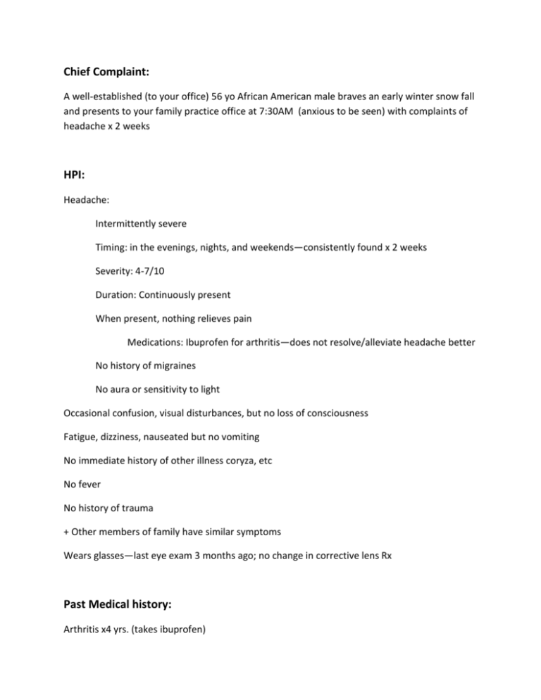
Chief Complaint: A well-established (to your office) 56 yo African American male braves an early winter snow fall and presents to your family practice office at 7:30AM (anxious to be seen) with complaints of headache x 2 weeks HPI: Headache: Intermittently severe Timing: in the evenings, nights, and weekends—consistently found x 2 weeks Severity: 4-7/10 Duration: Continuously present When present, nothing relieves pain Medications: Ibuprofen for arthritis—does not resolve/alleviate headache better No history of migraines No aura or sensitivity to light Occasional confusion, visual disturbances, but no loss of consciousness Fatigue, dizziness, nauseated but no vomiting No immediate history of other illness coryza, etc No fever No history of trauma + Other members of family have similar symptoms Wears glasses—last eye exam 3 months ago; no change in corrective lens Rx Past Medical history: Arthritis x4 yrs. (takes ibuprofen) Personal History: Occupation: Salesperson –5 days a week; Monday through Friday; no weekends Allergies: none Not necessarily physically active—yard work on weekends in summer and fall, etc but otherwise too busy. Married, two children ages 16 and 17; living in same house Family history: Father died of lung cancer (smoker for 40 years); see HPI Social history: Smoker – ½ ppd Occasional drink: social drinking (2/week) Denies illicit drug use Physical Exam BP: 120/80 HR: 90 bpm RR: 20 Temp: 98.7 Ht/Wt: 6’0 220 lbs (BMI 29.8) overweight No nuchal rigidity—no meningitis Heart—normal S1 and S2 heart sound; no murmurs, no rub; no distant heart sounds; peripheral vascular normal Lung—normal; vesicular sounds bilaterally; no pleural friction rub; no stridor, wheezes, ronchi/rales Eye—PERRLA; negative fundoscopic exam; Snellen visual acuity with glasses 20/20 on exam today Neuropsychiatric exam—normal/negative: Students should ask for a complete Neuro exam! Skin: normal (see note below) Other systems: Negative / Normal FYI: Patients typically present with tachycardia and tachypnea, and may complain of headache, nausea and vomiting. However, patients rarely have the classic findings of cyanosis, retinal hemorrhage and cherry-red lips.—Shows up on boards so might cover at end of case! Symptom severity ranges from mild (constitutional symptoms) to severe (coma, respiratory depression and hypotension) and is not associated with serum levels of carboxyhemoglobin, although duration of exposure is an important factor. Not all patients exhibit signs and symptoms immediately after exposure. In some patients, neuropsychiatric symptoms, including cognitive and personality changes, may develop anywhere from three days to eight months after exposure. The mechanism for these conditions is unknown, but hypoxia alone is not sufficient to explain the observed clinical manifestations. Labs CBC—look for anemia, infection, cancer—all normal; At the conclusion of the case, ask the students if they would suspect a secondary polycythemia in a chronic exposure to carbon monoxide? An acute exposure? Physiologically explain it. - WBC 7,000 (4-10,000); Hbg 18 (13-18); Hematocrit 62% (45-62) ; RBC 6.1 million cells/mcL (4.7-6.1) ; platelet 150,000 (150-400,000) MCV 88 (80-100); MCHC 35% (32-36); RDW 13 (11-15) Arterial carboxyhemoglobin saturation slightly elevated. – 16% elevated. Must have a degree of suspicion of carbon monoxide poisoning in order to know to obtain this lab—they may go right past this…and have to come back to it in the end. Arterial blood gas testing and pulse oximetry are not useful because they give falsely normal Pao2 and oxyhemoglobin saturation determinations. A newer pulse oximetery devise, the pulse CO-oximeter is capable of distinguishing oxyhemoglobin from carboxyhemoglobin. The diagnosis of CO poisoning is based upon a compatible history and physical exam in conjunction with an elevated carboxyhemoglobin level measured by cooximetry of a blood gas sample. Nonsmokers may have up to 3 percent carboxyhemoglobin at baseline; smokers may have levels of 10 to 15 percent. Levels above these respective values are consistent with CO poisoning. Once the diagnosis of CO intoxication is confirmed, it is recommended to obtain an electrocardiogram (ECG); cardiac biomarker evaluation is warranted in patients with ECG evidence of ischemia or a history of cardiac disease. Additionally, evaluation of family members should be performed. Electrolytes normal Arterial Acid Base Normal EKG normal—too early to have students interpret; however, ask them why they might be interested in obtaining one when diagnosis established. End organ disease secondary to ischemia… Kidney function tests—normal; BUN 14 (7-18) R/O End organ disease secondary to ischemia… Computed tomography (CT) of the head is usually helpful only to rule out other causes of neurologic decompensation. Conclusion The most important interventions in the management of a CO-poisoned patient are prompt removal from the source of CO – correlate symptoms of “while home, patient has symptoms; while at work, no symptoms.” Institution of 100 percent oxygen by nonrebreathing face mask or endotracheal tube. We recommend initial treatment with 100 percent normobaric oxygen for all suspected victims of CO poisoning, regardless of pulse oximetry or arterial PO2 Determination of the mechanism of exposure is critical in cases of accidental poisoning in order to limit the risk to others. Evaluate functioning of furnace/ ensure no use of kerosene heaters in enclosed spaces, functioning stoves, heaters, etc. “emphasize use of furnace (onset of use with correlation of symptoms) Evaluation of family members should be performed. Installation of carbon monoxide detectors are inexpensive and widely available, but there are no standard recommendations regarding their use in the home or the workplace. Consider follow up appointments to switch arthritis medication to: consider Acetaminophen for arthritis and Encourage lifestyle modifications:-- exercise, stop smoking. Enlarge by clicking on algorithm and dragging corners. Because carbon monoxide poisoning has no pathognomonic signs or symptoms, a high level of suspicion is needed to confirm the diagnosis. Measuring the level of carbon monoxide in the exhaled air of a patient can be diagnostic, but a blood sample should be obtained to measure carboxyhemoglobin levels and any coexisting acidosis. A detailed neurologic examination, including psychological testing, is recommended to document any abnormal findings, which are often subtle. The most important steps in treatment are removing the patient from the source of poisoning and administering 100 percent oxygen. Oxygen should be given until the patient's carboxyhemoglobin level returns to normal. Hospitalization should also be considered for patients with severe poisoning or serious underlying medical problems. Indications for hyperbaric oxygen therapy are not clear, except that it is undisputedly indicated in unconscious patients. Carbon monoxide (CO) is an odorless, tasteless, colorless, nonirritating gas formed by hydrocarbon combustion. About 600 accidental deaths from carbon monoxide poisoning occur each year, making it one of the most common causes of morbidity from poisoning in the United States. About five to 10 times as many intentional deaths from carbon monoxide are reported. Smoke inhalation is responsible for most inadvertent cases of CO poisoning. Other potential sources of CO include poorly functioning heating systems, improperly vented fuel-burning devices (eg, kerosene heaters, charcoal grills, camping stoves, gasoline-powered electrical generators), and motor vehicles operating in poorly ventilated areas (eg, ice rinks, warehouses, parking garages). Exposure occurs from a variety of sources, particularly motor vehicle exhaust. The number of accidental deaths caused by carbon monoxide poisoning from motor vehicle exhaust is higher in the North and peaks during the winter months. PATHOPHYSIOLOGY — Carbon monoxide (CO) diffuses rapidly across the pulmonary capillary membrane and binds to the iron moiety of heme (and other porphyrins) with approximately 240 times the affinity of oxygen. The degree of carboxyhemoglobinemia (COHb) is a function of the relative amounts of CO and oxygen in the environment, duration of exposure, and minute ventilation. Nonsmokers may have up to 3 percent carboxyhemoglobin at baseline; smokers may have levels of 10 to 15 percent. Severe chronic obstructive pulmonary disease can cause a modest but significant elevation in carboxyhemoglobin levels, even among patients without exposure to tobacco smoke. The mechanism and clinical significance of this finding is unclear. Once CO binds to the heme moiety of hemoglobin, an allosteric change occurs that greatly diminishes the ability of the other three oxygen binding sites to off-load oxygen to peripheral tissues. This results in a deformation and leftward shift of the oxyhemoglobin dissociation curve, and compounds the impairment in tissue oxygen delivery. CO also interferes with peripheral oxygen utilization. Approximately 10 to 15 percent of CO is extravascular and bound to molecules such as myoglobin, cytochromes, and NADPH reductase, resulting in impairment of oxidative phosphorylation at the mitochondrial level. The half-life of CO bound to these molecules is longer than that of COHb. The importance of these nonhemoglobin-mediated effects has been best documented in the heart, where mitochondrial dysfunction due to CO can produce myocardial stunning despite adequate oxygen delivery Because of their higher oxygen utilization and higher minute ventilation, young children may develop signs and symptoms of carbon monoxide poisoning before older children and adults who experience the same exposure (eg, family members living in a house with a faulty furnace). Enlarge by clicking on table and dragging corners.
