Accelerated progression of vascular calcification in children with
advertisement
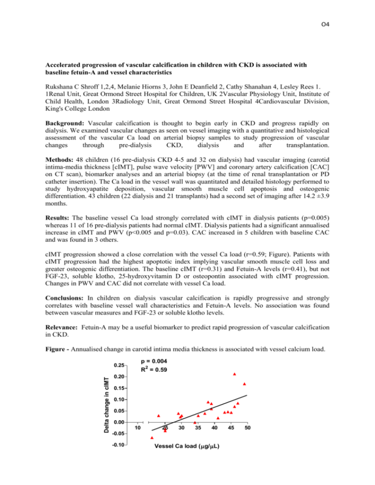
O4 Accelerated progression of vascular calcification in children with CKD is associated with baseline fetuin-A and vessel characteristics Rukshana C Shroff 1,2,4, Melanie Hiorns 3, John E Deanfield 2, Cathy Shanahan 4, Lesley Rees 1. 1Renal Unit, Great Ormond Street Hospital for Children, UK 2Vascular Physiology Unit, Institute of Child Health, London 3Radiology Unit, Great Ormond Street Hospital 4Cardiovascular Division, King's College London Background: Vascular calcification is thought to begin early in CKD and progress rapidly on dialysis. We examined vascular changes as seen on vessel imaging with a quantitative and histological assessment of the vascular Ca load on arterial biopsy samples to study progression of vascular changes through pre-dialysis CKD, dialysis and after transplantation. Methods: 48 children (16 pre-dialysis CKD 4-5 and 32 on dialysis) had vascular imaging (carotid intima-media thickness [cIMT], pulse wave velocity [PWV] and coronary artery calcification [CAC] on CT scan), biomarker analyses and an arterial biopsy (at the time of renal transplantation or PD catheter insertion). The Ca load in the vessel wall was quantitated and detailed histology performed to study hydroxyapatite deposition, vascular smooth muscle cell apoptosis and osteogenic differentiation. 43 children (22 dialysis and 21 transplants) had a second set of imaging after 14.2 ±3.9 months. Results: The baseline vessel Ca load strongly correlated with cIMT in dialysis patients (p=0.005) whereas 11 of 16 pre-dialysis patients had normal cIMT. Dialysis patients had a significant annualised increase in cIMT and PWV (p<0.005 and p=0.03). CAC increased in 5 children with baseline CAC and was found in 3 others. cIMT progression showed a close correlation with the vessel Ca load (r=0.59; Figure). Patients with cIMT progression had the highest apoptotic index implying vascular smooth muscle cell loss and greater osteogenic differentiation. The baseline cIMT (r=0.31) and Fetuin-A levels (r=0.41), but not FGF-23, soluble klotho, 25-hydroxyvitamin D or osteopontin associated with cIMT progression. Changes in PWV and CAC did not correlate with vessel Ca load. Conclusions: In children on dialysis vascular calcification is rapidly progressive and strongly correlates with baseline vessel wall characteristics and Fetuin-A levels. No association was found between vascular measures and FGF-23 or soluble klotho levels. Relevance: Fetuin-A may be a useful biomarker to predict rapid progression of vascular calcification in CKD. Figure - Annualised change in carotid intima media thickness is associated with vessel calcium load. p = 0.004 R2 = 0.59 Delta change in cIMT 0.25 0.20 0.15 0.10 0.05 0.00 10 25 30 35 40 -0.05 -0.10 Vessel Ca load (g/L) 45 50
