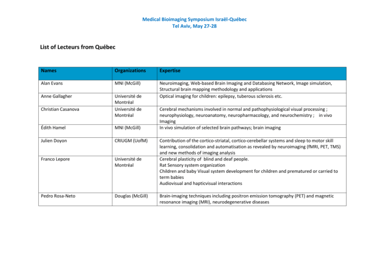
List of Lecteurs from Québec
advertisement

Medical Bioimaging Symposium Israël-Québec Tel Aviv, May 27-28 List of Lecteurs from Québec Names Organizations Expertise Alan Evans MNI (McGill) Anne Gallagher Université de Montréal Université de Montréal Neuroimaging, Web-based Brain Imaging and Databasing Network, Image simulation, Structural brain mapping methodology and applications Optical imaging for children: epilepsy, tuberous sclerosis etc. Christian Casanova Édith Hamel MNI (McGill) Julien Doyon CRIUGM (UofM) Franco Lepore Université de Montréal Cerebral mechanisms involved in normal and pathophysiological visual processing ; neurophysiology, neuroanatomy, neuropharmacology, and neurochemistry ; in vivo Imaging In vivo simulation of selected brain pathways; brain imaging 1. 2. 3. 4. Pedro Rosa-Neto Douglas (McGill) Contribution of the cortico-striatal, cortico-cerebellar systems and sleep to motor skill learning, consolidation and automatisation as revealed by neuroimaging (fMRI, PET, TMS) and new methods of imaging analysis Cerebral plasticity of blind and deaf people. Rat Sensory system organization Children and baby Visual system development for children and prematured or carried to term babies Audiovisual and hapticvisual interactions Brain-imaging techniques including positron emission tomography (PET) and magnetic resonance imaging (MRI), neurodegenerative diseases Medical Bioimaging Symposium Israël-Québec Tel Aviv, May 27-28 Lecteurs from Israel Names Organizations Expertise Assaf Tal assaf.tal@weizmann.ac.il Weizmann Instituteof Science Avi Karni Avi.karni@yahoo.com University of Haifa My lab develops new magnetic resonance imaging (MRI) methodologies to better understand the human body. Most atomic nuclei possess an intrinsic magnetic moment. By shaping the electromagnetic fields inside the MRI scanner we interact with these moments, with which we can observe the structure, physiology and biochemistry of the human body in vivo and non-invasively. Developing new ways of manipulating the nuclear spins can provide us not only with important tools to diagnose disease, but also with a better understanding of the basic biological processes which govern us. Using behavioral (psychophysics) and brain imaging (fMRI) techniques, the lab focuses on studying what drives the ability of the human brain to change with experience and establish effective long-term memory: Work relates to neuro-behavioral constraints on learning and skill (procedural memory) acquisition, sleep, development and neurological rehabilitation. Yael Mardor Sheba Medical Center Brain MRI (depiction of subtle BBB disruption, tissue characterization, response to therapy), Brain tumors (imaging and therapy), drug delivery into the CNS Tel Aviv University The main research interest of the lab is measuring brain micro-structure with magnetic resonance imaging (MRI). The general work hypothesis is that brain structure, function and behavior are linked. Under this hypothesis we study the anatomical characteristics of brain tissue as well as structural aspects of neuroplasticity and behavioral correlates. Neurology, Neurophysiology (Cellular, neuronal network, system and clinical neuroelectrophysiology), Cellular Imaging, Molecular and Cellular Biology yael.mardor@sheba.health.gov.il Yaniv Assaf assafyan@tauex.tau.ac.il Sheba Medical nicola.maggio@sheba.health.gov.il Center Nicola Maggio Hayit Greenspan hayit@eng.tau.ac.il Tel Aviv University •MRI Brain Image Analysis; affiliated as a Faculty member in the Sagol school of Brain research and Neuroscience ◦Augmenting resolution in MRI; Medical Bioimaging Symposium Israël-Québec Tel Aviv, May 27-28 ◦Neurological diseases, Multiple-Sclerosis; •CT lesion analysis; ◦Liver lesions – detection, segmentation, characterization in time (Collaborations with Radiology Dept, Stanford & Head of CT Abdomen Unit at Sheba medical center, Israel) ◦Mammmography - microcalcification analysis, using deep learning methodologies ◦Interstitial Lung Disease - characterization in CT •Uterine-cervix image analysis and indexing for cancer screening; •Medical image retrieval from large archives (PACS). The role of blood-brain barrier in pathophysiology of brain disorders: structural and functional brain imaging. Alon Friedman Alonf@bgu.ac.il Ben Gurion University Ilan Distein dinshi@bgu.ac.il Ben Gurion University Functional and structural MRI and EEG in autism and ADHD. Identification of early markers for autism diagnosis in toddlers. Gadi Goelman gadig@hadassah.org.il Hadassah Medical Center Functional MRI studies of a rat model of Parkinson's disease. Functional characterization of motor cortical areas of a rat with Development of new approaches for mapping neuronal functional Information transfer and plasticity in the rat barrel field using Theoretical analysis of neuronal communication and the MRI signal. Development of new MRI contrast based on spatial correlation Testing the effect of FTS on recovery from head trauma We conduct parallel research in vision research: On one hand (I.) psychophysically, functional fMRI and computational aspects on several topics such as adaptation of different visual properties, induction, completion of lines (Kanizsa illusion). On the other hand (II.) developing generic algorithms which are yielded from the above computational models and in addition developing specific medical applications of these algorithms for medical images. It occurs also that the computational models yield ideas for new experimental studies and to discovery of new visual effects (such as chromatic Mach bands and specific spatial properties of adaptation effects). Hedva Spitzer Hedva@eng.tau.ac.il Medical Bioimaging Symposium Israël-Québec Tel Aviv, May 27-28


