Sorting Best Practices
advertisement
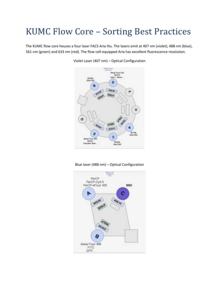
KUMC Flow Core – Sorting Best Practices The KUMC flow core houses a four laser FACS Aria IIIu. The lasers emit at 407 nm (violet), 488 nm (blue), 561 nm (green) and 633 nm (red). The flow cell-equipped Aria has excellent fluorescence resolution. Violet Laser (407 nm) – Optical Configuration Blue laser (488 nm) – Optical Configuration Green Laser (561 nm) – Optical Configuration Red Laser (633 nm) – Optical Configuration Fluorochromes and Dyes: If you are unsure about which fluorochromes or dyes will work in your experiment, please consult the Becton Dickinson Biosciences Spectrum Viewer available at this link: http://www.bdbiosciences.com/research/multicolor/spectrum_viewer/index.jsp#search=(spectrum) Or consult the KUMC LSR II and FACSAria IIIu Dye Selection Guide available at this link: http://www.kumc.edu/flow/dye-selection-guide.html Important Notes: The flow core is serious about bio-safety. All new users and current users initiating sorts with new cell lines or primary cells must have their PI fill out the last two pages of the Cell sorter SOP form. Cell sorting is a highly complex process encompassing many variables effecting sort time, yield, purity and viability. All sorts are experiments. Your cells may respond poorly to the sorting process. We may have to alter the sorting conditions for better survivability. The Aria is not in a bio-safety cabinet. The instrument is sterilized by running an aseptic sort protocol. Your tubes are opened on the bench top. Microbial contamination of your sorted sample is a possibility, therefore, we take precautions to maintain the sterility of your samples. We run DNase-free, RNasefree, protease-free, 0.2 µm filtered phosphate buffered saline through the instrument. Each night, we clean the sample lines with detergent and 70% EtOH. We will run 70% EtOH between sorts to prevent cross-contamination. How the Aria sorts: The Aria sorts by incorporating cells from the sample tube into a stream of sterile PBS. The stream is interrogated by lasers at the flow cell. The instrument electronics know the whereabouts of each cell as the cell passes through the laser intercepts and determine the specific cells meeting the sort criteria. A transducer in the instrument vibrates the stream inducing droplet formation. Cells in the stream are incorporated into drops. If a cell meeting the sorting criteria is in the last drop before the break off, the instrument will charge that drop and that charged droplet will be deflected into the proper collection tube by the charge plates. Cell Size and Morphology: All shapes and sizes of cells can be sorted. Small, spheroid cells sort best, as they will easily be incorporated into a droplet. Large, irregular or elongated cells are much more difficult to sort efficiently as they may not be adequately packaged into a droplet. Large particles may cause problems at the droplet break-off. We carefully calculate the droplet break-off point, if the break-off is drifting the side streams may fan, causing the cells to miss the collection tube, contaminating other collection tubes or the break-off distance may change and negatively impact sample purity. Cell Size, Nozzle Choice, and Sort Pressure: We sort with four different nozzle sizes (70, 85, 100, and 130 um). A good rule of thumb is to choose the nozzle 5x the diameter of the cell to be sorted. For each nozzle, the optimal sheath pressure is: 70 um = 70 PSI, 85 um = 45 PSI, 100 um = 20 PSI, and 130 um = 11 PSI. We have reliably sorted lymphocytes at speeds > 15,000 evts/sec. Lymphocytes are viable after being sorted at 70 PSI, unfortunately many primary cells and certain cell lines are significantly less viable after high speed, high pressure sorting. For these cell lines, we recommend sorting with the 130 um nozzle at 11 PSI. 10-11 PSI sorting can be problematic, as we are at the sorter pressure regulator’s lower boundary and slower stream velocities can give rise to random and uneven droplet formation. The 130 um nozzle requires extra set up time as the nozzle needs 30 minutes or more for the stream to stabilize, and of course, we sort much slower with the 130 um nozzle. Cell Preparation Methods: Fluorescent activated cell sorting works best when cells are in a single cell suspension. Keeping cells in a single cell suspension during the entire sort is critical for successful sorting. Clumps, debris and doublets will lower the efficiency and purity of a sort and may clog the instrument. If the instrument clogs, significant time is wasted cleaning the nozzle and recalibrating the laser delay and drop delay profile. Note: we have experienced a nozzle clog so severe the instrument required a service call. We will filter your cells through a 35 µm nylon mesh before sorting them. The recommended staining buffer is: 1x Phosphate buffered saline (Ca/Mg2+ free) or Hank’s Balanced Salt solution (HBSS) 1 mM EDTA 25 mM Hepes 1% BSA or FCS For adherent cells, the EDTA concentration should be 5 mM. Adherent cells are typically dissociated using trypsin or some type of EDTA treatment. Adherent cells can be a problem when sorted. Debris-free, single cell suspensions sort best. Often when cells are trypsinized, the trypsin is inactivated by adding fetal calf serum. This addition of fetal calf serum adds back divalent cations enabling the cells to clump. Consider using a trypsin inhibitor, in place of serum. Often cells can be killed in the trypsinization process, DNAse I (100 µg/ml, or 10 units/ml) or II can be added to prevent free DNA sticking to cells causing them to aggregate. To prevent your cells from clumping, please centrifuge your cells at the slowest speed possible in a round bottom tube. On all sorts, we run an analysis template that is designed to exclude doublets (cells passing through the laser intercept simultaneously). We recommend two products from Innovative Cell Technologies for cell dissociation. Accutase gently detaches confluent cells from plastic ware, does not have to be neutralized like trypsin and preserves epitopes that can be stained by flow cytometry. Accumax breaks up aggregates in suspension culture. http://www.innovativecelltech.com/index.html Please do not bring your cells in media. Media containing phenol red will increase background fluorescence. Most auto-fluorescence is generated by the 488 nm (blue) laser line. If you suspect that your cells will have high background fluorescence, we recommend that you use red-excitable (633 nm laser line) fluorochromes such as APC or APC-Cy7 because cells tend to demonstrate less autofluorescence when emission is measured past 650 nm. High protein concentrations in sample buffer may create a refractive bias with our protein-free sheath buffer because of the increased optical density and refractive index of the buffer containing protein. Laser light passing through buffers having different indices of refraction will be bent causing distortion of the light scatter signals. Best results will be obtained using just enough protein to keep your cells happy but at low enough concentrations to not significantly affect light scattering. Note: You know the optimal conditions for keeping your cells healthy and alive. If you expect your cells will do better in their culture media, by all means, please bring your cells in media. Sorting dead or unhealthy cells is a waste of time, reagents and money! Ideal Cell Concentrations: We want to sort cells as quickly and efficiently as possible. If your sample is too dilute or concentrated, these conditions may adversely affect the yield and purity of your sort. The sorter keeps track of where your cells are as they pass through the laser intercepts and get incorporated into the last attached droplet. If your cell preparation is too concentrated, cells will be in close proximity and the instrument may decline to sort your cell of interest because the sort electronics have determined that an undesired cell is too close to the sorted drop and may contaminate the sort. This is called a coincidence abort. We can adjust the sorting criteria to bias the sort towards purity by sacrificing yield, or vice versa. If your cell preparation is too dilute, we may have to speed up the sample acquisition rate. This widens the core stream which increases the CVs (coefficient of variation) and lowers the fluorescence sensitivity of the instrument. Lower sensitivity is detrimental to the quality of your sort, especially if your cells are not brightly stained for your sort marker. Recommended cell concentrations: Nozzle size Cell Type Concentration 70 um Lymphocytes, small cells 7.5 -12 x 106 cells/ml 85-100 um <15 um in diameter cell lines 5 - 7.5 x 10⁶ cells/ml 130 um* >15 um in diameter cell lines 3 - 5 x 10⁶ cells/ml *If your cells tend to aggregate, you may want to further lower your concentration to prevent clumping. We can control the temperature of the sort chamber and we will agitate your cells to keep them from aggregating. Controls: Please bring the proper experimental controls. We cannot gate properly without adequate controls. If you are doing a multi-color experiment, you must bring compensation controls or design your experiment with fluorochromes that do not spectrally overlap. Sorting Dim Populations: Marginally fluorescent populations are difficult to sort. Sorting works best when the population to be sorted is much brighter than its negative control. We advise setting very tight gates on dim cells. Dimly stained cells have fewer attached fluorochromes for the lasers to excite, leading to fewer photons gathered by the light collecting optics. Finally, this cascade ends with fewer photons generating photo-electrons at the photo-multiplier tubes. Also dim cells tend to have larger standard deviations, so a cell that appears positive in the sort window may appear negative in a postsort analysis. We recommend biasing these sorts toward purity over yield. If you suspect that your marker of interest is present in low levels on your cells, use PE or APC as they are the brightest fluorochromes. You may also try staining your primary antibody with a labeled secondary antibody to amplify fluorescence. Excluding Dead Cells: If you suspect that your cell preparation has a significant number of dead cells, adding Propidium iodide (0.5 – 2.0 µg/ml) to your preparation may allow us to gate out dead cells. PI does overlap with PE, Texas Red, PerCP, Cy3… but PI is very bright (we have the 610/20 filter) and can be used with proper controls. IF PI is a problem in your experiment consider 7-AAD (ex 488, em 647), or DAPI (ex 405, em 461) as viability reagents. The figure below demonstrates PI leaking into the PE, PE-Cy5 and PE-Cy7 channels. Recovering sorted cells: The Aria is equipped with a chiller that can cool the collection media and sorted cells. Be advised, some cells do poorly when sorted into 4 C media. We get better recovery when sorting into polypropylene tubes. We have both 12 x 75 mm and 15 ml conical polypropylene tubes in the flow core. Polystyrene can hold a charge and can deflect charged droplets containing your sorted cells. We recommend sorting into cell culture media. Generally, cells will do better post-sort if sorted into media with higher than normal concentrations of serum or protein. Remember your sorted cells are in droplets of PBS and this PBS will dilute out the protein in your collection media. It is good practice to pre-coat your collection tubes with media or buffer containing protein. We have 15 ml and 5 ml tubes in the core that have been pre-coated with 1% BSA in PBS. Pre-coating collection tubes with protein eliminates the charge on the plastic collection tubes. Because the droplet incorporating the sorted cell is charged, the droplet may become attracted to the charge of the plastic. This results in the droplet hitting and sticking to the side of the tube wall, and the cell dying as the small volume of liquid evaporates. Adding HEPES buffer will buffer the CO2 in the collection media. We recommend a concentration of 10 mM HEPES. The addition of antibiotics to your recovery media will cut down on the risk of microbial contamination. If you are sorting out a rare population, it may be faster to sort twice, once for enrichment and secondly, for purity. Post-sort Yield, Purity and Viability: A common question asked is ‘How many cells will I get after sorting and how pure will they be?’ The answer is: it depends. Factors influencing sort recovery are: the stringency of the sort criteria (purity vs. yield, see the document KUMC Setting BD FACSAria Sort Criteria) and gating strategy, speed the sample is run at, quality of the sample preparation, durability of your cells, the brightness of your marker of interest and the set up and operation of the instrument. We check post-sort purity on the instrument, unless you direct us not to. If post-sort cell death is an issue, we will add Propidium iodide to assay viability. Terry Hoy, PhD, University of Wales College of Medicine, wrote a beautiful explanation detailing the condition of cells post-sort: ‘Other possible sources of error arise from the nature of sorted droplets. These droplets will all have identical electrical charges and, therefore, have a tendency to repel one another if sorted into an empty tube. Being extremely small, they evaporate quickly, and cells may be lost as a consequence. Even when sorting into a volume of liquid, the liquid will eventually acquire a charge, causing some droplets to be repelled before coalescing with the liquid. Apparently low recoveries and yields may well result from these losses, especially if a small number of particles have been sorted. It is noticeable that when sorting “hard” particles, such as fluorescent calibration beads, recoveries can be significantly better than when sorting cells. It is worth remembering that after immune-fluorescent labeling involving several incubations, centrifugations and washings, the cells are then subjected to: ■ Pressures up to 70 psi ■ Rapid acceleration to 20 m/sec ■ Exiting a small orifice ■ Returning to atmospheric pressure ■ Passing through laser beams ■ Charging to a few hundred volts ■ Passing through electric field of several Kilovolts/cm ■ Hitting a liquid surface still travelling at 20 m/sec It should, therefore, come as no surprise that not all cells survive this process. Once again low recoveries and yields will be apparent, but a further phenomenon can affect the calculation of the purity. A single cell may fragment into several pieces, and care must be taken not to count these particles as recovered cells. This is best accomplished by slightly increasing the scatter threshold when reanalyzing sorted material. It is also advisable to check any regions used to define the sorting conditions as it is possible that 1) the fluorescence may have faded or become photo-bleached during the primary sort, and 2) if “tight” gates have been applied, material that is inside a gate on one pass may be measured as outside on a subsequent pass for statistical reasons. Hence, slightly larger regions are advisable for reanalyzing sorted material.’
