Etiology and Pathogenesis
advertisement
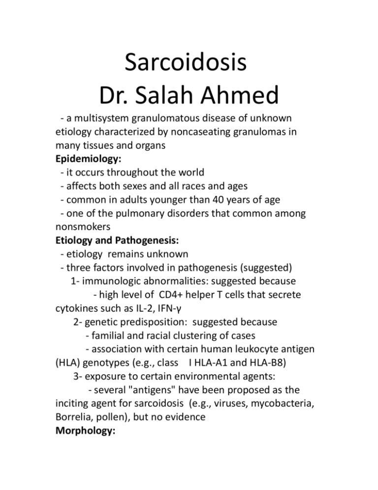
Sarcoidosis Dr. Salah Ahmed - a multisystem granulomatous disease of unknown etiology characterized by noncaseating granulomas in many tissues and organs Epidemiology: - it occurs throughout the world - affects both sexes and all races and ages - common in adults younger than 40 years of age - one of the pulmonary disorders that common among nonsmokers Etiology and Pathogenesis: - etiology remains unknown - three factors involved in pathogenesis (suggested) 1- immunologic abnormalities: suggested because - high level of CD4+ helper T cells that secrete cytokines such as IL-2, IFN-γ 2- genetic predisposition: suggested because - familial and racial clustering of cases - association with certain human leukocyte antigen (HLA) genotypes (e.g., class I HLA-A1 and HLA-B8) 3- exposure to certain environmental agents: - several "antigens" have been proposed as the inciting agent for sarcoidosis (e.g., viruses, mycobacteria, Borrelia, pollen), but no evidence Morphology: - noncaseating epithelioid granuloma: collection of epithelioid cells rimmed by an outer zone of CD4+ T cells. also multinucleated giant cell, fibroblasts - Two other microscopic features are sometimes seen in the granulomas: (1) Schaumann bodies, laminated concretions composed of calcium and proteins (2) asteroid bodies, stellate inclusions enclosed within giant cells Organs involvement: Lungs Upper airways Skin Eyes Liver Bones Salivary gland Heart system Joints Endocrine Kidneys glands Breast Uterus Lymph nodes Spleen Nervous Lacrimal 1- Lungs: - granulomas involve the interstitium rather than airspaces (around bronchioles and pulmonary venules and in the pleura) - In 5% to 15% of cases, granulomas are replaced by fibrosis 2- Lymph nodes: a- hilar and paratracheal lymph nodes b- peripheral lymph nodes: - occurs in one third of patients - the node "nonmatted" and do not ulcerate (TB) 3- Skin: - encountered in approximately 25% of patients 1- Erythema nodosum: - the hallmark of acute sarcoidosis - consists of raised, red, tender nodules - on the anterior aspects of the legs 2- painless subcutaneous nodules 3- raised painless plaques 4- lupus pernio:- indurated plaques - in nose, cheeks, and lips 4- Eye and lacrimal glands: - in the form of conjunctivitis, iritis, retinitis - suppression of lacrimation (sicca syndrome) - complicated by: corneal opacities, glaucoma, and total loss of vision 5- Salivary glands: - parotitis (painful enlargement with xerostomia) 6- Spleen, liver: enlargement 7- CNS: - Cranial nerves, and peripheral nerves can be involved - 7th nerve facial palsy is most common - Hereford's syndrome: facial palsy accompanied by fever, uveitis, and enlargement of the parotid gland 8- Heart: - involved in about 5% of cases - in the form of: 1- conduction abnormalities (arrhythmias) 2- cardiomyopathy 3- sudden death 9- Musculoskeletal: - acute polyarthritis with fever is common - chronic destructive bone disease with deformity is rare - polymyositis and chronic myopathy 10- Kidneys: - granulomatous interstitial nephritis Clinical Course: either: - asymptomatic: discovered on routine chest X-ray (as bilateral hilar adenopathy) - symptomatic: - peripheral lymphadenopathy, cutaneous lesions, eye involvement, splenomegaly, or hepatomegaly may be presenting manifestations - respiratory symptoms (shortness of breath, dry cough, vague substernal discomfort) - constitutional signs and symptoms (fever, fatigue, weight loss, anorexia, night sweats) Diagnosis: 1- Imaging studies (CT scan, X-ray ) 2- Biopsies: lung, lymph node,……… 3- Hypercalcemia and hypercalciuria: - not related to bone destruction - caused by increased calcium absorption secondary to production of active vitamin D by the mononuclear phagocytes in the granulomas - sarcoidosis is a diagnosis of exclusion (must rule out other granulomatous diseases) Treatment: - steroid therapy Prognosis: - 65% to 70% of affected individuals recover - 20% develop permanent lung dysfunction or visual impairment - 10% to 15% develop progressive pulmonary fibrosis and cor pulmonale Thank you
