Lambda Red Recombinase System for mutagenesis of
advertisement
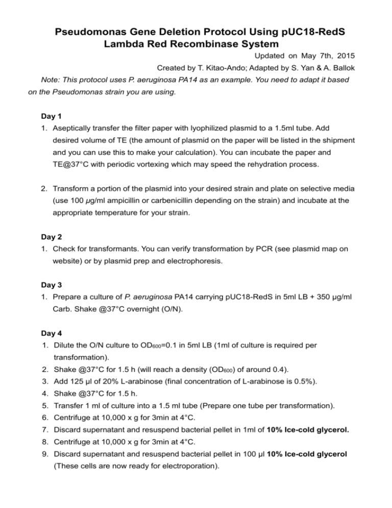
Pseudomonas Gene Deletion Protocol Using pUC18-RedS Lambda Red Recombinase System Updated on May 7th, 2015 Created by T. Kitao-Ando; Adapted by S. Yan & A. Ballok Note: This protocol uses P. aeruginosa PA14 as an example. You need to adapt it based on the Pseudomonas strain you are using. Day 1 1. Aseptically transfer the filter paper with lyophilized plasmid to a 1.5ml tube. Add desired volume of TE (the amount of plasmid on the paper will be listed in the shipment and you can use this to make your calculation). You can incubate the paper and TE@37°C with periodic vortexing which may speed the rehydration process. 2. Transform a portion of the plasmid into your desired strain and plate on selective media (use 100 μg/ml ampicillin or carbenicillin depending on the strain) and incubate at the appropriate temperature for your strain. Day 2 1. Check for transformants. You can verify transformation by PCR (see plasmid map on website) or by plasmid prep and electrophoresis. Day 3 1. Prepare a culture of P. aeruginosa PA14 carrying pUC18-RedS in 5ml LB + 350 µg/ml Carb. Shake @37°C overnight (O/N). Day 4 1. Dilute the O/N culture to OD600=0.1 in 5ml LB (1ml of culture is required per transformation). 2. Shake @37°C for 1.5 h (will reach a density (OD600) of around 0.4). 3. Add 125 µl of 20% L-arabinose (final concentration of L-arabinose is 0.5%). 4. Shake @37°C for 1.5 h. 5. Transfer 1 ml of culture into a 1.5 ml tube (Prepare one tube per transformation). 6. Centrifuge at 10,000 x g for 3min at 4°C. 7. Discard supernatant and resuspend bacterial pellet in 1ml of 10% Ice-cold glycerol. 8. Centrifuge at 10,000 x g for 3min at 4°C. 9. Discard supernatant and resuspend bacterial pellet in 100 µl 10% Ice-cold glycerol (These cells are now ready for electroporation). 10. Add at least 1 µg of DNA fragment consisting of two ~500-bp flanking regions and antibiotic cassette to the competent cells and incubate 10 min on ice (DNA volume is up to 10 µl). 11. Transfer competent cells into electroporation cuvette (Bio-Rad #165-2086, 0.2 cm). 12. Electroporate cells with a voltage of 2.5 kV (Ec2 mode on Biorad micropulser). 13. Add 900 µl of LB into cuvette, resuspend cell well, and transfer contents to a 1.5 ml tube. 14. Shake tube @30°C for at least 1h (During this step, prepare a dry LB agar plate(s) containing the appropriate antibiotic). 15. Centrifuge bacterial cells at 10,000 x g for 3min. 16. Discard 800 µl supernatant. 17. Resuspend pellet by pipetting and spread onto LB agar plate containing appropriate antibiotic. 18. Incubate plate @30°C O/N. Day 3 1. Check for transformant colonies (should appear after 20-24hrs). Note: 1. We found that a 500bp flanking region gives the best efficiency. Shorter flanking regions can reduce efficiency, but it depends on the region used for recombination. 2. We use 10% ice-cold glycerol instead of 10% sucrose because we found that this has a higher efficiency. 3. After electroporation, we incubate cells at 30°C because the lower temperature may increase the chance of getting colonies. 4. For long-term storage of the plasmid in bacteria, we prefer E. coli rather than P. aeruginosa, as storage in P. aeruginosa can increase the risk in genetic drift of the lambda red system.
