Hydrocephalus - English - Vietnam Health Improvement Project
advertisement
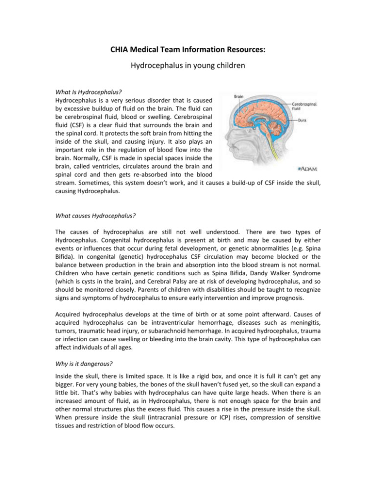
CHIA Medical Team Information Resources: Hydrocephalus in young children What Is Hydrocephalus? Hydrocephalus is a very serious disorder that is caused by excessive buildup of fluid on the brain. The fluid can be cerebrospinal fluid, blood or swelling. Cerebrospinal fluid (CSF) is a clear fluid that surrounds the brain and the spinal cord. It protects the soft brain from hitting the inside of the skull, and causing injury. It also plays an important role in the regulation of blood flow into the brain. Normally, CSF is made in special spaces inside the brain, called ventricles, circulates around the brain and spinal cord and then gets re-absorbed into the blood stream. Sometimes, this system doesn’t work, and it causes a build-up of CSF inside the skull, causing Hydrocephalus. What causes Hydrocephalus? The causes of hydrocephalus are still not well understood. There are two types of Hydrocephalus. Congenital hydrocephalus is present at birth and may be caused by either events or influences that occur during fetal development, or genetic abnormalities (e.g. Spina Bifida). In congenital (genetic) hydrocephalus CSF circulation may become blocked or the balance between production in the brain and absorption into the blood stream is not normal. Children who have certain genetic conditions such as Spina Bifida, Dandy Walker Syndrome (which is cysts in the brain), and Cerebral Palsy are at risk of developing hydrocephalus, and so should be monitored closely. Parents of children with disabilities should be taught to recognize signs and symptoms of hydrocephalus to ensure early intervention and improve prognosis. Acquired hydrocephalus develops at the time of birth or at some point afterward. Causes of acquired hydrocephalus can be intraventricular hemorrhage, diseases such as meningitis, tumors, traumatic head injury, or subarachnoid hemorrhage. In acquired hydrocephalus, trauma or infection can cause swelling or bleeding into the brain cavity. This type of hydrocephalus can affect individuals of all ages. Why is it dangerous? Inside the skull, there is limited space. It is like a rigid box, and once it is full it can’t get any bigger. For very young babies, the bones of the skull haven’t fused yet, so the skull can expand a little bit. That’s why babies with hydrocephalus can have quite large heads. When there is an increased amount of fluid, as in Hydrocephalus, there is not enough space for the brain and other normal structures plus the excess fluid. This causes a rise in the pressure inside the skull. When pressure inside the skull (intracranial pressure or ICP) rises, compression of sensitive tissues and restriction of blood flow occurs. In the picture on the left, you can see a normal brain, and a brain that has hydrocephalus. The compression of brain tissue and the restriction of blood flow can cause permanent brain damage, resulting in developmental delay, seizure disorders, hearing loss, impaired vision and muscle weakness. What are the symptoms? There are several signs that someone has hydrocephalus. Signs and symptoms can differ depending on what age the child is when hydrocephalus begins. System Infants Toddlers and Children Physical appearance of head An unusually large head that rapidly increases in size A bulging or tense soft spot on the top of the head Eyes fixed downwards ‘sunsetting eyes’ Sleepiness & irritability Seizures A high pitched cry Abnormal enlargement of the head Vision and eyes Neurological Memory and concentration Blurred or double vision Irritability and change in personality Seizures Extreme sleepiness Headache* Problems with attention Decline in school performance Musculoskeletal Deficits in muscle tone and strength, repsonsiviness to touch and expected growth Delays in walking and talking Problems with previously acquired skills (e.g. walking) Unstable Balance and poor coordination Gastrointestinal Vomiting** Poor feeding Nausea or vomiting** Poor appetite Genitourinary Young and middle aged adults Impaired vision Difficulty in remaining awake or waking up Headache* Decline in memory, concentration and other thinking skills that may affect job performance Loss of coordination or balance Memory loss Progressive loss of other thinking or reasoning skills Loss of bladder control or frequent urge to urinate Loss of bladder control or frequent urge to urinate *Headache – the pain is normally in the morning and worse after lying down **Vomiting – usually in the morning and without associated nausea Images for identification of hydrocephalus: ‘Sunset eyes’ – eyes that are fixed downwards Older adults Enlarged head Difficulty walking, often described as a shuffling gait or the feeling of being stuck Poor coordination or balance Slower than normal movements How is hydrocephalus diagnosed? Sometimes they can diagnose hydrocephalus based on how someone looks. This is particularly the case for infants who have large heads, or sunset eyes. Hydrocephalus is diagnosed through clinical neurological evaluation and by using cranial imaging techniques such as ultrasonography, computed tomography (CT), magnetic resonance imaging (MRI), or pressure-monitoring techniques. How is hydrocephalus treated? Hydrocephalus is most often treated by surgically inserting a shunt system. This system diverts the flow of CSF from the CNS to another area of the body where it can be absorbed as part of the normal circulatory process. A shunt system consists of the shunt, (a flexible plastic tube), a catheter, and a valve. One end of the catheter is placed within a ventricle inside the brain or in the CSF outside the spinal cord. The other end of the catheter is commonly placed within the abdominal cavity (called a Ventricular Peritoneal Shunt of VP Shunt), but may also be placed at other sites in the body such as a chamber of the heart or areas around the lung where the CSF can drain and be absorbed. A valve located along the catheter maintains one-way flow and regulates the rate of CSF flow. Ventricular Peritoneal shunt treatment is the most common form of treatment in Vietnam. It is normally a very quick surgery, taking about an hour to insert. The first 24 hours after surgery are very important, and the patient should be monitored very closely. Are there any complications from this surgery? People who have shunts can have complications days or even years after the initial operation. 1. Shunt Malfunction – is usually a problem with a partial or complete blockage of the shunt. The fluid backs up from the site of obstruction and if the blockage is not corrected will almost always result in symptoms of hydrocephalus. Shunts are very durable, but parts of the shunt can break as a result of wear as a child grows, or move from where they were initially placed. 2. Shunt Obstruction – a part of the shunt becomes blocked. Can occur in any part of the shunt 3. Shunt Infection – usually a shunt infection is caused by the bacteria that normally live inside a person’s body. Infections are most likely to occur 1-3 months after the placement of the shunt. People who have VP shunts can get a shunt infection after they have a gastrointestinal infection. Shunt infection must be treated immediately to avoid life threatening illness and/or brain damage. Symptoms of a shunt infection are: a. Fever b. Poor feeding/low appetite c. Nausea and vomiting d. Neck stiffness – where the child can’t look up to the sky or down to the ground e. Unusual tiredness – sleeping more than normal, difficult to wake up f. Changes in personality or behaviour g. Abdominal (belly) pain 4. Overdrainage – this is a rare complication where the shunt drains too much fluid away from the brain, which causes the ventricles to decrease in size and the brain can pull away from the skull. This can cause damage to the blood vessels in the brain, which can cause bleeding in the brain which is very dangerous. 5. Underdrainage – the shunt is not draining enough fluid so the shunt does not treat the hydrocephalus. If this happens, it might be necessary to place a new shunt Early Detection and Prognosis: Identifying hydrocephalus early is very important. Hydrocephalus can cause brain damage, which is irreversible, if it is left too long. This can be a significant burden for the family, and can decrease the quality of life of the child. If you have a child who has Spina Bifida, Dandy Walker Syndrome or Cerebral Palsy, they should be monitored closely for signs of hydrocephalus. You should educate parents about how to check for signs of hydrocephalus, and the importance of taking their children to the hospital. The prognosis for individuals diagnosed with hydrocephalus is difficult to predict, although there is some correlation between the specific cause of the hydrocephalus and the outcome. Prognosis is further complicated by the presence of associated disorders, the timeliness of diagnosis, and the success of treatment. The degree to which relief of CSF pressure following shunt surgery can minimize or reverse damage to the brain is not well understood Early treatment for hydrocephalus can be quite successful. Sadly, it cannot undo neurological damage that may have occurred before diagnosis. Early diagnosis and treatment improves the chance of a good recovery. CHIA FOLLOW UP PROCEDURE FOR HYDROCEPHALUS: After initial contact/assessment: Call one week after initial contact/assessment to ensure that all tests have been done, doctors consulted, appointments made etc Ask family to bring copies of hospital/surgical assessments to CHIA office Determine whether it is feasible for CHIA to assist this child Ask family to call after each assessment, or doctors appointment and/or CHIA staff call if they are aware of further assessments/doctors appointments. After surgical treatment: Make contact with family as soon as possible after surgical intervention to determine success of surgery and child’s well being Call one week after surgery to see if there have been any complications (it is important at this stage to check whether the symptoms of hydrocephalus have gone away, because if they haven’t, this could be evidence of a shunt malfunction) Assess in person one month after surgery to check for any complications, particularly shunt infection and malformation Assess in clinic with HAF 6 months after surgery If there are no complications, assess child 1 year after surgery If there are complications, follow-up with family regularly until issue is resolved Further continuing care will depend on the cause of hydrocephalus, prognosis of child and presence of complications Continuing care: Educate the family about signs of shunt complications – particularly malfunction and obstruction (risk of infection decreases significantly 6 months after surgery) Ensure that family knows to call CHIA if there are any further health issues complications Depending on the cause of hydrocephalus, refer to Therapy Team (e.g. refer cases that have Spina Bifida or Cerebral Palsy) If there are no further complications, then appropriate cases can be managed by the therapy team Questions to ask on follow-up: If you suspect hydrocephalus: 1. 2. 3. 4. 5. 6. Has there been an accident lately where the child hit their head? Has the child been sleepier than normal, or more difficult to wake up? How has the child been feeding? Has the child had poor concentration/trouble in school lately? (for a baby) Were there any problems during pregnancy? Assess whether the child has any symptoms of hydrocephalus (see above). If yes to any of these questions, then the child should be seen as soon as possible by a doctor to get a CT scan or an MRI. In the first 6 months after treatment: 1. Have the child’s symptoms gone away? (if no, advise to see a doctor) 2. Has the child had any fevers, or been sick in the tummy? Or any symptoms of a shunt infection? (if yes, advise to see a doctor) 3. Is the child sleepier than normal, or had any changes in their personality? (If yes advise to see a doctor) 6+ months after treatment: 1. Does the child have any symptoms of hydrocephalus? 2. How is the child going at school? 3. How is the child’s appetite? 4. Is the child gaining weight? 5. Has the child had any illnesses? References: www.childrenshospital.org/clinicalservices/Site1592/mainpageS1592P7.html http://www.ninds.nih.gov/disorders/hydrocephalus/detail_hydrocephalus.htm#113403125
