Electronic Supplemental Material Richmond JQ, Wood DA, Stanford
advertisement
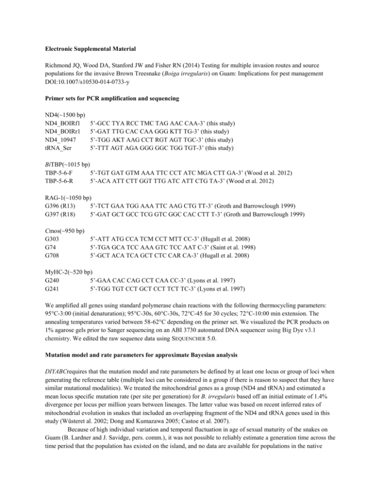
Electronic Supplemental Material Richmond JQ, Wood DA, Stanford JW and Fisher RN (2014) Testing for multiple invasion routes and source populations for the invasive Brown Treesnake (Boiga irregularis) on Guam: Implications for pest management DOI:10.1007/s10530-014-0733-y Primer sets for PCR amplification and sequencing ND4(~1500 bp) ND4_BOIRf1 ND4_BOIRr1 ND4_10947 tRNA_Ser 5’-GCC TYA RCC TMC TAG AAC CAA-3’ (this study) 5’-GAT TTG CAC CAA GGG KTT TG-3’ (this study) 5’-TGG AKT AAG CCT RGT AGT TGC-3’ (this study) 5’-TTT AGT AGA GGG GGC TGG TGT-3’ (this study) BiTBP(~1015 bp) TBP-5-6-F 5’-TGT GAT GTM AAA TTC CCT ATC MGA CTT GA-3’ (Wood et al. 2012) TBP-5-6-R 5’-ACA ATT CTT GGT TTG ATC ATT CTG TA-3’ (Wood et al. 2012) RAG-1(~1050 bp) G396 (R13) 5’-TCT GAA TGG AAA TTC AAG CTG TT-3’ (Groth and Barrowclough 1999) G397 (R18) 5’-GAT GCT GCC TCG GTC GGC CAC CTT T-3’ (Groth and Barrowclough 1999) Cmos(~950 bp) G303 5’-ATT ATG CCA TCM CCT MTT CC-3’ (Hugall et al. 2008) G74 5’-TGA GCA TCC AAA GTC TCC AAT C-3’ (Saint et al. 1998) G708 5’-GCT ACA TCA GCT CTC CAR CA-3’ (Hugall et al. 2008) MyHC-2(~520 bp) G240 5’-GAA CAC CAG CCT CAA CC-3’ (Lyons et al. 1997) G241 5’-TGG TGT CCT GCT CCT TCT TC-3’ (Lyons et al. 1997) We amplified all genes using standard polymerase chain reactions with the following thermocycling parameters: 95°C-3:00 (initial denaturation); 95°C-30s, 60°C-30s, 72°C-45 for 30 cycles; 72°C-10:00 min extension. The annealing temperatures varied between 58-62°C depending on the primer set. We visualized the PCR products on 1% agarose gels prior to Sanger sequencing on an ABI 3730 automated DNA sequencer using Big Dye v3.1 chemistry. We edited the raw sequence data using SEQUENCHER 5.0. Mutation model and rate parameters for approximate Bayesian analysis DIYABCrequires that the mutation model and rate parameters be defined by at least one locus or group of loci when generating the reference table (multiple loci can be considered in a group if there is reason to suspect that they have similar mutational modalities). We treated the mitochondrial genes as a group (ND4 and tRNA) and estimated a mean locus specific mutation rate (per site per generation) for B. irregularis based off an initial estimate of 1.4% divergence per locus per million years between lineages. The latter value was based on recent inferred rates of mitochondrial evolution in snakes that included an overlapping fragment of the ND4 and tRNA genes used in this study (Wüsteret al. 2002; Dong and Kumazawa 2005; Castoe et al. 2007). Because of high individual variation and temporal fluctuation in age of sexual maturity of the snakes on Guam (B. Lardner and J. Savidge, pers. comm.), it was not possible to reliably estimate a generation time across the time period that the population has existed on the island, and no data are available for populations in the native range. This is likely due to shifts in prey resource availability on Guam, which has changed dramatically over time. Thus, we calculated a range of mutation rates based on a low and high estimate of generation time. These were as follows: 2.2 yr generation time = 8.1 x 10⁻⁶ mutations/site/generation; 6.0 yr generation time = 2.9 x 10 6 mutations/site/generation. To account for the differences in rates, we used a broad uniform prior for the mean mutation rate (1 x 10⁻⁸ – 1 x 10⁻⁶)and assumed an HKY mutation model for sequence evolution. The questions we attempted to address for the Guam snake population have occurred over a time scale that is essentially irrelevant to mutation (~65 years). Rather, the current population genetics are more reflective of the random sampling of gene copies from the source population, the founder event, and the subsequent fixation or elimination of those gene copies due to genetic drift once the population became established. DNA sequence variability Levels of sequence divergence varied substantially among loci, with the mitochondrial ND4 gene having the highest number of informative substitutions (Table 1). The nuclear genes varied as follows (in decreasing order for number of informative characters); BiTBP, MyHC-2, RAG-1 and Cmos. We detected heterozygous individuals for all nuclear loci for length and or site-specific polymorphisms, with MyHC-2 showing the highest heterozygosity within individuals. Length variants were most common in the BiTBP gene, with a few deletions being parsimony informative in B. irregularis. Nearly every B. irregularis in the dataset had a 300 base pair insertion within the BiTBP intron that none of the outgroup taxa possessed – only one B. irregularis individualfrom Indonesia (Pulau Tahulandang, Sulawesi) lacked this insertion and retained the ancestral gene length recovered in all outgroup taxa. Several snakes from the D’Entrecasteaux Islands (Fergusson and Normanby Islands) also had a synapomorphic deletion in the BiTBP gene not found in any other B. irregularis. Site-specific ambiguities that could not be resolved with a posterior >0.90 were conservatively scored as missing data. This mainly affected the outgroup species, as the accuracy of phase inference decreased as the amount of divergence among terminal taxa grew larger, as expected for this type of analysis. Even with this conservative approach, we still gained considerable phylogenetic information by teasing out the different allelic phases within individuals. In contrast to the complete lack of mtDNA variation on Guam, we detected some allelic differences for each of the nuclear genes; however, the differences involved single-site substitutions and only 2-4 alleles per locus were recovered from all of the Guam snakes combined. These allelic polymorphisms reflect the retention of ancestral variation from the source population rather than in situ evolution on Guam, given that ‘Guam alleles’ were detected in the Admiralties for all nuclear genes. In fact, incomplete lineage sorting in the nuclear genes was a general pattern observed across all populations in the dataset – this was demonstrated by the relatively high nuclear diversity overall, but with few shared derived characters for diagnosing regional groups. Table S1. Summary data for the various genetic markers used in this study. Data are shown for all taxa (outgroup species &B. irregularis) and for B. irregularis only. Column headers are as follows: No. Characters = number of total nucleotide sites (nt) in the gene sequence; VPU = variable parsimony uninformative characters; PI = parsimony informative characters. ‘Indel’ refers to nucleotide insertions or deletions. Marker ND4 + tRNAs RAG-1 Cmos MyHC2 MyHC2 indel Bi-TBP Bi-TBP indel No. Characters (nt) All taxa VPU 1561 1054 954 561 6 1014 21 189 44 6 12 0 25 6 PI 457 38 47 85 6 94 15 B. irregularis VPU 89 9 4 6 0 17 2 PI 202 10 7 14 0 26 3 Literature Cited Dong S andKumazawa Y (2005) Complete mitochondrial DNA sequences of six snakes: phylogenetic relationships and molecular evolution of genomic features. J MolEvol 61:12-22 Groth JG and Barrowclough GF(1999) Basal divergences in birds and the phylogenetic utility of the nuclear RAG-1 Gene. Mol Phylo Evol 12:115-123. Hugall AF, FosterR, HutchinsonM, and Lee MSY (2008) Phylogeny of Australasian agamid lizards based on nuclear and mitochondrial genes: implications for morphological evolution and biogeography. Biol J Linn Soc 93:343-358. LyonsLA, LaughlinTF, CopelandNG, JenkinsNA, WomackJE, and O'BrienSJ(1997) Comparative anchor tagged sequences (CATS) for integrative mapping of mammalian genomes. Nat Genet 15:47-56. Saint KM, AustinCC, Donnellan SC, and Hutchinson MN(1998) C-mos, a nuclear marker useful for squamate phylogenetic analysis. Mol Phylo Evol 10:259-263. WoodDA, Vandergast AG, Lemos Espinal JA, Fisher RN, and Holycross AT(2012) Refugial isolation and divergence in the Narrowheaded Gartersnake species complex (Thamnophis rufipunctatus) as revealed by multilocus DNA sequence data. Mol Ecol 20:3856-3878. Wüster W, da GracaSalomão M, Quijada-Mascareñas JA, Thorpe RS, and Butantan-British BothropsSystematics Project (2002) Origin and evolution of the South American pitviper fauna: evidence from mitochondrial DNA sequence data. In:Schuett GW, Höggren M, Douglas ME, Greene HW (eds) Biology of the Vipers. Eagle Mountain Publishing, Salt Lake City, UT, pp. 111-128


