Subacute Ruminal Acidosis - Utrecht University Repository
advertisement
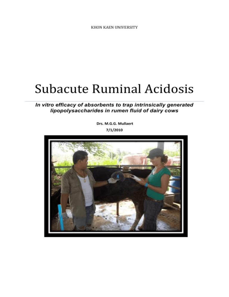
KHON KAEN UNIVERSITY Subacute Ruminal Acidosis In vitro efficacy of absorbents to trap intrinsically generated lipopolysaccharides in rumen fluid of dairy cows Drs. M.G.G. Mullaert 7/1/2010 Subacute Ruminal Acidosis 2010 Drs. M.G.G. Mullaert Studentnr. 0461636 Juli-oktober 2010 Supervisors: Dr. J.T. (Thomas) Schonewille Department of Farm Animal Health Faculty of Veterinary Medicine, Utrecht University Dr. Anantachai Chaiyotwittayakun Department of Veterinary Medicine Faculty of Veterinary Medicine, Khon Kaen University Drs. Rittichai Pilachai Department of Veterinary Medicine Faculty of Veterinary Medicine, Khon Kaen University Acknowlegdement We gratefully thank Drs. D de Haan from Trouw International for his kind donation of the clay minerals NovasilTM Plus and TOX-OTM . Research Project Drs. M.G.G. Mullaert Page 2 Subacute Ruminal Acidosis 2010 Abstract Laminitis is a common health problem in dairy cows in Thailand nowadays. In order to maintain high production, dairy cows need to be fed with a diet high in grain and low in forage. Consequently, the diet contains a high amount of rapid fermentable carbohydrates and insufficient fiber. This results in the production of a large quantity of volatile fatty acids, and causes a depression on the rumen pH and associated with rumen acidosis. (Kleen et al, 2009). Subacute ruminal acidosis (SARA), the condition were the rumen pH<5.6 for at least 180 min/d (Gozho et al., 2005), is suggested to play an important role in the cascade. It has been suggested that the pathogenic mechanism of laminitis may involve the activation of systemic inflammatory responses due to the lysis of gram-negative bacteria. Rumen pH depression during SARA increases free LPS concentration of the rumen (Emmanuel et al., 2008; Gozho et al., 2007) and may result in the transfer of LPS from the gut into blood circulation, and activation of an inflammatory response (Khafipour et al., 2009). Thus, materials that can bind LPS in the rumen and can prevent the transfer of endotoxin (LPS) to the blood are potentially of interest to the prevention of laminitis. It is know that montmorillonite have binding affinity for LPS (Pilachai et al., 2010, unpublished data). This trial was conducted to evaluate the efficacy of three adsorbents; montmorillonite, hydrate sodium calcium aluminosilicate (HSCAS) and modified HSCAS, to trap intrinsically generate lipopolysaccharides (LPS) in rumen fluid collected from SARA induced cows. In vitro, we added the three absorbents at three levels (0, 0.5 and 1% (w/v) to the rumen fluid from SARA cows and measured the LPS concentration. It appear that the sequestering agents tested here had no significant absorbing effect on the LPS in the rumen fluid of the SARA induced cows. Research Project Drs. M.G.G. Mullaert Page 3 Subacute Ruminal Acidosis 2010 Contents Abstract ................................................................................................................................................... 3 Contents .................................................................................................................................................. 4 Introduction............................................................................................................................................. 5 Materials and Methods ......................................................................................................................... 12 Results ................................................................................................................................................... 14 Discussion .............................................................................................................................................. 16 Conclusion ............................................................................................................................................. 16 Acknowledgement................................................................................................................................. 17 References ............................................................................................................................................. 18 Research Project Drs. M.G.G. Mullaert Page 4 Subacute Ruminal Acidosis 2010 Introduction Laminitis (pododermatitis aseptic diffusa) is a common problem in dairy cattle in Thailand. Laminitis is defined as the inflammation of the dermal layers inside the foot (Nocek, 1997). The cause of laminitis should be considered as a combination of predisposing factors leading to an altered vascular reactivity and microcirculation and disruption of normal cell differentiation and horn formation in the clay epidermis (Mulling, 2002). There are multiple causes of lameness such as infectious diseases of the interdigital space for example foot rot or digital dermatitis. Traumatic damage to the sole can also occur when the cattle is housed in stables with hard or rough flours or when they have to walk long distances over rough roadways. Embedded stones for example can cause white line abcesses to form. Laminitis is a condition where the bloodflow to the extremities is restricted which causes damage to the claws. Laminitis can be divided in three forms. Acute, subacute and subclinical (Greenough 2007). Acute laminitis is a consequence of a grain overload where the cow has eaten a large amount of grain and the pH of the rumen drops rapidly below 5.0. Affected animals show signs rapidly after overloading and an increase in heart rate, respiratory rate and liquid, light colored feces can be observed. Cows can become recumbent or crawl on their knees. Subacute laminitis is a more subtle disorder were the only signs are that of a mild discomfort with cows shifting weight from leg to leg and they place their feet on the ground very carefully. Subacute laminitis is also associated with nutritional changes which can cause vasoactive substances to be released into the bloodstream that alter the bloodflow in the claw causing damage and discomfort. Subclinical laminitis or pododermatitis aseptica diffuse is a disorder without clinical signs at the moment that structures in the claw are damaged but creates specific lesions that will show after a period of time. These lesions are caused by two different pathological processes. 1) The claw horn is weakened structurally and functionally and no longer able to endure loading and more vulnarabule to environmental agents. 2) the support system of the pedal bone and the integrity of the suspensory apparatus of the digit are weakened (Greenough 2007). Systemic vasoactive substances change the bloodpressure in the extremities and arteriovenous shunts start to form. This increases the bloodpressure in the claws and the vessels start to leak and are damaged and edema and hemorraghing occurs. The opening of the shunts also causes ischemia to occur in the dermis (Nocek et al 1997, Greenough 2007). As a result of the increased pressure and ischemia in the dermis the degeneration of the corium and the formation of horn is impaired. This can be detected in the sole 4 to 8 weeks after the initial damage is done as sole hemorraghes, white line defects, dorsal ridging of the wall or double soles (Nocek et al 1997). If the damage to the epidermis is severe the laminar layer can separate completely which will cause the pedal bone to shift. This creates even more damage to the soft tissue and necrosis can occur. The claw is tilted slightly which can be observed. It is very difficult to distinguish between sole hemorraghes caused by subclinical laminitis or caused by traumatic damage to the sole. Housing should always be considered when sole bruising is found as a herd problem. Laminitis has been associated with a reduced pH of rumen fluid: i.e. rumen acidosis. This condition occurs both in dairy cattle as well as in beef cattle. The drop of the pH in the rumen, the formation of endotoxins (lipopolysaccharides) and damage to the rumen wall can have a systemic effect that could possibly have an effect on the bloodflow in the extremitities and play a role in the occurence of laminitis. See figure 1. Research Project Drs. M.G.G. Mullaert Page 5 Subacute Ruminal Acidosis 2010 Figure 1. Progression of physiological events that link acidosis with laminitis. CHO = Carbohydrate. (Modified from Nocek, 1997) Ruminal acidosis is a bovine disease that affects feedlot as well as dairy cattle. It’s caused by the ingestion of greater than normal quantities of ruminally fermentable carbohydrates. This results in a decreased ruminal pH. The cascade effects of this acidosis depend upon the intensity and duration of the insult. Most critical is the pH threshold, which not only relates to microbial growth rates and shifts in ruminal populations but also significantly influences the systemic metabolic state (Nocek, 1997). Rumen acidosis can be divided into an acute and a subacute form. The subacute form is also known as SARA: Subacute Rumen Acidosis. Acute acidosis results in very sick cows and is characterized by a ruminal pH < 5.0. (Nocek, 1997), its generally linked to lactic acid production (figure 1). There are many definitions of when to speak of SARA based on method and location of taking rumen fluid samples. Samples taken by stomach tube, rumenocentesis or through a fistula from the ventral sac of the rumen differ in pH value. Samples taken by stomach tube can be diluted by saliva and are on average 0,3 point higher than samples taken by rumenocentesis (Plaizier et al 1998). The pH of the rumen fluctuates during the day and the most accurate way to measure the pH is by using an indwelling pH probe in the ventral sac of the rumen. SARA is characterized by intermittent periods of depressed rumen pH each day, e.g. <5.6 for at least 180 min/day (Kleen et al., 2003; Stone, 2004; Gozho et al., 2005). The onset of SARA is marked by the intake of a diet low in structure and high in energy, while the ruminal environment is not yet adapted to ferment and later absorb the arising shortchain fatty acids (SCFA) adequately enough to keep the ruminal pH within physiological borders. This diet will result in reduced amount of time spent chewing and thus less saliva production. Saliva contains inorganic buffers that contribute to the neutralization of the organic acids produced during fermentation in the rumen (Church, 1988). The ruminal pappilae are of crucial importance in the absorption of SCFA; the proliferation of the papillae is promoted by SCFA arising from the fermentation. If the ruminal mucosa is not adapted, which especially is the case at the shift from a dry-period to an early lactation diet, the papillae are too short and the resorbing surface too small to deal with the sudden increase of SCFA-levels (Nordlund et al.,1995). Research Project Drs. M.G.G. Mullaert Page 6 Subacute Ruminal Acidosis 2010 Also the microbial population, which has to metabolize the lactic acid arising from the fermentation of abundant carbohydrates of Streptococcus bovis or Dasytricha spp., is insufficiently developed (Slyter, 1976; Dawson and Allison, 1988; Nordlund et al., 1995). These factors together cause a transient fall of ruminal pH. The balance between production and absorption of fermentation end products is disturbed (Garrett, 1996). In conclusion, rumen acidosis is linked to lactic acid production and in the case of SARA, VFA production plays an important role. That’s the difference between these two. See figure 2. Figure 2. Step-by-step microbial mechanisms of occurence of acidosis in the rumen. (Modified from rumenhealth.com) Thus, subacute ruminal acidosis is a common disease in high yielding dairy cows that receive highly digestible diets, and has a high economic impact (Plaizier et al., 2009). Especially because its most of the time subclinical. Oetzel (2004) found that 20% of cows from commercial dairy herds sampled by rumenocentesis during clinical herd investigations had ruminal pH of <5.5. Also Garrett et al. 1997 concluded that SARA is present in a large number of dairy herds. Costs of SARA resulting from lost production alone were estimated to be 1.12/d per cow in a herd diagnosed with SARA (Stone, 1999). Subacute ruminal acidosis (SARA), is often difficult to detect. The clinical signs are variable and not specific to SARA. The related clinical signs in dairy cows can be diarrhea, rumen mucosal damage, laminitis, inflammation throughout the body, and liver abscesses (Nocek, 1997; Kleen et al., 2003; Stone, 2004; Alzahal et al., 2007). Herds with a high prevalence of SARA may have a high culling rate, increased death loss, and decreased milk production (Nocek, 1997, Stone, 1999).We can speak of two major critical situations were SARA can develop, in other words, where the non-adaptated rumen is likely to meet a concentrate-based diet. The most important one is the early post-partum period: a rumen non-adapted to concentrates and a possibly high concentrate-uptake around parturition, during general low-feed intake in combination with a considerable impact of stress and a more or less sever negative energy balance. And the midlactation period: mostly linked to managerial factors, like feeding frequency, processing of feed and housing (Kleen, 2003). There is substantial evidence that grain-based SARA challenge increases the content of free LPS in the rumen due to the increase in lysis of gram-negative bacteria (Gozho et al., 2007; Nagaraja and Lechtenberg, 2007; Plaizier et al., 2008). This increase in luminal LPS could increase permeability of the gut for LPS (Chin et al., 2006). Also, the barrier function of rumen epithelium may be compromised by the parakeratosis, rumenitis, and abscesses of the rumen wall that result from high rumen acidity (Kleen et al., 2003). See figure 3. Research Project Drs. M.G.G. Mullaert Page 7 Subacute Ruminal Acidosis 2010 Figure 3. Proposed relationships between route of endotoxin exposure, hepatic function, biological reponse and clinical response in bovine endotoxicossis. (Modified from Nocek, 1997) Additionally, the high rumen osmolality (by increase of VFA (Bennink et al., 1978)) that is seen during SARA can cause swelling and rupture of ruminal papillae, which will also reduce the barrier function of the rumen (Plaizier et al., 2009). This results in translocation of immunogenic compounds into circulation, such as free LPS. Endotoxin, also known as LPS, is a cellular component of gram-negative bacteria and is an extremely potent toxin (Andersen et al., 2000). See figure 4. LPS interacts with a specific acute phase protein, LPS binding protein (LBP), an increase in LBP in peripheral circulation facilitate binding of LPS to cell membrane receptors, which then triggers a cascade of events toward a local or systemic inflammatory response and release of other acute phase proteins (APP) into the blood (Khafipour 2009). The main stimulators of APP production are the inflammation-associated cytokines IL-1, IL-6, tumor necrosis factor (TNF)-α, and IFN-γ which are released during inflammatory processes (Gabay and Kushner, 1999). Two APP, serum amyloid A (SAA) and LPS-binding protein (LBP), directly participate in the detoxification and removal of endotoxin during an acute phase response. (Emmanuel et. al., 2008) Figure 4. Endotoxins (lipopolysaccharides) are structural parts of the bacterial cell wall and in general comprise 3 major regions: the side chain, the core polysaccharides and lipid A (Modified from Rietschel 1996). Research Project Drs. M.G.G. Mullaert Page 8 Subacute Ruminal Acidosis 2010 The link between acidosis and laminitis appears to be associated with altered hemodynamics of the peripheral microvasculature. The primary target of endotoxins appears to be vascular arterial tissues, which affects the release of vasoactive mediators, thus inducing an acute inflammation of the dermis and associated vascular responses. Histamine causes impedance of blood flow and pressure building within the capillaries forces fluid through the vessels into the interstitial tissue spaces setting up ischemia. When the microvasculature of the corium is affected by vasoactive substances, vascular destruction is inevitable. When blood is not returned to circulation by the musculature of the vascular system, seepage and hemorrhage can result. The anatomical features of the corium vascular network play a very important role in the development of various stages of laminitis. (Nocek., 1997). See also figure 6. To prevent problems around laminitis, caused by SARA in dairy herds, it’s important to concentrate on two points: prevention of a fermentation disorder or regulation of the fermentation process by direct intervention. Alternatively, there are other options regarding the tecnonology of feedstuffs and certain substances or organisms added. Materials that can bind LPS in the rumen and can prevent the transfer of endotoxin to the blood. Those materials, like microbials, buffers or lactate are potentially of interest to the prevention of SARA and thus laminitis. It is well know that absorbent materials such as clay minerals have a high absorbing capacity for some toxins, the layered structure can load inorganic antibacterial cations (Ag, Cu, Zn etc.) within the interlayer space by an ion exchange reaction (Dakovic, 2008). Indeed, in vitro studies have shown that clay minerals have the ability to bind aflatoxin B1 under different pH conditions (Diaz et al, 2003; Spotti, 2005; Moschini, 2008). In addition, clay minerals also have a high capacity to bind mycotoxin in rumen fluid (Diaz et al, 2003; Spotti et al, 2005). Clay minerals can bind mycotoxins, despite this, there is no evidence that they can also bind endotoxins produced during SARA-conditions. Recently, Pilachai et al. (2010) (unpublished data) demonstrated that both activated charcoal and montmorrilonite are capable of absorbing LPS in a buffer as well in rumen fluid. Furthermore, it appeared that pH had no effect on the efficacy of the clay minerals to trap LPS. In the study of Pilachai et al. (2010) (unpublished data) montmorillonite (MONT) was the most promising clay mineral because it showed the highest sequestering efficacy in rumen fluid. Therefore, montmorillonite was also used in the current experiment. Montmorillonite in the pure form isn’t yet used in animals, so we also want to evaluate hydrate sodium calcium aluminosilicate (HSCAS) and modified HSCAS, because they are already used in animals. Montmorillonite, a smectite clay, has a 2:1 layered structure with the inner layer composed of an octahedral sheet that shares oxygen atoms between two tetrahedral sheets. In smectites, the substitution of Al3+ for Si4+ in tetrahedral sheets and substitution of divalent cations such as Mg2+ and Fe2+, for trivalent cations such as Al3+ and Fe3+ in octahedral sheets give rise to a deficiency of the positive charge in the framework. This charge is balanced by monovalent and divalent cations (usually Na+, K+, Ca2+ and Mg2+), and is expressed as the cation exchange capacity (CEC). These cations positioned mainly in the interlamellar space, can easily be exchanged with other inorganic and/or organic cations under aqueous conditions (Dakovic., 2008). Research Project Drs. M.G.G. Mullaert Page 9 Subacute Ruminal Acidosis 2010 Figure 5. Zn-MONT structure. ( Modified from Taylor et al., 2000) Because it was demonstrated that clay mineral could trap added LPS under in vitro conditions, it was hypothesized that clay mineral could also trap intrinsically generated LPS in rumen fluid of cows suffering from rumen acidosis. The objective in this in vitro study is to evaluate the efficacy of three adsorbents; montmorillonite, hydrate sodium calcium aluminosilicate (HSCAS) and modified HSCAS, to trap intrinsically generate lipopolysaccharides (LPS) in rumen fluid collected from SARA induced cows. Research Project Drs. M.G.G. Mullaert Page 10 Subacute Ruminal Acidosis 2010 Figure 6. The phasic progression of laminitis development. Phase 1, initial activation; phase 2, local mechanical damage; phase 3, local metabolic insult; and phase 4, progressive local damage of the bone structure. AV = Arteriovenous. (Modified from Nocek, 1997) Research Project Drs. M.G.G. Mullaert Page 11 Subacute Ruminal Acidosis 2010 Materials and Methods Animals, experimental treatment and feeding Cows used in this study were housed in individual 3 × 3 m2 pens with concrete floors and natural ventilation in the Ruminant Research Unit at Udon Thani Rajabhat University, Thailand, in accordance with the guidelines of the Animal Ethics Committee of Khon Kaen University, based on the Ethics of Animal Experimentation of National Research Council of Thailand. Four ruminally fistulated, adult, non-pregnant and non-lactating cows were used in this study. The cows were used during two experimental periods. These consisted of a 21 day run-in period, followed by a 7 day rumen acidosis induction according to the method of Pilachai et al. (2010) (unpublished). Briefly, cows were fed 6 kg of concentrate and rice straw ad libitum during the run-in period of 21 days (no SARA). Then, SARA was induced with increasing the amount of concentrate to 10.5 kg DM/cow/day in combination with a restriction of the amount of rice straw to 1.5 kg DM/cow/day. The ration was offered daily in two equal portion at 08:00 and 17:00 h and the animals had unlimited access to water throughout the experiment. Table 1 Ingredient composition of experimental rations Ingredients, % of DM Experimental ration Rice straw 12.5 Rice bran 15.0 Soybean meal 30.5 Ground cassava chips 40.5 a Dairy premix minerals 1.5 a Dairy premix minerals consisted per kg DM; 6 g Zn, 8 g Mn, 10 g Fe, 1.6 g Cu, 0.10 g I, 0.02 g Co, 0.06 g Se, 10 g Mg, 2,000,000 IU vitamin A, 400,000 IU vitamin D and 3,000 IU vitamin E (TS Dairy mix®, Thailand) Absorbents Three mineral clays (product 1, 2 and 3), were used in this study. Product 1: montmorillonite was purchased from Sigma-aldrich inc.. Product 2: Hydrated Sodium Calcium Aluminosilicate (NovasilTM Plus) was donated by Trouw Nutrition inc., and product 3: a modified Hydrated Sodium Calcium Aluminosilicate (TOX-OTM) was also donated by Trouw Nutrition inc. Product 3 consisted of a combination of montmorillonite and Yeast Cell Walls (raw material). The three absorbents were sterilized by using UV light for 10 min. In vitro experimental design Three absorbents (product 1, 2 and 3) at three levels (0, 0.5 and 1% (w/v)) were conducted in triplicate to evaluate the capacity of them to bind LPS in rumen fluid during acidosis challenge. In vitro trial Rumen fluid was collected from 4 different cows 7 days after the induction of SARA. Rumen fluid samples were collected from the ventral sac of the rumen at 4 h after the morning feeding, using the cannula tube fitted with a strainer and a 20-ml syringe. Immediately after collection, pH of ruminal fluid was recorded (Mettler-Toledo GmBh 8603, Schwerzenbach, Switzerland) whereafter the rumen content was filtrated through four layers of cheesecloth and transferred into 1.8-ml pyrogen free tubes. The samples were kept on ice until transported to the laboratory for the initial processing. Immediately after arriving at the lab, the samples were centrifuged at 10,000×g at 4 ºC for 15 min and autoclaved at 121 ºC for 20 minutes. Research Project Drs. M.G.G. Mullaert Page 12 Subacute Ruminal Acidosis 2010 Ten-ml of the sterile supernatant, was placed into 15-ml pyrogen-free tubes with 0, 50 and 100 mg to reach final concentrations 0, 0.5 and 1 % (w/v) of each adsorbent and then thoroughly mixed by using a vortex mixer. The solution was incubated at 39 ºC for 1 h. Each 15 min the tubes were gently turned over to prevent too much deposit and so achieve a totally mixed solution. After incubation 1.8-ml of the solution was transferred into 1.8-ml pyrogen-free tube and centrifuged for 15 min at 10,000×g at 4 ºC to deposit any sequestering absorbent-endotoxin complex as a pellet. The supernatant was aspirated gently to prevent its mixing with the pellet and passed through a disposable 0.22 µm sterile filter (Sartorius Stedim Biotech Minisart syringe filter) and stored in 1.8 ml pyrogen-free tube at -20 ºC for subsequent LPS analysis. Throughout the experiment all cows were clinically examined daily for any signs related to sub-acute laminitis. They were observed for hearth rate, respiration rate, rectal temperature, rumen contractions, fecal score and claw inflammation and foot pain according to the definition of subacute laminitis described by Greenough et al. (2007). Weight shifting was defined as a shifting of weight laterally from one leg to another in a monotonous manner as 0 = no reaction, and 1 = marked reaction. LPS analysis The concentration of free LPS in the rumen fluid was determined by a chromogenic kinetic Limulus amebocyte lyase (LAL) according to Ghozo et al. (2005). Table 2 Experimental design Treatment Positive control; sample at day 7, SARA Product 1 Product 2 Product 3 0 3 0 0 0 Absorbent concentration, % 0.50 0 3 3 3 1 0 3 3 3 Statistic analysis and calculations LPS concentrations were logarithmically transformed before statistical analysis. The data were subjected to one-way ANOVA with cow as factor (SPSS® , version 16). Fishers least-significantdifference-test was used to identify treatment with different effects. Throughout, significance was declared at P<0.05. Research Project Drs. M.G.G. Mullaert Page 13 Subacute Ruminal Acidosis 2010 Results In vivo After a 21-day run-in period in which the cows were fed 6 kg of concentrates, concentrate supply was abruptly increased to 12 kg / cow /day (equal to 10.5 kg DM). At the same time the supply of rice straw was changed from ad. Lib. to a restricted amount of 2 kg/cow/day. The first days a sharp decline in especially the concentrate intake . After four days the feed intake increases again, but the level of feed intake doesn’t reach the same amount as before the induction period. Figure 7. Mean feed intake during the last 7 days of the experimental period. (DMI= dry matter intake) The pH of the rumen fluid collected on day 7 of each cow is shown in Table 3 and it is clear that the cows suffered from rumen acidoses. Table 3 Clinical examination, d7 Heart Respiration Rumen Rec.Temp, rate, rate, contraction, Cow ID ºC beats/min breaths/min /5 min 1 3 5 6 38.8 38.6 38.6 39.7 92 76 75 96 44 43 37 79 9 6 8 6 Faeces score 4 3 2 3 Weight pH shifting rumenfluid 0 2 0 2 4.84 4.75 5.33 4.56 During the trial, the cows were also clinically evaluated on day 7, the sampling day (Table 3). Cow 6 suffered from a light fever, an increased heart rate and respiration rate and shows some signs of weight shifting. Thus, this cow showed clinical signs of laminitis. Research Project Drs. M.G.G. Mullaert Page 14 Subacute Ruminal Acidosis 2010 In vitro The outcome of the in-vitro experiment is shown in figure 10. Although there are some suggestive trends, the data clearly show that the LPS concentrations in rumen fluid were not significantly affected by the various treatments. It is clear that the analysis of LPS in rumen fluid is subject to high variation. Figure 10 Measured LPS concentrations in rumen fluid (EU/ml) after supplementation of the various absorbents. (Bar represents mean +/- SD) Figure 11 shows the relationship between the log-transformed LPS concentration and rumen pH (n = 25). It appears that the variation in rumen pH accounted for 60.3% of the observed variation in LOG LPS values ( P < 0.001). Clearly, the LPS values measured at pH 5 can be considered as outlier. Figure 11 Relation between Log LPS and pH 6,000 Log LPS 5,000 4,000 3,000 2,000 1,000 0,000 4,40 4,60 4,80 5,00 5,20 5,40 Rumen pH Research Project Drs. M.G.G. Mullaert Page 15 Subacute Ruminal Acidosis 2010 Discussion It is clear that we had to reject our hypothesis that intrinsically generated LPS could be trapped by clay minerals. The observed mean LPS concentrations were subject to a high variation which may be caused by the fact that conditions were not optimal during sample collection. Indeed, in the air and on our clothes, there is LPS. While collecting the rumen fluid we didn’t wear special clothes. After collecting the samples we brought the samples to the office to centrifuge, again wearing the same clothes and not in a special room. These conditions can play a role in the scattered results. The LPS concentration in the positive control does not match the values found in the literature (Plaizier et al 1998). These should be much higher. Clearly high LPS concentrations were not prevented by the pH value of the rumen fluid. Cows were clearly suffering from rumen acidois. Interstingly, the highest LPS were found in combination with the highest rumen pH values, although the pH values was lower than 5.5. Therefore, it can be concluded that there is large between- animal variation in rumen LPS. Another thing that strikes, is the high LPS concentration in the sample with 1% of Montmorillonite. That does not match our expectations. This is most likely due the fact that while mixing the clay minerals with the rumen fluid in the laboratories, it was very difficult to dilute the 1% of Mont. It was almost impossible to dilute all of the Mont and after a couple of minutes it precipitated again. So it’s possible that there wasn’t enough time and absorption surface to bind the LPS. During this trial one cow suffered from laminitis. However, it seems unlikely that the occurrence of laminitis was related to the LPS concentrations in the rumen because LPS concentration was around 11.500 EU/ml. Thus, it could be speculated that the occurrence of laminitis is associated with other causative agents, such as histamine (Greenough 2007). Conclusion In conclusion, our results indicate that the three adsorbents we tested; montmorillonite, hydrate sodium calcium aluminosilicate (HSCAS) and modified HSCAS, do not significantly trap intrinsically generated LPS in rumen fluid collected from SARA induced cows. Montmorillonite in a concentration of 0.5% appears to have the most promise as it shows the highest absorption capacity in rumen fluid. Nevertheless, further in vitro and in vivo studies are required to test the absorption capacity from these clay minerals. Research Project Drs. M.G.G. Mullaert Page 16 Subacute Ruminal Acidosis 2010 Acknowledgement I am heartly thankful to dr. Rittichai Pillachai for his effort, guidance and support and the opportunity to fulfill my research. He was a great help and I enjoyed working together with him. I would also like to thank him for everything he arranged for me and made my stay very pleasant. I would also like to thank dr. Anantachai Chaiyotwittayakun for the opportunity to do my research project at the university of Khon Kaen. His effort to make this project and my stay in Khon Kaen to a success. I thank dr. Thomas Schonewille for his backup during my stay abroad and his advice. Besides, I would like to thank the Asian Link Project for providing the facilities for carrying out the work. I express my gratitude to the University of Khon Kaen for providing a place to work and sleep. I would like to express my gratitude to the workers on the farm in Udon Thani, to help us with the practical side of the experiment and taking care of the cows. They were great people to work with and I enjoyed playing soccer with them a lot. I am grateful to the students at the university of Khon Kaen and especially Oat, Joe and Souk for their great help and pleasant times. Their hospitality was great and therefore I had a very nice time working and living in Khon Kaen. I would like to express my gratitude to my friends and family for contacting me in all kind of ways while my stay abroad. It’s very pleasant to hear from the people at home. Finally I would like to thank my parents, Hugo en Gonny Mullaert, my boyfriend, Frank van de Vijver and my best friend, Maartje Timmers, for their encouragement and support throughout my studies, anywise. Research Project Drs. M.G.G. Mullaert Page 17 Subacute Ruminal Acidosis 2010 References Andersen, P.H., Bergelin, B., Christensen, K.A., (1994) Effect of feeding regimen on concentration of free endotoxin in ruminal fluid of cattle. J Anim Sci 1994. 72:487-491. Andersen, P.H., (2003) Bovine Endotoxicosis – Some Aspects of relevance to Production Diseases. A Review* Acta vet. scand. 2003, Suppl. 98, 141-155. Butkeraitis, P, (2007) Evaluation of modified montmorillonites in poultry diets. Cooper, R., Klopfenstein, T.J. Stock, R., Parrot, C., Herold, D., (1997) Effect of Rumensin and Intake Variation on Ruminal pH. Nebruska Beef Report. 49-52 Feed Dakovic, A., Matijasevic, S., Rottinghaus, G.E., Ledoux, D.R., Butkeraitis, P., Sekulic, Z., (2008) Aflatoxin B1 adsorption by natural and copper modified montmorillonite. Colloids and Surfaces B: Biointerfaces 66 (2008) 20-25 Diaz, D.E., Hagler Jr., W.M., Hopkins, B.A., Whitlow, L.W. (2003). Aflatoxin binders I: In vitro binding assay for aflatoxin B1 by several potential sequestering agents. Mycopathologia 156, 223–226 Emmanuel, D.G.V., Madsen, K.L., Chruchill, T.A., Dunn, S.M., Ametaj, B.N. (2007). Acidosis and lipopolysaccharide from Escherichia coli B:055 cause hyperpermeability of rumen and colon tissues. Journal of Dairy Science. 90, 5552–5557 Emmanuel, D.G.V., Dunn, S.M., Ametaj, B.N. (2008). Feeding high proportions of barley grain stimulates an inflammatory response in dairy cows Journal of Dairy Science. 91, 606–614 Gallay, P.,Heumann, D., Le Roy, D., Barras, C., Glauser, M.P.,(1994) Mode of action of antilipopolysaccharide-binding protein antibodies for prevention of endotoxemic shock in mice Proc. Natl. Acad. Sci. USA Vol. 91, pp. 7922-7926 Garret, E.F., Pereira, M.N., Nordlund, K.V., Armentano, L.E., Goodger, W.J., Oetzel, G.R. (1999) Diagnostic Methods for the Detection of Subacute Ruminal Acidosis in Dairy Cows, Journal of Dairy Science 82, 1170–1178 Goad, D.W., Goad, C.L., Nagaraja, T.G., (1998) Ruminal microbial and fermentative changes associated with experimentally inducedsubacute acidosis in steers Journal of Animal Science 76, 234241 Gozho, G.N., Plaizier, J.C., Krause, D.O., Kennedy, A.D., Wittenberg, K.M. (2005). Subacute ruminal acidosis induces ruminal lipopolysaccharide endotoxin release and triggers an infammatory response Journal of Dairy Science. 88, 1399–1403 Gozho, G.N., Krause, D.O., Plaizier, J.C., (2006). Rumen lipopolysaccharide and inflammation during grain adaptation and subacute ruminal acidosis in steers Journal of Dairy Science. 89, 4404–4413 Greenough, P. R. (2007). Bovine laminitis and lameness: a hands-on approach. Philadelphia, USA. Research Project Drs. M.G.G. Mullaert Page 18 Subacute Ruminal Acidosis 2010 Jiang, D., Huang, Q., Cai, P., Rong, X., Chen, W., (2006) Adsorption of Pseudomonas putida on clay minerals and iron oxide. Colloids and Surfaces B: Biointerfaces 54 (2007) 217-221 Khafipour, E., Krause, D.O., Plaizier, J.C., (2009a). Alfalfa pellet-induced subacute ruminal acidosis in dairy cows increases bacterial endotoxin in the rumen without causing inflammation Journal of. Dairy Science. 92, 1712–1724 Khafipour, E., Krause, D.O., Plaizier, J.C., (2009b) A grain-based subacute ruminal acidosis challenge causes translocation of lipopolysaccharide and triggers inflammation Journal of. Dairy Science. 92, 1060–1070 Khafipour, E., Shucong, L., Krause, D.O., Plaizier, J.C., (2009) Rumen Microbiome Composition Determined Using Two Nutritional Models of Subacute Ruminal Acidosis APPLIED AND ENVIRONMENTAL MICROBIOLOGY, Vol. 75, No. 22, p. 7115–7124 Kleen J.L., Hooijers, G.A., Rehage, J., Noordhuizen, J.P.T.M., (2003) Subacute Ruminal Acidosis (SARA): a Review, Journal of Veterinary Medicine. A 50, 406–414 Kleen J.L., Hooijers, G.A., Rehage, J., Noordhuizen, J.P.T.M., (2009) Subacute ruminal acidosis in Dutch dairy herds. Veterinary Record (2009) 164, 681-684 Krause, K.M., Oetzel, G.R., (2006) Inducing Subacute Ruminal Acidosis in Lactating Dairy Cows. J. Dairy Sci. 88:3633–3639 Krause, K.M., Oetzel, G.R., (2006) Understanding and preventing subacute ruminalacidosis in dairy herds: A review, Animal Feed Science and Technology 126, 215–236 Moschini, M., Gallo, A., Piva, G., Masoero, F., (2007) The effects of rumen fluid on the in vitro aflatoxin binding capacity of different sequestering agents and in vivo release of the sequestered toxin. Animal Feed Science and Technology 147 (2008) 292–309 Nocek, J. E. (1997). Bovine acidosis: Implications on laminitis. Journal of Dairy Science 80, 1005-1028. Piebes. A.H.G., van de Griend, B., Rijnders, M.J.W.,de Vries, IJ.R., van der Weijden, G.C., Crowe, M.A., Gruys, E., (2009) Subacute pensacidose, klauwproblemen en verminderde vruchtbaarheid bij (hoogproductieve) melkkoeien. Tijdschrift voor Diergeneeskunde, 134, 11 Plaizier, J.C., Krause, D.O., Gozho, G.N., McBride, B.W., (2009) Subacute ruminal acidosis in dairy cows: The physiological causes, incidence and consequences, The Veterinary Journal 176, 21–31 Osborne, J.K., Mutsvangwa, T., Alzahal, O., Duffield, T.F., Bagg, R., Dick, P., Vessie, G., McBrid, B.W., (2004) Effects of Monensin on Ruminal Forage Degradability and Total Tract Diet Digestibility in Lactating Dairy Cows During Grain-Induced Subacute Ruminal Acidosis. J. Dairy Sci. 87:1840–1847 Owens, F.N., Secrist, D.S., Hill, W.J., Gill, D.R., (1998) Acidosis in cattle: a review . Journal of Animal Science 76, 275-286 Sabater-Vilar, M., Malekinejad, H., Selman, M.H.J., van der Doelen, M.A.M., Fink-Gremmels, J., (2007) In vitro assessment of adsorbents aiming to prevent deoxynivalenol and zearalenone mycotoxicoses. Mycopathologia 163:81–90 Research Project Drs. M.G.G. Mullaert Page 19 Subacute Ruminal Acidosis 2010 Spotti, M., Fracchiolla, M.., Arioli, F., Caloni, F., Pompa, G. (2005) Aflatoxin B1 binding to sorbents in bovine rumen fluid. Veterinary researsch communication 29, 507-515 Stone, W.C., (2004) Nutritional approaches to minimize subacute ruminal acidosis and laminitis dairy cattle, Journal of Dairy Science 87,(E. Suppl.) E13–E26 in Ruminal Acidosis – understandings, prevention and treatment A review for veterinarians and nutritional professionals. by the Reference Advisory Group on Fermentative Acidosis of Ruminants (RAGFAR). June 2007 Research Project Drs. M.G.G. Mullaert Page 20
