Lab #1- Sponges
advertisement
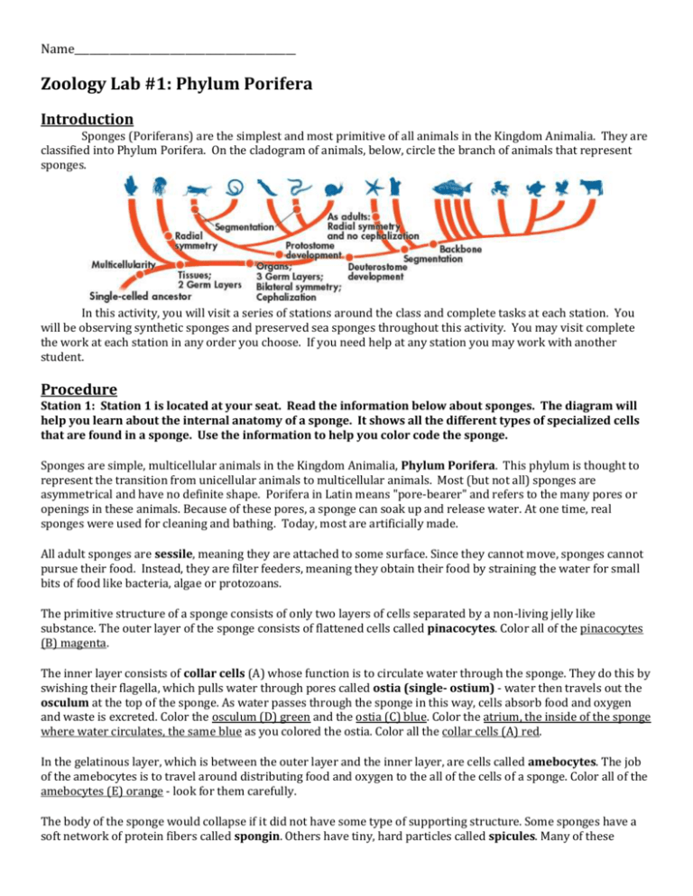
Name____________________________________________ Zoology Lab #1: Phylum Porifera Introduction Sponges (Poriferans) are the simplest and most primitive of all animals in the Kingdom Animalia. They are classified into Phylum Porifera. On the cladogram of animals, below, circle the branch of animals that represent sponges. In this activity, you will visit a series of stations around the class and complete tasks at each station. You will be observing synthetic sponges and preserved sea sponges throughout this activity. You may visit complete the work at each station in any order you choose. If you need help at any station you may work with another student. Procedure Station 1: Station 1 is located at your seat. Read the information below about sponges. The diagram will help you learn about the internal anatomy of a sponge. It shows all the different types of specialized cells that are found in a sponge. Use the information to help you color code the sponge. Sponges are simple, multicellular animals in the Kingdom Animalia, Phylum Porifera. This phylum is thought to represent the transition from unicellular animals to multicellular animals. Most (but not all) sponges are asymmetrical and have no definite shape. Porifera in Latin means "pore-bearer" and refers to the many pores or openings in these animals. Because of these pores, a sponge can soak up and release water. At one time, real sponges were used for cleaning and bathing. Today, most are artificially made. All adult sponges are sessile, meaning they are attached to some surface. Since they cannot move, sponges cannot pursue their food. Instead, they are filter feeders, meaning they obtain their food by straining the water for small bits of food like bacteria, algae or protozoans. The primitive structure of a sponge consists of only two layers of cells separated by a non-living jelly like substance. The outer layer of the sponge consists of flattened cells called pinacocytes. Color all of the pinacocytes (B) magenta. The inner layer consists of collar cells (A) whose function is to circulate water through the sponge. They do this by swishing their flagella, which pulls water through pores called ostia (single- ostium) - water then travels out the osculum at the top of the sponge. As water passes through the sponge in this way, cells absorb food and oxygen and waste is excreted. Color the osculum (D) green and the ostia (C) blue. Color the atrium, the inside of the sponge where water circulates, the same blue as you colored the ostia. Color all the collar cells (A) red. In the gelatinous layer, which is between the outer layer and the inner layer, are cells called amebocytes. The job of the amebocytes is to travel around distributing food and oxygen to the all of the cells of a sponge. Color all of the amebocytes (E) orange - look for them carefully. The body of the sponge would collapse if it did not have some type of supporting structure. Some sponges have a soft network of protein fibers called spongin. Others have tiny, hard particles called spicules. Many of these spicules also stick out of the epidermis and provide the sponge with protection. Most sponges have a combination of spicules and spongin, the ratio often determines how soft or hard the sponge is. Search for and color all the pointy spicules (F) brown. Station 2: Station 2 is located on the front screen. A series of photos will be shown throughout the class. You will be looking at photos of a synthetic (man-made) sponge and a silk sponge (preserved). Your task for this station is to guess which photos are real sponges and which are synthetic. You will also briefly research the materials that makeup sponge skeletons. For every photo shown, draw what you see as best as you can below. Once all the photos have been shown, answer the questions after the diagrams. Photo #1 _________________ Photo #2 _________________ Photo #3 _________________ Photo #4 _________________ 1. Label each diagram you made as either living, preserved sponge or synthetic sponge. 2. Spicules and spongin are the two main types of molecules that sponge skeletons are made of. Which material is found in sponges harvested for cleaning purposes? What type of sponge skeleton were we looking at above? Why is that material mainly seen in cleaning sponges? Research these questions, and write your answers below. Station 3: Station 3 is located at the front lab table. Working with other members of the class, test whether there is a significant difference in how once living, preserved sponges soak up water versus synthetic sponges. Use the chart below as a guide of what to do. 1. Which sponge do you predict will soak up the most water? __________________________________ 2. Use the scale and measure the mass of a synthetic sponge and a silk sponge. These two measurements are your initial mass. 3. Place the sponges into the cups of water for 60 seconds. Time this using your phone or the stopwatch on the table. 4. Carefully remove the sponge from the water. Try not to squeeze it and let some of the extra water on the surface to drain off. 5. Measure each sponge’s mass again. Complete the rest of the chart to answer this station’s main question! 6. When you are done with both sponges, gently squeeze out the water from them and place them in their original dish. Type of Sponge Initial Mass (g) Final Mass, mass after soaking for 60 seconds (g) Change in Mass = Final Mass – Initial Mass (g) % Change in Mass = (Change in Mass ÷ Initial Mass) x 100 (%) Synthetic Silk sea sponge Wool sea sponge According to the data you collected, which sponge type soaked up the most water? (Hint: which had the biggest % change in mass) Based on your answer above, what type of sponge should be the best choice for household use? Station 4: Station 4 is found along the side of the classroom. The compound microscopes contain sides of actual sponges. These are all cross section pictures. Imagine that you cut a slice of a sponge so that you cut it leaving the top and bottom portions of the sponge. Each microscope is showing something different. Some contain slides of a Scypha sponge at different magnifications. Other microscopes contain slides of a commercial sponge at different magnifications. Look through all of the microscopes but look for the 4 microscopes below and draw what you see in the space below. Then answer the question afterwards. DO NOT touch the microscopes, as they are set. Scyphia- Low Power (40x) Scyphia- High Power (400x) Commercial Sponge- Low Power (40x) Commercial Sponge- High Power (400x) Do you think the commercial sponge is synthetic (man-made) or preserved? Why do you think that? Station 5: Station 5 is found in the back of the classroom. Look at the samples of sea wool sponges, silk sea sponges, and synthetic sponges. Use the space below to draw samples of each. Draw each type of sponge and label each diagram. For the preserved sponge, can you identify and label the ostia and oscula? Is there a big difference between synthetic sponges and real sponges? (Think of look, structure, texture, etc.) Is it difficult to identify the ostia and osculum of the real sponge? Why do you think that is?
