Data S1. SFRP5 expression in squamous cell carcinoma (SCC) and
advertisement
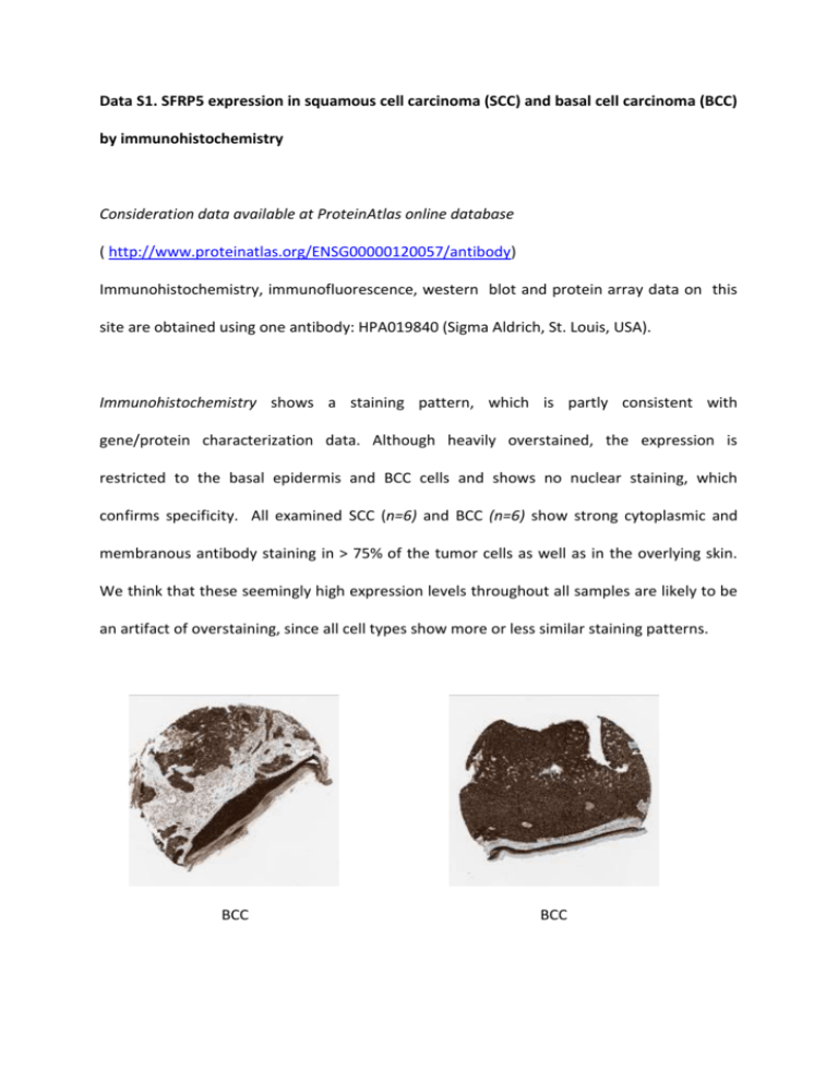
Data S1. SFRP5 expression in squamous cell carcinoma (SCC) and basal cell carcinoma (BCC) by immunohistochemistry Consideration data available at ProteinAtlas online database ( http://www.proteinatlas.org/ENSG00000120057/antibody) Immunohistochemistry, immunofluorescence, western blot and protein array data on this site are obtained using one antibody: HPA019840 (Sigma Aldrich, St. Louis, USA). Immunohistochemistry shows a staining pattern, which is partly consistent with gene/protein characterization data. Although heavily overstained, the expression is restricted to the basal epidermis and BCC cells and shows no nuclear staining, which confirms specificity. All examined SCC (n=6) and BCC (n=6) show strong cytoplasmic and membranous antibody staining in > 75% of the tumor cells as well as in the overlying skin. We think that these seemingly high expression levels throughout all samples are likely to be an artifact of overstaining, since all cell types show more or less similar staining patterns. BCC BCC Immunofluorescence of a human cell line, A-431 (epidermoid carcinoma), shows fluorescence in nucleus, cytoplasm and vesicles. Western blot analysis shows a band at the predicted height, corresponding to an MW of 35.6 kDa. Protein array confirms specific binding of the antibody to its antigen.

