case scenario 1
advertisement
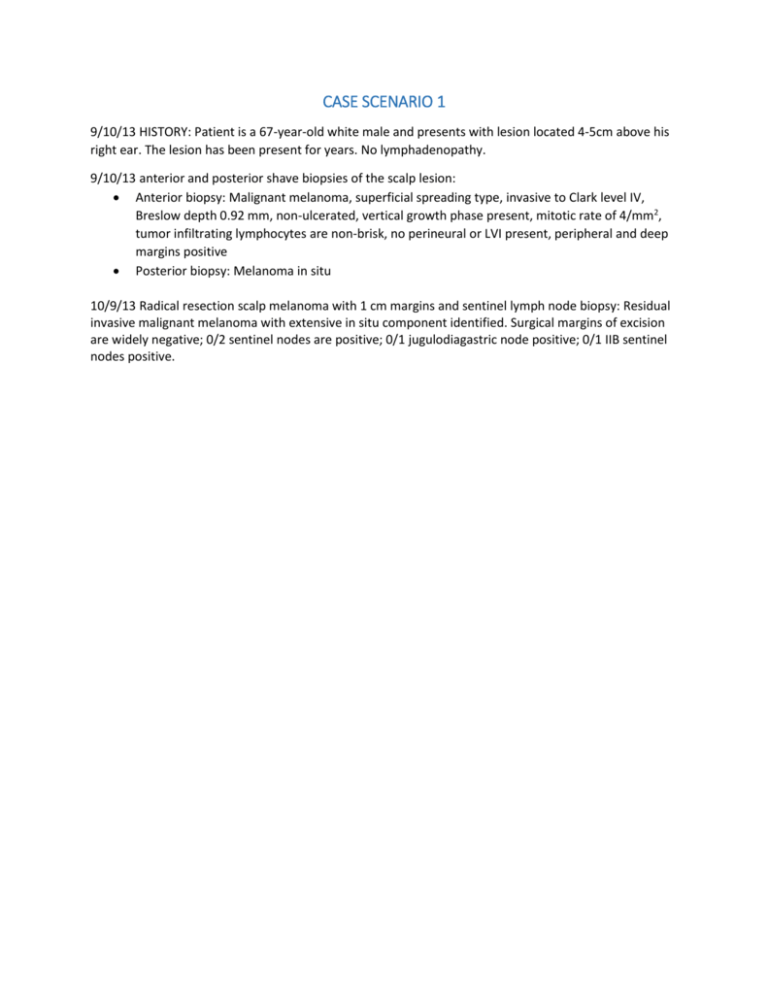
CASE SCENARIO 1 9/10/13 HISTORY: Patient is a 67-year-old white male and presents with lesion located 4-5cm above his right ear. The lesion has been present for years. No lymphadenopathy. 9/10/13 anterior and posterior shave biopsies of the scalp lesion: Anterior biopsy: Malignant melanoma, superficial spreading type, invasive to Clark level IV, Breslow depth 0.92 mm, non-ulcerated, vertical growth phase present, mitotic rate of 4/mm2, tumor infiltrating lymphocytes are non-brisk, no perineural or LVI present, peripheral and deep margins positive Posterior biopsy: Melanoma in situ 10/9/13 Radical resection scalp melanoma with 1 cm margins and sentinel lymph node biopsy: Residual invasive malignant melanoma with extensive in situ component identified. Surgical margins of excision are widely negative; 0/2 sentinel nodes are positive; 0/1 jugulodiagastric node positive; 0/1 IIB sentinel nodes positive. Case Scenario 1 Worksheet Primary Site C__ __.__ Morphology __ __ __ __/__ __ Laterality __ Stage/ Prognostic Factors CS Tumor Size CS Extension CS Tumor Size/Ext Eval CS SSF 9 CS SSF 10 CS SSF 11 988 988 988 CS Lymph Nodes CS Lymph Nodes Eval Regional Nodes Positive Regional Nodes Examined CS Mets at Dx CS Mets Eval CS SSF 1 CS SSF 2 CS SSF 3 CS SSF 4 CS SSF 5 CS SSF 6 CS SSF 7 CS SSF 8 Summary Stage 1 – Localized CS SSF 12 CS SSF 13 CS SSF 14 CS SSF 15 CS SSF 16 CS SSF 17 CS SSF 18 CS SSF 19 CS SSF 20 CS SSF 21 CS SSF 22 CS SSF 23 CS SSF 24 CS SSF 25 Derived AJCC TNM Stage 988 988 988 988 988 988 988 988 988 988 988 988 988 988 Clinical AJCC TNM Stage Pathologic AJCC TNM Stage (indicate c or p in the space before the T, N, or M) Treatment Diagnostic Staging Procedure Surgery Codes Surgical Procedure of Primary Site Scope of Regional Lymph Node Surgery Surgical Procedure/ Other Site Systemic Therapy Codes Chemotherapy Hormone Therapy Immunotherapy Hematologic Transplant/Endocrine Procedure Systemic/Surgery Sequence Radiation Codes Radiation Treatment Volume Regional Treatment Modality Regional Dose Boost Treatment Modality Boost Dose Number of Treatments to Volume Reason No Radiation Radiation/Surgery Sequence CASE SCENARIO 2 6/21/13 HISTORY: 66 year-old white female presented for her yearly skin exam. She had a suspicious lesion on her right lower calf. No lymphadenopathy present. 6/21/13 Shave biopsy of right lower calf: Lentigo maligna melanoma; Breslow depth 0.17 mm; Clark level II; no ulceration; no regression; mitotic rate less than 1/mm2; no LVI or perineural invasion; lateral margins positive. 7/3/13 Wide local excision of right lower calf lesion with excision margin just over 3 cm: Malignant melanoma, lentigo maligna type; tumor invades to Clark level II and Breslow depth 0.55 mm; nonulcerated and RGP present; mitotic rate is 0/mm2; no regression, no microsatellite tumor nodules; negative margins. 7/3/13 It was recommended that patient see an oncologist post-op, but the patient declined. Case Scenario 2 Worksheet Primary Site C__ __.__ Morphology __ __ __ __/__ __ Laterality __ Stage/ Prognostic Factors CS Tumor Size CS Extension CS Tumor Size/Ext Eval CS SSF 9 CS SSF 10 CS SSF 11 988 988 988 CS Lymph Nodes CS Lymph Nodes Eval Regional Nodes Positive Regional Nodes Examined CS Mets at Dx CS Mets Eval CS SSF 1 CS SSF 2 CS SSF 3 CS SSF 4 CS SSF 5 CS SSF 6 CS SSF 7 CS SSF 8 Summary Stage CS SSF 12 CS SSF 13 CS SSF 14 CS SSF 15 CS SSF 16 CS SSF 17 CS SSF 18 CS SSF 19 CS SSF 20 CS SSF 21 CS SSF 22 CS SSF 23 CS SSF 24 CS SSF 25 Derived AJCC TNM Stage 988 988 988 988 988 988 988 988 988 988 988 988 988 988 (indicate c or p in the space before the T, N, or M) Clinical AJCC TNM Stage Pathologic AJCC TNM Stage Treatment Diagnostic Staging Procedure Surgery Codes Surgical Procedure of Primary Site Scope of Regional Lymph Node Surgery Surgical Procedure/ Other Site Systemic Therapy Codes Chemotherapy Hormone Therapy Immunotherapy Hematologic Transplant/Endocrine Procedure Systemic/Surgery Sequence Radiation Codes Radiation Treatment Volume Regional Treatment Modality Regional Dose Boost Treatment Modality Boost Dose Number of Treatments to Volume Reason No Radiation Radiation/Surgery Sequence CASE SCENARIO 3 8/9/2013 HISTORY: 64 year-old white female presents with a 2 month history of a lesion on the right upper arm. No palpable regional lymph nodes. 8/9/2013 Pathology Report: Excisional biopsy of skin of right upper arm: 3.0 cm nodule; malignant melanoma, nodular type; Breslow measurement 11.80 mm; Clark level 4; extensive ulceration; no regression; mitotic index 10/mm2; no LVI; no satellite lesions; positive margin (deep margin); unknown vertical growth phase. 8/10/2013 LDH: 351 8/15/2013 LDH: 357 Normal Range-Female: 46-100 IU/L Male: 46-232 IU/L Normal Range-Female: 46-100 IU/L Male: 46-232 IU/L 9/10/2013 PET/CT: 2.0 cm subcutaneous nodule on right chest revealing abnormal FDG accumulation (SUV 5.8). This is highly suspicious for malignancy. Subcutaneous nodule in left lower abdomen, probably non-malignant etiology (no increased FDG uptake). 10/1/2013 CT Abdomen/Pelvis: Subcutaneous nodule in left lower abdomen is unchanged, but it may represent enlarged lymph nodes. No other abnormalities are noted. 10/1/2013 CT of brain: No metastatic disease. 10/21/2013 Pathology Report: Wide local excision of right upper arm lesion with 2.5 cm margins, excision of subcutaneous lesion on the right chest wall, and right axillary sentinel lymph node biopsy Wide excision right upper arm: Residual deep dermal subcutaneous mass of malignant melanoma measuring 3.4mm. A 2.5 cm margin of healthy tissue is present. Right chest wall excision: 5 cm lesion subcutaneous lesion, metastatic malignant melanoma. Positive deep margin. Sentinel node biopsy: axillary soft tissue metastasis widely free from peripheral/deep margins; 0/8 lymph nodes, negative IHC. ONCOLOGY: Interferon started 11/26/13 Case Scenario 3 Worksheet Primary Site C__ __.__ Morphology __ __ __ __/__ __ Laterality __ Stage/ Prognostic Factors CS Tumor Size CS Extension CS Tumor Size/Ext Eval CS SSF 9 CS SSF 10 CS SSF 11 988 988 988 CS Lymph Nodes CS Lymph Nodes Eval Regional Nodes Positive Regional Nodes Examined CS Mets at Dx CS Mets Eval CS SSF 1 CS SSF 2 CS SSF 3 CS SSF 4 CS SSF 5 CS SSF 6 CS SSF 7 CS SSF 8 Summary Stage CS SSF 12 CS SSF 13 CS SSF 14 CS SSF 15 CS SSF 16 CS SSF 17 CS SSF 18 CS SSF 19 CS SSF 20 CS SSF 21 CS SSF 22 CS SSF 23 CS SSF 24 CS SSF 25 Derived AJCC TNM Stage 988 988 988 988 988 988 988 988 988 988 988 988 988 988 (indicate c or p in the space before the T, N, or M) Clinical AJCC TNM Stage Pathologic AJCC TNM Stage Treatment Diagnostic Staging Procedure Surgery Codes Surgical Procedure of Primary Site Scope of Regional Lymph Node Surgery Surgical Procedure/ Other Site Systemic Therapy Codes Chemotherapy Hormone Therapy Immunotherapy Hematologic Transplant/Endocrine Procedure Systemic/Surgery Sequence Radiation Codes Radiation Treatment Volume Regional Treatment Modality Regional Dose Boost Treatment Modality Boost Dose Number of Treatments to Volume Reason No Radiation Radiation/Surgery Sequence CASE SCENARIO 4 4/24/2013 HISTORY: 49 year-old white female presented with history of a mole-like lesion on her right upper back that is now increasing in size and bleeding. Axillary lymphadenopathy is not present. 4/24/2013 Pathology Report: Excisional biopsy of skin of back: Malignant melanoma, superficial spreading type; Clark level IV; Breslow thickness 1.32 mm; ulceration present; focal regression; mitotic index 1/mm2. 5/16/2013 Pathology Report: Sentinel lymph node biopsy and wide local excision of melanoma. Wide excision No residual melanoma; 2cm negative margins Sentinel lymph node biopsy: 1 sentinel node involved by melanoma, microscopic focus is 0.2 mm; IHC staining confirms sentinel lymph node micrometastasis. 5/30/2013 Pathology Report: Right axillary lymph node dissection: 0/20 lymph nodes positive. 6/14/13 CT of Abdomen/Pelvis: Indeterminate luceny in body of T2 (no uptake in PET/CT) and indeterminate left upper lobe lung nodule (no uptake on PET) as well as right adrenal gland nodule. 6/14/13 MRI Brain: No metastatic disease. 8/1/13 Oncology Consult: Patient is not a candidate for interferon or any type of clinical trial due to her comorbidities. Patient will be followed up with scans. Case Scenario 4 Worksheet Primary Site C__ __.__ Morphology __ __ __ __/__ __ Laterality __ Stage/ Prognostic Factors CS Tumor Size CS Extension CS Tumor Size/Ext Eval CS SSF 9 CS SSF 10 CS SSF 11 988 988 988 CS Lymph Nodes CS Lymph Nodes Eval Regional Nodes Positive Regional Nodes Examined CS Mets at Dx CS Mets Eval CS SSF 1 CS SSF 2 CS SSF 3 CS SSF 4 CS SSF 5 CS SSF 6 CS SSF 7 CS SSF 8 Summary Stage CS SSF 12 CS SSF 13 CS SSF 14 CS SSF 15 CS SSF 16 CS SSF 17 CS SSF 18 CS SSF 19 CS SSF 20 CS SSF 21 CS SSF 22 CS SSF 23 CS SSF 24 CS SSF 25 Derived AJCC TNM Stage 988 988 988 988 988 988 988 988 988 988 988 988 988 988 (indicate c or p in the space before the T, N, or M) Clinical AJCC TNM Stage Pathologic AJCC TNM Stage Treatment Diagnostic Staging Procedure Surgery Codes Surgical Procedure of Primary Site Scope of Regional Lymph Node Surgery Surgical Procedure/ Other Site Systemic Therapy Codes Chemotherapy Hormone Therapy Immunotherapy Hematologic Transplant/Endocrine Procedure Systemic/Surgery Sequence Radiation Codes Radiation Treatment Volume Regional Treatment Modality Regional Dose Boost Treatment Modality Boost Dose Number of Treatments to Volume Reason No Radiation Radiation/Surgery Sequence
