University оf Kragujevac, Faculty of Science CENTRE FOR PRE
advertisement
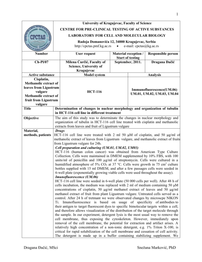
1 University оf Kragujevac, Faculty of Science CENTRE FOR PRE-CLINICAL TESTING OF ACTIVE SUBSTANCES LABORATORY FOR CELL AND MOLECULAR BIOLOGY Radoja Domanovića 12, 34000 Kragujevac, Serbia http://cpctas.pmf.kg.ac.rs e-mail: cpctas@kg.ac.rs Number User request Ch-Pl/07 Milena Ćurčić, Faculty of Science, University of Kragujevac Model system Material reception / Start of testing September, 2011. Responsible person Dragana Đačić Active substance Analysis Cisplatin, Methanolic extract of leaves from Ligustrum Immunofluorescence(UM.06) vulgare HCT-116 UM.01, UM.02, UM.03, UM.04 Methanolic extract of fruit from Ligustrum vulgare Title Determination of changes in nuclear morphology and organization of tubulin in HCT-116 cell line in different treatment The aim of this study was to determinate the changes in nuclear morphology and Objective organization of tubulin in HCT-116 cell line treated with cisplatin and methanolic extracts from leaves and fruit of Ligustrum vulgare Material, Drugs methods, patients HCT-116 cell line were treated with 2 ml 50 µM of cisplatin, and 50 µg/ml of methanolic extract of leaves from Ligustrum vulgare, and methanolic extract of fruits from Ligustrum vulgare for 24h. Cell preparation and culturing (UM.01, UM.02, UM03) HCT-116 (human colon cancer) was obtained from American Type Culture Collection. Cells were maintained in DMEM supplemented by 10% FBS, with 100 units/ml of penicillin and 100 µg/ml of streptomycin. Cells were cultured in a humidified atmosphere of 5% CO2 at 37 °C. Cells were growth in 75 cm2 culture bottles supplied with 15 ml DMEM, and after a few passages cells were seeded in 6-well plate (exponentially growing viable cells were used throughout the assay). Imunofluorescence (UM.06) HCT-116 cell line were seeded in 6-well plate (50 000 cells per well). After 48 h of cells incubation, the medium was replaced with 2 ml of medium containing 50 μM concentrations of cisplatin, 50 µg/ml methanol extract of leaves and 50 µg/ml methanol extract of fruit from plant Ligustrum vulgare. Untreated cells served as a control. After 24 h of tretmant we were observated changes by microscpe NIKON Ti. Imunofluorescence is based on usage of specificity of antibodies to their antigen to target fluorescent dyes to specific bimolecular targets within a cell, and therefore allows visualization of the distribution of the target molecule through the sample. In our experiment, detergent lysis is the most usual way to remove the cell membrane, thus exposing the cytoskeleton. However, immediately upon removal of the cell membrane, the potential for extraction and artifact arises. A relatively high concentration of a non-ionic detergent, e.g. 1% Triton X-100, is critical for rapid solubilization of the cell membrane and cessation of cell activity. The detergent is made up in a buffer containing stabilizing supplement. We Dragana Đačić, MSci Snežana Marković, PhD 2 Results Discussion Conclusion References Notes routinely add high molecular weight polyethileneglycol (PEG) for general preservation on the cytoskeleton. Phallioidin are added for the specific prevent of actin filaments, respectively. Extraction was made at room temperature. After extraction, washing and fixation with 0.2% glutaraldehide we added primary antibody for protein tubulin (mouse anti- tubulin). For visualization cell nuclear material we used DAPI staining. After incubation and washing we added secondary antibody anti mouse Cay3. Fixed cover slip with mounting media and viewed under Nikon inverted fluorescence microscope (Ti-Eclipse) at 600x magnification Our micrographs clearly showed the separate cells in the control. The analysis of micrographs showed that cells in control (untreated cells) not had affinity for associating in clusters. The analysis of images showed that all treatments had highly affinity for associating of cells compared with control cells as shown in Figures 1-4. When compared treatments with control (untreated) cells, we can observe that all treatments showed associating of cells. The analysis of treatments with methanol extracts of leaves and fruit from Ligustrum vulgare showed reorganization of cytoskeleton protein tubulin, who as part of cytoskeleton, have role in organization structure and position of cells organelles. That could be to mean that investigated active substances induced changes in cells metabolism and cells had highly communications between them. Both extracts and cisplatin showed highly affinity for compaction and associating of cells. We can conclude that cisplatin had effect on reorganization of tubulin in cell cytoskeleton . All treatments showed significant associating and compaction of cells compared to untreated cells and could be used in further studies. Immunocytochemistry Methods and Protocols. Edited by Lorette C. Javois, 2nd edition, 1999. Human Press. This report applies only to the tested substances. Responsible for the report, (accuracy and technical explanations of results) are researcher and manager and are considered the report's authors, which they have confirmed with their signature. The report should not be used or reproduced partially, except in its entirety in form of the publication of results as an integral part of the report. Publication of results based on this report must be approved by the authors. Sign Responsible person for testing Dragana Đačić Date 17.10.2011. Dragana Đačić, MSci Responsible person for Laboratory Dr Snežana Marković Snežana Marković, PhD 3 Figure 1. Imunofluorescence images of HCT-116 cell line untreated cells (control) a) protein tubulin Cy3staining, b) nuclear-DAPI staining Figure 2. Imunofluorescence image of HCT-116 cell line treated with 50 μM concentrations of cisplatin for 24 h a) protein tubulin -Cy3staining, b) nuclear-DAPI staining Figure 3. Imunofluorescence images of HCT-116 cell line treated with 50 μg/ml concentrations of methanolic extract of leaves from Ligustrum vulgare for 24 h a) protein tubulin -Cy3staining, b) nuclearDAPI staining Figure 4. Imunofluorescence images of HCT-116 cell line treated with 50 μg/ml concentrations of methanolic extract of fruits from Ligustrum vulgare for 24 h a) protein tubulin -Cy3staining, b) nuclearDAPI staining Dragana Đačić, MSci Snežana Marković, PhD 4 Figure 1. Dragana Đačić, MSci Snežana Marković, PhD 5 Figure 2. Dragana Đačić, MSci Snežana Marković, PhD 6 Figure 3. Dragana Đačić, MSci Snežana Marković, PhD 7 Figure 4. Dragana Đačić, MSci Snežana Marković, PhD
