concept of tissue expansion in reconstructive surgery
advertisement
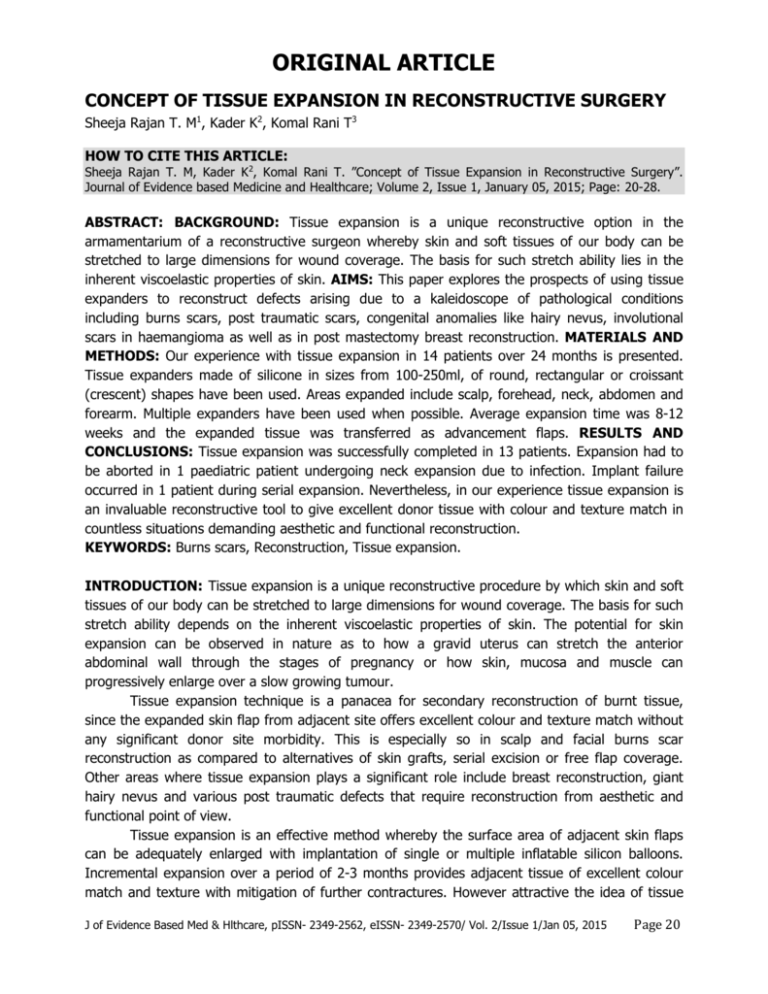
ORIGINAL ARTICLE CONCEPT OF TISSUE EXPANSION IN RECONSTRUCTIVE SURGERY Sheeja Rajan T. M1, Kader K2, Komal Rani T3 HOW TO CITE THIS ARTICLE: Sheeja Rajan T. M, Kader K2, Komal Rani T. ”Concept of Tissue Expansion in Reconstructive Surgery”. Journal of Evidence based Medicine and Healthcare; Volume 2, Issue 1, January 05, 2015; Page: 20-28. ABSTRACT: BACKGROUND: Tissue expansion is a unique reconstructive option in the armamentarium of a reconstructive surgeon whereby skin and soft tissues of our body can be stretched to large dimensions for wound coverage. The basis for such stretch ability lies in the inherent viscoelastic properties of skin. AIMS: This paper explores the prospects of using tissue expanders to reconstruct defects arising due to a kaleidoscope of pathological conditions including burns scars, post traumatic scars, congenital anomalies like hairy nevus, involutional scars in haemangioma as well as in post mastectomy breast reconstruction. MATERIALS AND METHODS: Our experience with tissue expansion in 14 patients over 24 months is presented. Tissue expanders made of silicone in sizes from 100-250ml, of round, rectangular or croissant (crescent) shapes have been used. Areas expanded include scalp, forehead, neck, abdomen and forearm. Multiple expanders have been used when possible. Average expansion time was 8-12 weeks and the expanded tissue was transferred as advancement flaps. RESULTS AND CONCLUSIONS: Tissue expansion was successfully completed in 13 patients. Expansion had to be aborted in 1 paediatric patient undergoing neck expansion due to infection. Implant failure occurred in 1 patient during serial expansion. Nevertheless, in our experience tissue expansion is an invaluable reconstructive tool to give excellent donor tissue with colour and texture match in countless situations demanding aesthetic and functional reconstruction. KEYWORDS: Burns scars, Reconstruction, Tissue expansion. INTRODUCTION: Tissue expansion is a unique reconstructive procedure by which skin and soft tissues of our body can be stretched to large dimensions for wound coverage. The basis for such stretch ability depends on the inherent viscoelastic properties of skin. The potential for skin expansion can be observed in nature as to how a gravid uterus can stretch the anterior abdominal wall through the stages of pregnancy or how skin, mucosa and muscle can progressively enlarge over a slow growing tumour. Tissue expansion technique is a panacea for secondary reconstruction of burnt tissue, since the expanded skin flap from adjacent site offers excellent colour and texture match without any significant donor site morbidity. This is especially so in scalp and facial burns scar reconstruction as compared to alternatives of skin grafts, serial excision or free flap coverage. Other areas where tissue expansion plays a significant role include breast reconstruction, giant hairy nevus and various post traumatic defects that require reconstruction from aesthetic and functional point of view. Tissue expansion is an effective method whereby the surface area of adjacent skin flaps can be adequately enlarged with implantation of single or multiple inflatable silicon balloons. Incremental expansion over a period of 2-3 months provides adjacent tissue of excellent colour match and texture with mitigation of further contractures. However attractive the idea of tissue J of Evidence Based Med & Hlthcare, pISSN- 2349-2562, eISSN- 2349-2570/ Vol. 2/Issue 1/Jan 05, 2015 Page 20 ORIGINAL ARTICLE expansion may seem, its high cost, multiplicity of procedures, intentional deformation for prolonged period that has to be tolerated by the patient are some of the factors that limit its use. MATERIALS AND METHODS: Over a 24 month period 14 patients who underwent tissue expansion procedures were chosen for this study. The expanders were inserted into forehead in 3 patients, scalp in 3, neck in 2, abdomen in 2, forearm in 3 and in multiple sites in 1. The indications, type of expanders, duration of expansion, resting period and the outcome in each case are illustrated in Table 1. Tissue expanders are inflatable devices with bio inert silicone envelope and a remote or integral injection port. They come in different capacities (40ml-500ml), shapes (rectangular, oval, croissant or contoured) and textures (smooth or textured).We have used expanders of 100-250 ml capacities, mostly smooth and seamless variety in rectangular, oval as well as croissant shapes. Injection ports were preferably kept remote with a connector tube and Luer lock to allow a one way flow of saline into the expander and for prevention of leaks. For our patients, two operative steps were involved, first for the insertion of the implant followed by 2 weeks of wound healing. Serial expansion of the device is then started, by saline injections at biweekly intervals for 2-3 months. After expansion, a further resting period was allowed, roughly equal to the total time taken for expansion. A second operation was then performed to remove the implant, excise the pathological tissues and advance the expanded tissue into the defect as a flap from the donor site. Later, 3 patients underwent scar revision procedures, 6 months after the second procedure. RESULTS: Tissue expansion was successfully completed in 13 patients. In all cases tissue expansion yielded excellent donor tissue, raised as advancement flaps. Resurfacing with tissue of similar colour and texture were particularly advantageous especially in scalp reconstruction providing hair bearing skin over areas of alopecia. There were no episodes of skin necrosis during or after transfer of expanded tissue. 1 paediatric patient with expander in the neck region developed infection and wound breakdown following an upper respiratory infection. The implant was removed and infection controlled. Re-expansion was attempted after 6 months but had to be discontinued following another episode of wound infection. In a patient with burns scar of scalp, expansion with a single croissant expander could resurface only 60% of the scar in the initial session and had to undergo serial expansion. Towards the end of expansion, implant failure by leak at the connecting tube occurred. However, sufficient expansion had already been achieved for the resurfacing of rest of the scarred area with hair bearing scalp. Spreading scars were noted in all patients and 3 patients with prominent scars over forehead and 1 over forearm underwent scar revision surgery after 6 months. 1 patient with expander over anterior abdominal wall developed wound dehiscence on the seventh postoperative day following trauma and had to undergo secondary suturing with antibiotic irrigation of implant pocket. Expansion was started 3 weeks after the re suturing and completed successfully. DISCUSSION: The first clinical report on the concept of tissue expansion was by Dr. Charles Neumann1 in 1957 when he used a latex balloon to reconstruct a part of external ear. However the credit for reintroducing and popularizing tissue expansion goes separately to Dr. Chedomir J of Evidence Based Med & Hlthcare, pISSN- 2349-2562, eISSN- 2349-2570/ Vol. 2/Issue 1/Jan 05, 2015 Page 21 ORIGINAL ARTICLE Radovan2 and Dr Eric Austad3 in 1979. Tissue expanders are silicone balloons molded into pre shaped prosthesis of circular, rectangular or crescent (croissant) shapes. Custom built and anatomical expanders like the Becker implant for breast reconstruction are also available. The expanders are inflated by filling with saline through a valve system. The port for injection is placed well away from the expander to avoid inadvertent puncture or introduction of infection. The port can be internal or external and is usually kept over a supportive surface in an accessible location. There should be meticulous attention to technique & proper patient selection to ensure favourable results. Generally, a psychologically stable patient, good quality well vascularised tissue at the donor site, meticulous attention to asepsis and good techniques are the prerequisites for any tissue expansion procedure. While choosing the expander, attention is given to the size of the defect, availability of donor tissues in the vicinity, the direction of advancement of the flap as well as to the position of the final scars. While Radovan2, Morgan and Edgerton3,4 suggest that the expander of base the same size as the defect can be chosen, Gibney5 chose expanders of base size 2.5 – 3 times the width of the defect. Van Rappard6 recommends round expanders with base diameters 2.5 times the size of the defect and rectangular and croissant expanders with base surface area 2.5 times as large as the defect. Manders7 preferred to use largest possible expander to fit the donor site. Theoretically rectangular implants produce more tissue gain of calculated surface area increase and make it easier to raise advancement flaps from the donor area. Sometimes multiple expanders simultaneously to cover larger defects are employed. Otherwise, re expansion of advanced flap after 6 months will have to be advocated. The incision for inserting the expander should be planned in such a way as to form the leading edge of the future advancement flap. An adequate pocket is dissected with the combination of blunt and sharp dissections to accommodate the expander without folds. The implant maybe placed in the subcutaneous, subgaleal, submuscular or subfascial planes8 as the situation demands. A connecting tunnel for placement of port in an accessible location is also made. Externalized ports to avoid pain of punctures during inflations are sometimes used in children but need constant monitoring to avoid complications. Inflation of the expander is done immediately after wound closure as is adequate to remove dead space and smooth out folds and sharp edges which may subsequently cause skin erosion from within. After 2-3 weeks, a regimen of weekly or biweekly inflation of sterile saline into the port using a 24 G butterfly needle, under strict aseptic precautions. End point of expansion is when patient complaints of discomfort due to expansion or by monitoring the capillary refill and has to be done with strict aseptic precautions.9 At the end of the desired expansion, a resting period roughly equal to the period required for inflation is allowed. During surgery for removal of expander, a final intra operative expansion is done and the capsule is incised to remove the expander and port. A trial advancement of the expanded flap over the defect and then the excision of the lesion and actual flap advancements are carried out. Since there is always a tendency for the stretched tissue to regress back, patient should be adequately warned that they may require scar management measures and revision procedures later on. This is especially true with scalp expansion and also whenever tissue is expanded in aesthetically important areas like the head and neck region.10,11,12 J of Evidence Based Med & Hlthcare, pISSN- 2349-2562, eISSN- 2349-2570/ Vol. 2/Issue 1/Jan 05, 2015 Page 22 ORIGINAL ARTICLE Unfavourable results with tissue expansion are most often the result of poor patient selection or faulty techniques. Fochtmann13 et al state that complications are higher in expansions of paediatric patients, lower extremities, and larger total body surface area of scars. Risk is also higher in expansion of unhealthy skin or with serial expansion. Bozkurt14 et al however, in a retrospective analysis found no statistical correlation between implant failure with age, gender, shape or number of implants. Major complications like infections, hematoma, extrusion, implant failures or flap ischemia may warrant removal of expander. Minor complications like pressure effects, neuropraxia or spreading scars allow expansion to continue but at a slower rate. We had major complications only in 2 patients warranting termination of expansion. Proper selection of patients and expanders, adequate cavity size to avoid wrinkles, seamless expanders to avoid sharp folds under the skin and meticulous attention to hemostasis to avoid hematomas and protection against inadvertent trauma are all significant factors that can influence the outcome of tissue expansion. Nevertheless, in our experience tissue expansion is an invaluable reconstructive tool to give excellent donor tissue with colour and texture match in countless situations. REFERENCES: 1. Neuman CG. The expansion of an area of skin by progressive distension of subcutaneous ballon. Plast Reconstr Surg.1957; 19: 124-9. 2. Radovan C. Tissue expansion in soft tissue reconstruction. Plast Reconstr Surg 1984; 74: 482-492. 3. Austad E. A self-inflating tissue expander. Paper presented at the Annual meeting of the American society of Plastic & reconstructive surgeons. Toronto; 1979. 4. Morgan RF, Edgerton MT. Tissue expansion in reconstructive hand surgery. J Hand Surg Am 1985; 10: 754-757. 5. Gibney J. Tissue expansion in reconstructive surgery. Paper presented at the Annual meeting of the American society of Plastic & reconstructive surgeons. Las Vegas; 1984. 6. Van Rappard JH, Sonneveld GJ, Boughouts JM. Geometric planning and shape of the expander. In van Rappard JH, editor. Tissue expansion in Facial Plastic Surgery. vol 5 New York: Thieme; 1988. 7. Manders EK, Schenden MJ, Furrey JA, Hetzler PT, Davis TS, Graham WP 3rd. Soft tissue expansion concepts and complications. Plast Reconstr Surg 1984; 74: 493-507. 8. Joao Madeiros, Tavares Filho, Manoel Belerique, Diogo Franco, Carlos Alberto Porchat, Talita Franco. Tissue expansion and burns sequelae repair.Burns. 2007; 33: 246-251. 9. Milind S Wagh, Varun Dixit. Tissue expansion concepts, technique and unfavourable results.Indian Journal of Plastic Surgery 2013; 6: 333-348. 10. Argenta LC, Watanabe MJ, Grabb WC. The use of tissue expansion in head and neck reconstruction. Ann Plast Surg 1983; 11: 31-7. 11. Norstrom RE, Devin JW. Scalp stretching with a tissue expander for closure scalp defects. Plast Reconstr Surg.1985; 75: 578-81. 12. Gurlek A, Alaybeyoglu N, Canser YD, et al. Aesthetic reconstruction of large scalp defects by sequential tissue expansion without interval. Aest Plast Surg. 2004; 28: 245-50. J of Evidence Based Med & Hlthcare, pISSN- 2349-2562, eISSN- 2349-2570/ Vol. 2/Issue 1/Jan 05, 2015 Page 23 ORIGINAL ARTICLE 13. Fochtmann A, Keck M, Mittibock, Rath Th. Tissue expansion for correction of scars due to burns and other causes: a retrospective comparative study of various complications.Burns.2013; 39: 984-989. 14. Bozkurt A, Groger, O Dey, Vogeler, Piatkowski, Fuchs, Pallu N. Retrospective analysis of tissue expansion in reconstructive burn surgery. Evaluation of complication rates. Burns 2008; 34: 1113-1118. Case 1: Scar hemiforehead of post involutional hemangioma. J of Evidence Based Med & Hlthcare, pISSN- 2349-2562, eISSN- 2349-2570/ Vol. 2/Issue 1/Jan 05, 2015 Page 24 ORIGINAL ARTICLE Case 2: Round expander in the scalp. Fig. 1 Case 3: Expander in the abdomen for elbow contracture release. Fig. 1 J of Evidence Based Med & Hlthcare, pISSN- 2349-2562, eISSN- 2349-2570/ Vol. 2/Issue 1/Jan 05, 2015 Page 25 ORIGINAL ARTICLE Case 4: Becker implant being expanded. Sl. no 1 Age/s ex Indication 18/F Post burn scar Scalp 24X 22 cm 2 22/F 3 25/M 4 5 6 15/F Post burn scar neck 18X 12 cm Cicatricial alopecia occipital scalp 10X9 cm Posburn scars anterior abdominal wall 25X 20cm Expander used Period of expansion Resting period Croissant 250 ml 12 weeks 12 weeks Croissant 200 ml 12 weeks 8 weeks Round 150 ml 8 weeks 6 weeks Round 200ml 12 weeks 8 weeks 13/M (R) postburn elbow contracture Rectangular 150ml in the abdomen 12 weeks 8weeks 19/F Post burn scars (R) forearm with wrist flexion contracture Rectangular 150ml in the abdomen and 100ml 12 weeks 12 weeks Outcome 60% scar area resurfaced in the first session Complete wound coverage Secondary procedures Re expansion after 6 months Scar revision Complete wound coverage Secondary suturing of traumatic wound dehiscence after a fall Complete wound coverage Complete wound coverage with expanded random abdominal flap Complete wound coverage with expanded random Flap division at 3 weeks post transfer J of Evidence Based Med & Hlthcare, pISSN- 2349-2562, eISSN- 2349-2570/ Vol. 2/Issue 1/Jan 05, 2015 Page 26 ORIGINAL ARTICLE in the forearm abdominal flap and forearm advancement flap 28/M Post-burn scar neck Round 150ml 8 18/F Post-surgical scar abdomen Rectangular 200ml 8 weeks 8weeks 9 10/F Hairy naevus forehead Rectangular 100 ml 12 weeks 8 weeks 20/F Unaesthetic skin graft over (R) hemiforehead Rectangular 150ml 18/F Postburn scar (R) forearm Rectangular 100ml 7 10 11 12 13 14 18/F 25/F 16/F Benign appendageal tumour parieto occipital scalp 20X 18cm Post mastectomy breast reconstruction expansion under previous Latissimus dorsi flap Scar of Involuted Hemangioma (L) hemiforehead Rectangular 150 ml with external port Becker implant 600 ml expansion Rectangular 150 ml 6 weeks - Wound infection, Implant removed Complete wound coverage Complete wound coverage 12 weeks 8 weeks Complete wound coverage 12 weeks 12 weeks Complete wound coverage 8 weeks 85% wound coverage, skin grafting in remaining area 12 weeks Aesthetically well matched contoured breast reconstruction 12 weeks 12 weeks 12 weeks 8 weeks Re expansion after 6 months aborted due to wound infection at 3 weeks Complete wound coverage Scar revision after 6 months Scar revision with readvancement of flap after 6 months TABLE 1: Patients undergoing tissue expansion from June 2011-May 2013 J of Evidence Based Med & Hlthcare, pISSN- 2349-2562, eISSN- 2349-2570/ Vol. 2/Issue 1/Jan 05, 2015 Page 27 ORIGINAL ARTICLE AUTHORS: 1. Sheeja Rajan T. M. 2. Kader K. 3. Komal Rani T. PARTICULARS OF CONTRIBUTORS: 1. Assistant Professor, Department of Plastic Surgery, Calicut Medical College. 2. Assistant Professor, Department of Plastic Surgery, Government Medical College, Kozhikode. 3. Former HOD, Department of Plastic Surgery, Government Medical College, Kozhikode. NAME ADDRESS EMAIL ID OF THE CORRESPONDING AUTHOR: Dr. Sheeja Rajan T. N, Shrirang, Opp. Chevayur Tel. Exchange, P.O. Chevayur, Calicut - 673017. E-mail: sheejarajantm@gmail.com Date Date Date Date of of of of Submission: 22/12/2014. Peer Review: 23/12/2014. Acceptance: 30/12/2014. Publishing: 01/01/2015. J of Evidence Based Med & Hlthcare, pISSN- 2349-2562, eISSN- 2349-2570/ Vol. 2/Issue 1/Jan 05, 2015 Page 28
