Supplementary Information (docx 146K)
advertisement
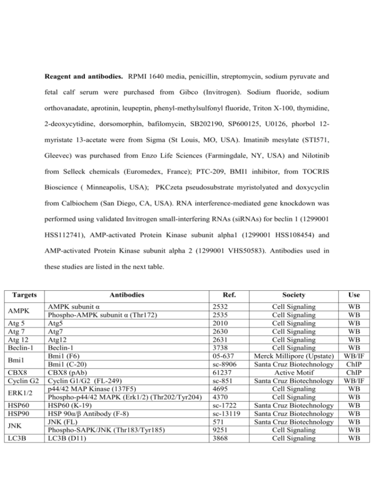
Reagent and antibodies. RPMI 1640 media, penicillin, streptomycin, sodium pyruvate and fetal calf serum were purchased from Gibco (Invitrogen). Sodium fluoride, sodium orthovanadate, aprotinin, leupeptin, phenyl-methylsulfonyl fluoride, Triton X-100, thymidine, 2-deoxycytidine, dorsomorphin, bafilomycin, SB202190, SP600125, U0126, phorbol 12myristate 13-acetate were from Sigma (St Louis, MO, USA). Imatinib mesylate (STI571, Gleevec) was purchased from Enzo Life Sciences (Farmingdale, NY, USA) and Nilotinib from Selleck chemicals (Euromedex, France); PTC-209, BMI1 inhibitor, from TOCRIS Bioscience ( Minneapolis, USA); PKCzeta pseudosubstrate myristolyated and doxycyclin from Calbiochem (San Diego, CA, USA). RNA interference-mediated gene knockdown was performed using validated Invitrogen small-interfering RNAs (siRNAs) for beclin 1 (1299001 HSS112741), AMP-activated Protein Kinase subunit alpha1 (1299001 HSS108454) and AMP-activated Protein Kinase subunit alpha 2 (1299001 VHS50583). Antibodies used in these studies are listed in the next table. Targets AMPK Atg 5 Atg 7 Atg 12 Beclin-1 Bmi1 CBX8 Cyclin G2 ERK1/2 HSP60 HSP90 JNK LC3B Antibodies AMPK subunit α Phospho-AMPK subunit α (Thr172) Atg5 Atg7 Atg12 Beclin-1 Bmi1 (F6) Bmi1 (C-20) CBX8 (pAb) Cyclin G1/G2 (FL-249) p44/42 MAP Kinase (137F5) Phospho-p44/42 MAPK (Erk1/2) (Thr202/Tyr204) HSP60 (K-19) HSP 90α/β Antibody (F-8) JNK (FL) Phospho-SAPK/JNK (Thr183/Tyr185) LC3B (D11) Ref. 2532 2535 2010 2630 2631 3738 05-637 sc-8906 61237 sc-851 4695 4370 sc-1722 sc-13119 571 9251 3868 Society Use Cell Signaling Cell Signaling Cell Signaling Cell Signaling Cell Signaling Cell Signaling Merck Millipore (Upstate) Santa Cruz Biotechnology Active Motif Santa Cruz Biotechnology Cell Signaling Cell Signaling Santa Cruz Biotechnology Santa Cruz Biotechnology Santa Cruz Biotechnology Cell Signaling Cell Signaling WB WB WB WB WB WB WB/IF ChIP ChIP WB/IF WB WB WB WB WB WB WB Myc Tag PKC zeta Myc Tag (4A6) PKC zeta (C-20) Phospho-PKC zeta (Thr 410) SQSTM1 (H-290) Tet Repressor 05-724 sc-216 sc-12894 25575 AB3541 Merck Millipore Santa Cruz Biotechnology Santa Cruz Biotechnology Santa Cruz Biotechnology Merck Millipore WB WB/IP WB WB WB SQSTM1 Tet repressor PP2Aa Protein Phosphatase 2A subunit a (81G5) 2041 Cell Signaling WB PP2Ac Protein Phosphatase 2A subunit c (52F8) 2259 Cell Signaling WB/IP WB: Western Blot ; IF: ImmunoFluorescence ; ChIP: Chromatin ImmunoPrecipitation ; IP: ImmunoPrecipitation BMI1 and CCNG2 overexpression. K562 cells mRNA was extracted and reversetranscribed by RT-PCR. BMI1 cDNA was specifically amplified by PCR with the following sequences: 5’-GAGATATCCATCGAACAACGAGAATCAA-3’ and 5’- TCTCGAGTCAACCAGAAGAAGTTGCCTGAT -3’ before cloning into the pcDNA.3 vector (Invitrogen) using the EcoRI and XhoI restriction sites. The CCNG2 gene was extracted from pEGFP-N1 vector (30) and inserted in pcDNA4-TetOn (Invitrogen) by using EcoRI and SacII restriction sites (CCNG2-TO). Lentiviral products. Lentiviral vector-expressing human CCNG2 (SV40-eGFP-IRESpuromycin with human CCNG2 gene) and lentiviral vector control (SV40-eGFP-IRESpuromycin with luciferase gene) were purchased from LabOmics S.A (Nivelles, Belgium) (LP-Q0134-Lv201-Pr and LPP-HLUC-Lv201-025). Lentiviral vector-expressing short hairpins against human BMI1 (pLKO.1shBMI1) and scrambled lentiviral vector (pKO.1Scr) were a kind gift from Dr Schuringa (University Medical Center Groningen, Hematology, Groningen, The Netherlands) (Rizo, 2009). CCNG2 silencing. A mammalian expression vector (pBluGFP) was constructed to simultaneously express a siRNA species against CCNG2 as well as GFP (30). K562 and derivatives were transfected with either pBluGFP or pBluGFP-CCNG2siRNA together with a pGL4 vector carrying a puromycin resistance gene before selection with puromycin (0.5 mg/ml). WST-1 assay. Cell metabolism was determined using a WST-1 based Colorimetric assay (Roche Diagnostics). Cells (20x103 cells/100µl) were incubated in a 96-wells plate with effectors. The WST-1 reagent added to each well is transformed into an orange formazan dye (490 nm) by metabolically active cells. Each assay was performed in quadruplicate. DNA Synthesis Assay. DNA synthesis was determined using an ELISA bromodeoxyuridine (BrdU) kit according to the manufacturer's instructions (Roche Diagnostics). Cells (20x10 3 cells/100 µl) were incubated in a 96-well plate with effectors before fixing, denaturation, addition of anti-BrdU antibody and monoclonal antibody anti-BrDu conjugated with peroxidase before colorimetric detection at 450 nm. Colony formation assay. Cells were grown in semi solid methyl cellulose medium (3x10³ cells; MethoCult H4230; StemCell Technologies Inc). Colonies were detected after 7 days by adding 1 mg/ml of MTT reagent and scored by the Image J software. Each condition was done in quadruplicates. For re-plating experiments, one well was dissolved in RPMI 10%SVF before centrifugation at 1200 rpm. Cells were then resuspended in the same medium, numerated, re-plated at 3x10³ cells/ml in Methocult for a new period of 7 days. Xenograft growth assay. K562 variants (5.106 in 0.2ml PBS, 40% matrigel) were subcutaneously injected to NMRI female nude mice (6 weeks old, Janvier) that were randomized into two groups homogeneous groups, according to tumors size, of 12 animals. No blinding was done to the group allocation. The day before, mice from one group received a 100µl intraperitoneal injection (three times a week) of doxycycline (1µg/ml) which was also added to the drinking water (100µg/ml). Tumors were measured with a digital caliper and volumes calculated using the formula: (a × b2)/2, where a and b are, respectively, the larger and smaller diameters. Animal care and procedures have been approved by the Animal Committee at the U1065/C3M. Confocal microscopy. K562 cells were stably transfected with a mammalian expression vector to express LC3-RFP protein fusion (19). Cells were spun on microscope slides 10min at 800g. After air drying cells are fixed with paraformaldehyde 4% and permeabilized with Triton X-100 0.1%. Cells were then hybridized with specific conjugated antibodies, mounted on coverslips and analyzed by confocal microscopy on a Zeiss LSM 510 Meta at the C3M cell imaging facility (INSERM U1065, Nice, France). May-Grünwald Giemsa (MGG) staining. Cells were spun onto a microscope slide 10min at 800g in a Cytospin 4 apparatus (Shandon Thermo Electron Corp). After air drying, slides were stained with MGG reagents from Sigma (St Louis, MO, USA). Western blotting. Cell extracts were made in lysis buffer (50mM HEPES pH 7.4, 150mM NaCl, 20mM EDTA, 100µM NaF, 10mM Na3VO4, 1% Nonidet P-40 and protease inhibitors). Proteins (40µg) were separated on 10% SDS-PAGE gels. Western blotting was performed using standard methods and ECL detection (Amersham Pharmacia). Light signals were recorded using a Luminescent Image Analyzer (LAS-4000, FujiFilm) and analyzed with the Multi Gauge software (FujiFilm). ImmunoPrecipitation. CCNG2-TO cells stimulated or not with 1µg/ml for 48h were lysated in a weakly stringent buffer (40mM Tris pH 7.5, 150mM NaCl, 0.2% Nonidet P-40, 20mM EDTA, 1mM DTT and phosphatase/protease inhibitors). 1mg of lysate was combined with 25µl of protein A/G magnetic beads (Fisher Scientific), pre-coupled with 2µg of different antibody, over night at 4°C. After washing, proteins complexes bound by antibody/beads were fragmented by incubation with Laemmli buffer for 5 minutes at 95°C. Protein interaction was analyzed by western blotting. Real-time quantitative PCR. After reverse transcription following manufacturer's instructions (Invitrogen) real-time quantitative polymerase chain reaction (RTQ-PCR) analysis was performed on a StepOne Plus Real Time PCR system. Complementary DNAs were amplified in quadruplicate using the SYBR Green Master Mix reagent (Promega) and specifics primer. Primers against HPRT were designed using PRIMER Express Software (Applied Biosystems), and sequences are available upon request. Primers against BMI1 and CyclinG2 were purchased from OriGene. Semi-logarithmic plots were constructed of delta fluorescence versus cycle number. For analysis, a threshold was set for change in fluorescence at a point in the linear PCR amplification phase (Ct). The differences in the Ct values (ΔCT) between the transcript of interest and endogenous control (RPLP0) were used to determine the relative expression of the gene in each sample and the ΔΔCT method was used to calculate fold expression. Chromatin ImmunoPrecipitation assay. For one chromatin preparation, 4 x10⁷ K562 cells were cross-linked with 1% formaldehyde for 10 min at room temperature. Subsequently, nuclei were isolated by lysis of the cytoplasmic fraction, and chromatin was digested into fragment of 150-900 bp by micrococcal nuclease for 20 min at 37°C and nuclear membrane was disrupted by sonification (three times at 40% amplitude for 20 sec using a Vibra Cell 75022, Bioblock, Illkirsh, France). 5-10 µg of total chromatin was incubated overnight at 4°C with Pierce Protein A/G magnetic beads (88802, Thermo Scientific) pre-coupled with 4µg of the respective antibodies. Antibody-DNA complexes were eluted from the beads and digested by 40 µg of proteinase K for 2h at 65°C. Chromatin was purified by spin column-based. DNA bound was finally assessed by qRT-PCR. Triplicate biological repeats were measured each in triplicate. ChIP qPCR results obtained were compared with IgG ChIP by 2 (-ΔΔCT) method and normalized to human RPL30 Exon3 control (7014, Cell Signaling). The ccng2 promoter sequence (gene ID: 21826; Prom ID: 116051) was consulted on transcriptional regulatory database (TRED) (Zhao et al., 2005). qRT-PCR primers used to ChIP analysis were designed with Primer 3 (v0.4.0) software (Rozen and Skaletsky, 2000). Primer sequences are provided in the next table. Oligonucleotides used in this study, Related to ChIP assay in Figure 1. Primers for ChIP qPCR HoxD11 forward primer 5’-CATAAAGACCCGGGTTGAAG-3’ HoxD11 reverse primer 5’-CAGACCCCCAGCACACTTAT-3’ ChIP-A forward primer 5’-GAATACCTTCTCCGCTGCAC-3’ ChIP-A forward primer 5’-GATTCACCCTCGAATCTGGA-3’ ChIP-B forward primer 5’-CTGGGAGGAAGGGTCGGATG-3’ ChIP-B forward primer 5’-GGGCTTCCAAACCTGTCTCG-3’ ChIP-C forward primer 5’-GAGGTTGGTACCATGCGAGT-3’ ChIP-C forward primer 5’-ACCCCACGAGCATCTATCTG-3’ ChIP-D forward primer 5’-CTATTGTGCAACACCCCATC-3’ ChIP-D forward primer 5’-CTCTTGGCGTTTCACAAACA-3’ Affymetrix GeneChip analysis. RNA were isolated using the Qiagen RNeasy mini kit (Qiagen). RNA quality was first checked by using an Agilent Bioanalyser (Agilent Technologies). cDNA was generated from 5 μg of total RNA. Biotinylated cDNA was generated and fragmented according to the Affymetrix protocol and hybridized to HG-U133 Plus2 human arrays (Affymetrix, Santa Clara, CA, USA). After scanning of the GeneChip arrays, the expression values were normalized on a per-sample basis using the Affymetrix MAS5 algorithm. Supplemental References : Zhao F, Xuan Z, Liu L, Zhang MQ. TRED: a Transcriptional Regulatory Element Database and a platform for in silico gene regulation studies. Nucleic acids research. 2005;33(Database issue):D103-7. Epub 2004/12/21. Rizo, A., Olthof, S., Han, L., Vellenga, E., de Haan, G., and Schuringa, J.J. (2009). Repression of BMI1 in normal and leukemic human CD34(+) cells impairs self-renewal and induces apoptosis. Blood 114, 1498-1505. Rozen S, Skaletsky H. Primer3 on the WWW for general users and for biologist programmers. Methods Mol Biol. 2000;132:365-86. Epub 1999/11/05.


