Boot Camp Case Scenarios
advertisement
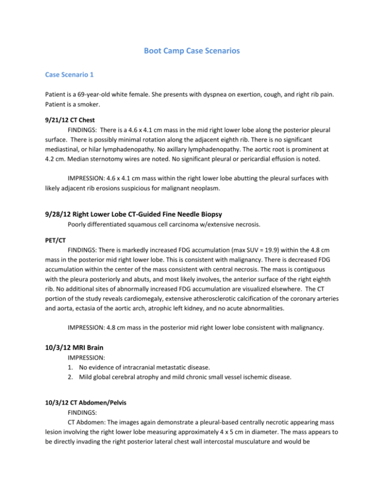
Boot Camp Case Scenarios Case Scenario 1 Patient is a 69-year-old white female. She presents with dyspnea on exertion, cough, and right rib pain. Patient is a smoker. 9/21/12 CT Chest FINDINGS: There is a 4.6 x 4.1 cm mass in the mid right lower lobe along the posterior pleural surface. There is possibly minimal rotation along the adjacent eighth rib. There is no significant mediastinal, or hilar lymphadenopathy. No axillary lymphadenopathy. The aortic root is prominent at 4.2 cm. Median sternotomy wires are noted. No significant pleural or pericardial effusion is noted. IMPRESSION: 4.6 x 4.1 cm mass within the right lower lobe abutting the pleural surfaces with likely adjacent rib erosions suspicious for malignant neoplasm. 9/28/12 Right Lower Lobe CT-Guided Fine Needle Biopsy Poorly differentiated squamous cell carcinoma w/extensive necrosis. PET/CT FINDINGS: There is markedly increased FDG accumulation (max SUV = 19.9) within the 4.8 cm mass in the posterior mid right lower lobe. This is consistent with malignancy. There is decreased FDG accumulation within the center of the mass consistent with central necrosis. The mass is contiguous with the pleura posteriorly and abuts, and most likely involves, the anterior surface of the right eighth rib. No additional sites of abnormally increased FDG accumulation are visualized elsewhere. The CT portion of the study reveals cardiomegaly, extensive atherosclerotic calcification of the coronary arteries and aorta, ectasia of the aortic arch, atrophic left kidney, and no acute abnormalities. IMPRESSION: 4.8 cm mass in the posterior mid right lower lobe consistent with malignancy. 10/3/12 MRI Brain IMPRESSION: 1. No evidence of intracranial metastatic disease. 2. Mild global cerebral atrophy and mild chronic small vessel ischemic disease. 10/3/12 CT Abdomen/Pelvis FINDINGS: CT Abdomen: The images again demonstrate a pleural-based centrally necrotic appearing mass lesion involving the right lower lobe measuring approximately 4 x 5 cm in diameter. The mass appears to be directly invading the right posterior lateral chest wall intercostal musculature and would be consistent with malignancy. The liver is normal in size. There is a 1.5 cm in diameter indeterminate lowattenuation lesion in the caudal tip of the right lobe of the liver. The gallbladder has been removed. There is no biliary duct dilatation. Spleen is normal in size and unremarkable. The pancreas and the adrenal glands are unremarkable. There is some focal atrophy and scarring involving the left kidney with mild compensatory hypertrophy of the right kidney. No significant focal renal mass lesions are demonstrated. Diffuse atherosclerotic calcifications are noted involving the abdominal aorta and its branches. No evidence of aneurysm. The IVC is unremarkable. No evidence of adenopathy. CT pelvis: No evidence of pelvic mass or adenopathy. No evidence of acute inflammatory change, abscess or ascites. Mild degenerative changes are noted in the lumbar spine. IMPRESSION: 1. Pleural-based mass lesion in the right lung lower lobe directly invading the right posterior lateral chest wall intercostal musculature consistent with malignancy. 2. Single indeterminate 1.5 cm right lobe of liver that does not appear to be hypermetabolic and favors benign etiology. Surgery 10/10/12 Bronchoscopy, right thoracotomy, right lower lobectomy and chest wall resection, including portions of 2 ribs with chest wall reconstruction; mediastinal node sampling. Pathology Final Diagnosis: A. Lymph node level X excision: Positive for metastatic poorly differentiated squamous cell carcinoma. B. Lung, right lower lobe and chest wall, lobectomy with chest wall resection: a. Histologic tumor type: Invasive poorly differentiated squamous cell carcinoma. b. Histologic tumor grade: Grade 3 (of 4). c. Tumor size: 7.0 x 6.0 x 5.5 cm. d. Tumor focality: Unifocal. e. Visceral pleura involvement: Tumor invades through the visceral and parietal pleura and into soft tissue of the chest wall and rib bone. f. Lymph-vascular invasion: Not identified. g. Margins: Surgical margins are negative for tumor. h. Pathologic stage: AJCC pT3 pN1. C. Lymph node, inferior pulmonary ligament excision: Negative for malignancy. D. Lymph node, periesophageal, excision: Negative for malignancy. E. Lymph node, subcarinal, excision: Negative for malignancy. F. Lymph node, Level XI, excision: Positive for metastatic poorly differentiated squamous cell carcinoma. 10/26/12 Radiation Oncology I am not recommending postoperative radiation. The PORT meta-analysis has actually shown a detrimental effect of postoperative radiation in the setting of N1 disease as this patient has. Since she has had an R0 resection, there would also be no reason to radiate the primary site. 10/26/12 Medical Oncology The patient is a 69-year-old lady with a history of stage IIIA non-small-cell lung cancer. She had R0 resection on October 10. She had slow recovery with complications including arrhythmia, pneumonia and CHF. She has significant comorbidities, and her performance status is at best between 2 and 3. I discussed that I have significant concerns regarding tolerability of chemotherapy. I made it clear to patient that risk of recurrence of her malignancy is high. However, she also has significant probability of dying from other causes in the next 5 years and overall benefit of adjuvant chemotherapy is relatively small. Patient decided against adjuvant chemotherapy. What is the primary site? What is the histology? Stage/ Prognostic Factors CS Tumor Size CS Extension CS Tumor Size/Ext Eval CS SSF 9 CS SSF 10 CS SSF 11 988 988 988 CS Lymph Nodes CS Lymph Nodes Eval Regional Nodes Positive Regional Nodes Examined CS Mets at Dx CS Mets Eval CS SSF 1 CS SSF 2 CS SSF 3 CS SSF 4 CS SSF 5 CS SSF 6 CS SSF 7 CS SSF 8 CS SSF 12 CS SSF 13 CS SSF 14 CS SSF 15 CS SSF 16 CS SSF 17 CS SSF 18 CS SSF 19 CS SSF 20 CS SSF 21 CS SSF 22 CS SSF 23 CS SSF 24 CS SSF 25 988 988 988 988 988 988 988 988 988 988 988 988 988 988 988 988 988 988 988 988 Treatment Diagnostic Staging Procedure Surgery Codes Surgical Procedure of Primary Site Scope of Regional Lymph Node Surgery Surgical Procedure/ Other Site Systemic Therapy Codes Chemotherapy Hormone Therapy Immunotherapy Hematologic Transplant/Endocrine Procedure Systemic/Surgery Sequence Radiation Codes Radiation Treatment Volume Regional Treatment Modality Regional Dose Boost Treatment Modality Boost Dose Number of Treatments to Volume Reason No Radiation Radiation/Surgery Sequence Case Scenario 2 History and Physical A 77 year old white male presented to my office with an elevated PSA of 4.6 on routine screening. A DRE was performed that revealed a palpable nodule on the right lobe of the prostate. The patient had a biopsy of the prostate on 2/6/12 that came back positive for acinar adenocarcinoma with a Gleason score 3+4=7. Tumor involved 4 of 12 cores. A CT of the abdomen and pelvis was performed on 3/2/12 and showed bulky retroperitoneal, mesenteric lymphadenopathy with associated splenomegaly and cardiophrenic and paraesophageal lymphadenopathy. No suspicious osseous lesions. No bowel obstruction. A retroperitoneal lymph node biopsy was done on 3/16/12 that was positive for mature B-cell non-Hodgkin lymphoma consistent with mantle cell lymphoma. A bone marrow biopsy and peripheral blood smear where negative for lymphoma. PET/CT Impression: 1. Extensive hypermetabolic lymphadenopathy both above and below the diaphragm compatible with patient's known lymphoma. 2. Splenomegaly with associated hypometabolism compatible with splenic involvement. Treatment Summary: Chemotherapy: The patient started a regimen of MODIFIED R-hyperCVAD- per Wisconsin Network 4/3/12. The regimen consisted of rituximab 375 mg/m2 administered on day 1 of all treatment cycles except cycle 1, cyclophosphamide 300 mg/m2 over 3 h, every 12 h days 1–3 (six doses, total dose 1800 mg/m2), doxorubicin 25 mg/m2/day as a continuous intravenous infusion over 48 h on days 1–2 (total dose 50 mg/m2), vincristine 2 mg i.v. push was administered on day 3, and dexamethasone 40 mg orally was given on days 1–4. Surgery 9/20/12: 1. ROBOTIC-ASSISTED LAPAROSCOPIC RADICAL PROSTATECTOMY. 2. ROBOTIC ASSISTED LAPAROSCOPIC BILATERAL PELVIC LYMPHADENECTOMY. Patient received maintenance Rituxan after prostatectomy. Pathology: Final Diagnosis: A) LYMPH NODES, ANTERIOR PROSTATIC FAT: - INVOLVEMENT BY MANTLE CELL LYMPHOMA (0.8 CM. LYMPH NODE). - NO EVIDENCE OF METASTATIC PROSTATE CARCINOMA, ONE (1) LYMPH NODE. B) LYMPH NODES, RIGHT PELVIC, RIGHT PELVIC LYMPH NODE DISSECTION: - INVOLVEMENT BY MANTLE CELL LYMPHOMA. - NO EVIDENCE OF METASTATIC PROSTATE CARCINOMA, SEVEN (7) LYMPH NODES. C) LYMPH NODES, LEFT PELVIC, LEFT PELVIC LYMPH NODE DISSECTION: - INVOLVEMENT BY MANTLE CELL LYMPHOMA. - NO EVIDENCE OF METASTATIC PROSTATE CARCINOMA, FOUR (4) LYMPH NODES. D) PROSTATE AND SEMINAL VESICLES, ROBOTIC-ASSISTED LAPAROSCOPIC RADICAL PROSTATECTOMY: HISTOLOGIC TUMOR TYPE: ADENOCARCINOMA. HISTOLOGIC TUMOR GRADE: GLEASON SCORE 3 + 4 = 7. TUMOR QUANTITATION: ADENOCARCINOMA INVOLVES RIGHT PROSTATE LOBE. THE ADENOCARCINOMA INVOLVES APPROXIMATELY 50% OF RIGHT PROSTATE LOBE AND INVOLVES APEX, MID AND BASE OF THE RIGHT PROSTATE LOBE. EXTRAPROSTATIC EXTENSION: IDENTIFIED. ADENOCARCINOMA EXTENDS INTO THE PERIPROSTATIC ADIPOSE TISSUE AT THE RIGHT POSTERIOR PROSTATE BASE. PERINEURAL INVASION: IDENTIFIED (PERINEURAL AND NERVE GANGLION INVASION IS PRESENT IN THE EXTRAPROSTATIC SOFT TISSUE ADJACENT TO THE RIGHT POSTERIOR PROSTATE BASE ). LYMPH-VASCULAR INVASION: NOT IDENTIFIED. SEMINAL VESICLE MUSCLE WALL INVASION: NOT IDENTIFIED. MARGINS: RIGHT AND LEFT APICAL MARGINS, RIGHT AND LEFT BLADDER NECK MARGINS, ALL PERIPHERAL SOFT TISSUE MARGINS AND RIGHT AND LEFT SEMINAL VESICLE AND VAS MARGINS ARE FREE OF TUMOR. PELVIC LYMPH NODES: INVOLVEMENT BY MANTLE CELL LYMPHOMA. NO EVIDENCE OF METASTATIC PROSTATE CARCINOMA, SEVEN RIGHT PELVIC LYMPH NODES, FOUR LEFT PELVIC LYMPH NODES, AND ONE "ANTERIOR PROSTATIC FAT" LYMPH NODE. PATHOLOGIC TNM STAGE: pT3a N0 What is the primary site? What is the histology? Stage/ Prognostic Factors CS Tumor Size CS Extension CS Tumor Size/Ext Eval CS SSF 9 CS SSF 10 CS SSF 11 CS Lymph Nodes CS Lymph Nodes Eval Regional Nodes Positive Regional Nodes Examined CS Mets at Dx CS Mets Eval CS SSF 1 CS SSF 2 CS SSF 3 CS SSF 4 CS SSF 5 CS SSF 6 CS SSF 7 CS SSF 8 CS SSF 12 CS SSF 13 CS SSF 14 CS SSF 15 CS SSF 16 CS SSF 17 CS SSF 18 CS SSF 19 CS SSF 20 CS SSF 21 CS SSF 22 CS SSF 23 CS SSF 24 CS SSF 25 Treatment Diagnostic Staging Procedure Surgery Codes Surgical Procedure of Primary Site Scope of Regional Lymph Node Surgery Surgical Procedure/ Other Site Systemic Therapy Codes Chemotherapy Hormone Therapy Immunotherapy Hematologic Transplant/Endocrine Procedure Systemic/Surgery Sequence Radiation Codes Radiation Treatment Volume Regional Treatment Modality Regional Dose Boost Treatment Modality Boost Dose Number of Treatments to Volume Reason No Radiation Radiation/Surgery Sequence 988 988 988 988 988 988 988 988 988 988 What is the primary site? What is the histology? Stage/ Prognostic Factors CS Tumor Size CS Extension CS Tumor Size/Ext Eval CS SSF 9 CS SSF 10 CS SSF 11 CS Lymph Nodes CS Lymph Nodes Eval Regional Nodes Positive Regional Nodes Examined CS Mets at Dx CS Mets Eval CS SSF 1 CS SSF 2 CS SSF 3 CS SSF 4 CS SSF 5 CS SSF 6 CS SSF 7 CS SSF 8 CS SSF 12 CS SSF 13 CS SSF 14 CS SSF 15 CS SSF 16 CS SSF 17 CS SSF 18 CS SSF 19 CS SSF 20 CS SSF 21 CS SSF 22 CS SSF 23 CS SSF 24 CS SSF 25 Treatment Diagnostic Staging Procedure Surgery Codes Surgical Procedure of Primary Site Scope of Regional Lymph Node Surgery Surgical Procedure/ Other Site Systemic Therapy Codes Chemotherapy Hormone Therapy Immunotherapy Hematologic Transplant/Endocrine Procedure Systemic/Surgery Sequence Radiation Codes Radiation Treatment Volume Regional Treatment Modality Regional Dose Boost Treatment Modality Boost Dose Number of Treatments to Volume Reason No Radiation Radiation/Surgery Sequence 988 988 988 988 988 988 988 988 988 988
