b a - Springer Static Content Server
advertisement

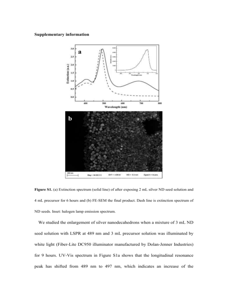
Supplementary information a b Figure S1. (a) Extinction spectrum (solid line) of after exposing 2 mL silver ND seed solution and 4 mL precursor for 6 hours and (b) FE-SEM the final product. Dash line is extinction spectrum of ND seeds. Inset: halogen lamp emission spectrum. We studied the enlargement of silver nanodecahedrons when a mixture of 3 mL ND seed solution with LSPR at 489 nm and 3 mL precursor solution was illuminated by white light (Fiber-Lite DC950 illuminator manufactured by Dolan-Jenner Industries) for 9 hours. UV-Vis spectrum in Figure S1a shows that the longitudinal resonance peak has shifted from 489 nm to 497 nm, which indicates an increase of the nanodecahedrons size. However, a broad peak in the longer wavelength region has emerged also. This peak is associated with silver nanoprisms and nanoplates as shown in Figure S1b. The reason for our results is straightforward. Since halogen light contains a wide spectrum particularly for the 500-750 nm region (as shown in the inset of Figure S1a), the mixture received a range of photon energies that can induce regrowth of silver NDs (LSP wavelength: 500-520 nm) as well as transformation of small silver nanoparticles into nanoprisms and nanoplate (500-750 nm). a b Figure S2. FDTD simulation results of electric field distribution around a 50 nm edge length silver nanodecahedron irradiated by 540 nm light of unit amplitude with electric polarization parallel with (a) longitudinal and (b) transverse of silver nanodecahedron. k is the direction of irradiation light. Figure S3. FE-SEM image of silver nanodecahedrons of large scale. Silver nanoprisms and nanoplates are labeled with red circles. 489 nm 516 nm 522 nm 550 nm 572 nm 590 nm Figure S4. Histograms showing the size distribution of silver nanodecahedrons that exhibit different extinction peaks. Figure S5. FE-SEM image of product irradiated by 578 nm LED.


