Supplementary Methods (doc 76K)
advertisement
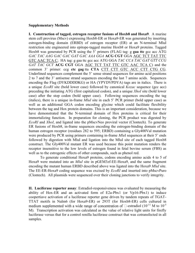
Supplementary Methods I. Construction of tagged, estrogen receptor fusions of Hoxb8 and Hoxa9. A murine stem cell provirus (Mscv) expressing Hoxb8-ER or Hoxa9-ER was generated by inserting estrogen-binding domain (ERBD) of estrogen receptor (ER) at an N-terminal MluI restriction site engineered into epitope-tagged murine Hoxb8 or Hoxa9 proteins. Tagged Hoxb8 was generated by PCR using the 5’ primers (FLAG tag: g gaa ttc gcc acc ATG GAC TAC AAG GAC GAC GAT GAC AAA GGA ACG CGT GGA AGC TCT TAT TTC GTC AAC TCA C; HA tag: g gaa ttc gcc acc ATG GGA TAC CCA TAC GAT GTT CCG GAT TAC GCT ACG CGT GGA AGC TCT TAT TTC GTC AAC TCA C) and the common 3’ primer: ccg ctc gag tta CTA CTT CTT GTC ACC CTT CTG CG. Underlined sequences complement the 5’ sense strand sequences for amino acid positions 2 to 7 and the 3’ antisense strand sequences encoding the last 7 amino acids. Sequences encoding the Flag (DYKDDDDKG) or HA (YPYDVPDYA) tags are in italics. There is a unique EcoRI site (bold lower case) followed by canonical Kozac sequence (gcc acc) preceding the initiating ATG (first capitalized codon), and a unique XhoI site (bold lower case) after the stop codon (bold upper case). Following sequences encoding the tag (italics), there is a unique in-frame MluI site in each 5’ PCR primer (bold upper case) as well as an additional GGA codon encoding glycine which could facilitate flexibility between the tag and Hox protein domains. This is an important consideration, because we have demonstrated that the N-terminal domain of Hox proteins is critical for their immortalizing function. In preparation for cloning, the PCR product was digested by EcoRI and XhoI, and ligated into the pMscvNeo proviral vector (Clontech). To generate ER fusions of Hoxb8, in-frame sequences encoding the estrogen-binding domain of the human estrogen receptor (residues 282 to 595; ERBD) containing a Gly400Val mutation were produced by PCR using primers containing in-frame MluI sequences at their 5’ ends followed by digestion with MluI and ligation into the MluI site of each tagged Hoxb8 construct. The Gly400Val mutant ER was used because this point mutation renders the receptor insensitive to the low levels of estrogen found in fetal bovine serum (FBS) as well as to the estrogenic effects of other compounds, such as phenol red. To generate conditional Hoxa9 proteins, codons encoding amino acids 4 to 5 of Hoxa9 were mutated into an MluI site in pGEM3zf-EE-Hoxa9, and the same fragment encoding the mutant human ERBD described above was ligated into the Hoxa9 MluI site. The EE-ER-Hoxa9 coding sequence was excised by EcoRI and inserted into pMscvPuro (Clontech). All plasmids were sequenced over their cloning junctions to verify integrity. II. Luciferase reporter assay: Estradiol-responsiveness was evaluated by measuring the ability of Hox-ER and an activated form of E2a-Pbx1 (or Vp16-Pbx1) to induce coopertiave activation of a luciferase reporter gene driven by tandem repeats of TGATTTAT motifs in Nalm6 (for Hoxa9-ER) or 293T (for Hoxb8-ER) cells cultured in -estradiol (10-11 M to 10-5 M). Transcription activation was calculated as the value of relative light units for firefly luciferase versus that for a control renilla luciferase construct that was cotransfected in all samples. III. Cell culture: Myeloid progenitor lines were maintained in a 37°C humidified incubator with 5% CO2. Progenitors were frozen as described below, and stored in liquid nitrogen. IV. Affymetrix array analysis: Gene expression profiling analysis was performed and analyzed using affymetrix mouse total genome array. Hybridization and quantitation of array signals were performed by the UCSD gene chip core laboratory, using the GeneChip Scanner 3000, enabled for high-resolution scanning, coupled with GeneChip operating software (GCOS). Array data was normalized to internal controls, and the overall chip signal intensity was normalized to the mean signal intensity across a group of 12 chips used to evaluate RNA levels in Hoxb8-ER progenitors undergoing neutrophil or macrophage differentiation, and in Hoxb8-ER macrophages stimulated with LPS or BLP. Data was analyzed using “perfect match minus mismatch” algorithms, and signature genes identified exhibited a minimum 5-fold difference in signal intensity between the averages of each groups of three samples. Normalization and processing of GCOS data was performed using dCHIP software. All data is normalized to a basal level of “1”, which was selected as a moderately low level of expression. V. Producing Retrovirus by CaPO4 transfection of 293T cells 1. Helper-free retrovirus is produced in 293T cells by CaPO4 co-transfection of the retroviral construct with an ecotropic or amphotropic packaging construct (CellGenesys). 2. We use Invitrogen’s CaPO4 Transfection Kit (#44-0052) a. Day 0 Seed 2 x 10⁶ 293T cells into a 10cm dish with 10ml DMEM (High glucose) + 10% FBS + penicillin/streptomycin/glutamine b. Day 1 Remove media and replace with 10ml of fresh, pre-warmed media. Cells should be at ~60-70% confluence Transfect 10µg of retroviral construct + 10µg of packaging construct as per protocol (remember to use ecotropic packaging construct for murine marrow, amphotropic for human cells) Incubate overnight c. Day 2 Remove media and replace with 6ml of fresh, pre-warmed media. d. Day 3 Harvest 6ml virus to a 15ml conical tube. Centrifuge briefly to pellet all cell debris. Freeze in 1-2ml aliquots in 2ml freezing tubes and store at –80°C. Alternatively, filter the virus supernatant and use immediately. Add another 6ml of fresh, pre-warmed media to transfected cells. e. Day 4 Harvest an additional 6ml virus. 3. At this point, it is a good idea to thaw a small (~250µl) aliquot for the purpose of determining the viral titer Depending on the size of the insert, we usually attain viral titers between 105 – 106/ml VI. Titering Retrovirus If this is the first time working with retrovirus, you should definitely determine the titer by infecting an “easily-infectable” cell line. Having determined that your protocol generates a good titer of virus, it is rarely necessary to titer each additional batch. Generally speaking, NIH3T3 cells are ideal for determining the titer of ecotropic retrovirus, while 293/293T (human embryonic kidney) cells are good for amphotropic virus. 1. 2. 3. 4. Day 1 A. Set up 150,000 NIH3T3 cells onto 6 60mm dishes B. NIH3T3 cells grow best in DMEM (high glucose) + 10% FBS + P/S/Glutamine Day 2 A. The cells should be ~20-30% confluent B. Prepare ~25ml of media. Add Lipofectamine 1:1000 (Can alternatively use polybrene at a final 8µg/ml) C. Thaw an aliquot of virus and prepare a series of 5 tubes of consecutive 1:10 dilutions, serially diluting 100µl of virus with an additional 900µl of media containing Lipofectamine D. Aspirate the media on the NIH3T3 cells and add 1.5ml of media with Lipofectamine (no virus yet) E. Add 500µl of each viral dilution and use 500µl of plain media for the control plate F. Allow the infection to proceed 4 hours at 37°C. G. Dilute with 5ml of fresh media without Lipofectamine Day 4 A. Aspirate the media B. Add fresh media with G418 (final concentration of active drug 1mg/ml) i. We use Gibco’s Geneticin (liquid, dissolved at 50mg/ml active drug, cat # 10131-035) ii. Of course, Puromycin selection (1µg/ml) works with vectors carrying the puroR cassette Days 6+ A. Change media every 2-3 days, maintaining G418 selection B. Don’t be deceived! After 5 or so days in G418, the cells are not yet dead! Don’t assume that those adherent cells are G418 resistant! Continue changing the medium on the cells for the full 10 to 14 days! C. By day 10-14, the cells on the control plate should be completely dead and detached and individual colonies should be visible on the other plates. If your virus is of good titer, the plates with the 1:10 and 1:100 dilutions of D. virus should be completely confluent. These can be discarded. Count the # of colonies on the other plates to determine the viral titer Counting the colonies can be done by staining with Giemsa stain (Sigma) i. Aspirate the media. Rinse 1x with PBS ii. Flood the dish gently with 80% methanol. Sit for 5 minutes at RT iii. Aspirate the methanol. Flood the dish gently with 20% Giemsa. Sit for 10’ at RT iv. Aspirate the Giemsa and wash the cells 1-2x with PBS v. The colonies should be easily visible and easy to count dilution actual virus (in 500µl) expected colonies (viral titer of 105/ml) expected colonies (viral titer of 106/ml) 1:10 50µl 5000 50,000 1:100 5µl 500 5000 1:1000 0.5µl 50 500 1:10,000 0.05µl 5 50 1:100,000 0.005µl 0.5 5 VII. Harvesting Marrow 1. Sacrifice female Balb/c (or other strain, see FAQs) mouse (generally 8-12 weeks, though older is OK) 2. Remove intact femurs and tibia into sterile dishes of PBS on ice 3. Cut off the ends of the bones 4. Use 10ml syringes (filled with RPMI 10% FBS) and 25G needles to shoot the marrow into a 50ml conical tube a. the bones should become translucent b. Use 1 x 50ml conical tube for each mouse 5. Top up to 50ml with PBS and pellet the cells 6. Resuspend well in 10ml ACK red blood cell lysis buffer. Incubate 5 min at RT. a. ACK RBC lysis buffer 150mM NH4Cl 10mM KHCO3 0.1mM Na2EDTA Adjust to pH 7.2-7.4 with 1N HCl Filter sterilize and store at 4°C 7. Top up to 50ml with PBS and pellet the cells. 8. Resuspend the cells in 4ml of PBS Using 5-Flurouracil prior to isolation of marrow and progenitors One can inject mice with 5-Flurouracil (5-FU) 3-5 days prior to harvesting the bone marrow. Injections are done at 100-150mg/kg I.P. The 5-FU will greatly reduce the total cellularity of the marrow with an increased % of progenitors (though we do not find a dramatic increase in total # myeloid CFU). The advantage of the 5-FU is that the marrow from more mice can be processed on the same Ficoll gradient and on the same StemCell Technology column (using less reagent). As mentioned above, if your retroviral infection/immortalization is of high efficiency (as we have observed for Hoxa9-ER, Hoxb8-ER, and E2a-ER-Pbx construct EPS∆578ER), you do not NEED to treat with 5FU or isolate marrow progenitors by negative selection. Infections can be done on “crude” marrow simply by removing the bone marrow cells and passing them over a Ficoll gradient before plating them under cytokine pre-stimulating conditions. NOTES: A. Cell numbers following purification with no 5-FU pre-treatment 1. One generally gets 15-30 million cells (post-Ficoll) per mouse. 2. Of these, ~2-3% are progenitors as isolated from the StemCell Technologies column. 3. After a 3 day stimulation in cytokine(IL3, IL6, SCF), these cells will usually double in number. B. Cell numbers following purification WITH 5-FU pre-treatment. 1. One generally gets 2-5 million cells (post-Ficoll) per mouse 2. Of these, 5-20% are progenitors, base on the fact that this fraction is isolated from the negative-selection column 3. In our experience, we do not see much of an increase in the total # of progenitors following 5-FU treatment (i.e. while the total cellularity decreases, the % of progenitors increases but the total # of progenitors does not increase all that much). However, it is very convenient to have fewer cells for the manipulations with the column and thus the use of 5-FU is recommended in my opinion. VIII. 1. 2. 3. 4. 5. 6. 7. 8. 9. Harvesting Fetal Liver Cells Sacrifice pregnant mouse Remove embryos (we’ve used them as early as day 11) Harvest fetal liver Using the plunger from a 5ml syringe, disperse the cells through a 70µ cell strainer (Falcon #352350) Pellet cells Resuspend well in 10ml of ACK red blood cell lysis buffer (use recipe in VII). Incubate 5 min at RT. Top up to 50ml with PBS and pellet the cells. Pellet cells and rinse 1x in PBS Resuspend the cells in 4ml of PBS IX. Negative selection using Stem Cell Technologies Murine Progenitor Isolation Cocktail 1. Wash cells derived from marrow or fetal liver 1x in PBS 2. Resuspend in PBS+FBS+Rat Serum at 2-8x10⁷cells/ml (~1-2mls is all) 3. Incubate in fridge at 4° for 15minutes 4. Proceed with Progenitor Cell Column Isolation as described in the Stem Cell Technology manual NOTES: 1. Using no treatment with 5-FU: a. One generally gets 15-30 million cells (post-Ficoll) per mouse (tibias and femers). b. Of these, ~2-3% are progenitors as isolated from the StemCell Technologies column. c. After a 3 day stimulation in cytokine (IL3, IL6, SCF), progenitors will double in number. 2. Using pre-treatment with 5-FU (see “5-FU pre-treatment” protocol in section III) a. One generally gets 2-5 million cells (post-Ficoll) per mouse b. Of these, 5-20% are progenitors, based on negative-selection by the StemCell Technologies column c. In our experience, we do not see much of an increase in the total # of progenitors following 5-FU treatment (i.e. while the total cellularity decreases, the % of progenitors increases but the total # of progenitors does not increase all that much). However, it is very convenient to have fewer cells for the manipulations with the column and thus the use of 5-FU is recommended in my opinion. Cytokine pre-stimulation of the cells For a good retroviral infection, progenitors subjected to negative selection are stimulated to enter the cell cycle by transferring them to a cytokine-rich media for 2 days. The media described below is very effective though others that include G-CSF, Flt3-ligand, etc. are likely to be equally effective. Cytokines are purchased from Peprotech as their activity is consistent and they are reasonably priced. Stem Cell Media IMDM (Iscove’s) + 15%FBS + 1%pen/strep/glutamine 10ng/ml murine IL-3 (5 ng/ml is probably sufficient) 20ng/ml murine IL-6 25ng/ml murine SCF (some protocols use up to 100 ng/ml) Notes on the cytokine stimulation: A. RPMI with 10-15% FBS can be substituted for Iscove’s media B. This is a “low concentration” cytokine cocktail. Many progenitor cocktails call for up to 100ng/ml as well as the addition of other cytokines such as Flt3 ligand or GCSF. At the same time, we have gone as low as 10ng/ml for all three cytokines (IL-3, IL-6, and SCF) and for a 3 day culture, this seems sufficient. C. The cells begin to divide actively after approximately 2 days in culture. Therefore, for a 2-3 day incubation, one expects about one cell doubling. After 3 days, however, the cells are actively dividing and can be expanded should one require more cells. We have not assayed or infected the cells past 4 days in culture, however, and are not sure for how long the cells retain their immature phenotype. During the culture, some mature monocytes will adhere to the dish and will not be harvested with the non-adherent cells used for the retroviral infection. X. Spin Infection Protocol – for infection in 12-well plates 1. Coat a non-TC treated plate with Fibronectin a. Falcon sells these plates (12-well #351153 or 6-well #351146) b. Human fibronectin is supplied as a 1mg/ml solution from Sigma (F-0895) c. Dilute the fibronectin 1:100 in PBS to a final 10µg/ml solution d. Aliquot 1ml into each well of a 12-well non-tissue treated plate (or 2ml per well in a 6-well plate). Apparently the Fibronectin coats non-TC treated plates more efficiently, though we have used both types of plates with success e. incubate at 37° for 1-4hrs or at 4° overnight 2. Set up cells a. Count cells and resuspend at 105-106/ml in “Progenitor Outgrowth Medium” Use this medium for the Hoxb8-ER virus. Estradiol is only necessary for use with ERfusion oncoproteins). We have not tested whether the BME is necessary for derivation of neutrophil progenitors described in the Nature Methods manuscript. b. Add 1µl of Lipofectamine per ml of cells. Or, add 1µl per ml of expected final volume if you are NOT going to add Lipofectamine separately to the virus. This shortcut is useful if one is using 250µl of cells and 1ml of virus in a 12-well plate c. Aspirate the fibronectin. d. Aliquot 250µl (~25,000 to 250,000 cells) into each well. Usually I try not to exceed the titer of the virus e. Place the plate at 37°C while thawing and preparing the virus 3. Virus a. Thaw the virus rapidly in a 37°C water bath b. Add 1-2ml of virus to each well of the 12-well plate. Make sure that the final Lipofectamine concentration is 1x (1:1000) 4. Spinoculation a. Wrap the plate(s) in Saran Wrap with an equivalent balance plate b. Spin the plate in plate carriers at 1500g for 60-90 minutes at 22°-32°. Forces up to 4500g have been tested with good results. This is easiest in Gernot Walter's Beckman JS5.2 rotor at 2800rpm (r=20cm, ~1300g) c. You do not need to remove the cells/virus from the 12-well plate. d. Dilute the Lipofectamine/Polybrene with 3ml of fresh “Progenitor Outgrowth Media” and incubate the cells at 37°C. Progenitor Outgrowth Medium OptiMem 10% FBS 1%PSG, 10ng/ml stem cell factor or 1% culture supernatant from an SCF-producing cell line 30uM beta mercapto ethanol (1ul neat into 500mls medium) 1uM estradiol XI. Progenitor purification using Ficoll-Hypaque centrifugation (For derivation of macrophage-committed progenitors immortalized by Hoxb8-ER or biphenotypic, neutrophil, or macrophage progenitors immortalizated by Hoxa9-ER). 1. In a 15ml conical tube, add 3ml of room-temperature Ficoll-Paque (Pharmacia, Piscataway, NJ) and layer 4ml of total marrow cells in PBS gently on top. a. try not to exceed 50 million cells per gradient b. usually this means one layer per mouse unless you used 5-FU (see 5-FU protocol and “Harvesting Total Progenitors” protocol) 2. Spin 30 minutes at 1500rpm @ 20° in a Sorvall 6000B rotor (450g) 3. Harvest cells from the interface and collect all supernatant to within ~0.5ml of the pellet c. Marrow progenitors will be scattered throughout the Ficoll along with lymphocytes d. The pellet will contain left-over RBCs and mature granulocytes 4. Dilute to 50ml in Myeloid Medium. Pellet and count cells. Proceed to spin infection protocol. I like to use around 100,000 to 200,000 cells per spin infection. One gets reliable immortalization at this level. Use myeloid medium throughout derivation of these progenitors. Note: In this protocol, the Ficoll-buoyant progenitors can be infected directly with retrovirus or pre-expanded in IL-3, IL-6, and SCF 48 hours prior to infection. Myeloid medium RPMI1640 10% FBS 1%Pen-Strep-Glut (PSQ, Gibco BRL, Rockville, MD) 20ng/ml GMCSF or 1% culture supernatant from a GM-CSF-producing cell line 1 uM -estradiol (Sigma). -estradiol was kept as 1,000x (1mM) or 10,000x (10mM) stocks in 100% ethanol and stored at -20°C. XII. Myeoid cell line derivation following retroviral infection A. All culture following the infection is done in RPMI + 10% FBS + GM-CSF + pen/strep/glutamine 1. When using ER-oncoprotein fusions, be sure to include 1µM ß-estradiol (Sigma E-2758). ß-estradiol can be purchased from Sigma (E-2758). It can be conveniently dissolved at 10mM in 100% ethanol (takes ~30 minutes of end over end at RT). This provides a 10,000x stock solution to be used at a final 1µM in cell culture. When rinsing the cells of ß-estradiol, it is a good idea to rinse 2x in 10ml PBS. 2. We used GM-CSF-conditioned media that contains 1-2ug GM-CSF/ml. We use this conditioned medium at a 1:100 dilution in our final medium, yielding a final GM-CSF concentration of 10-20ng/ml. B. Derivation procedure: 1. It is a good idea to keep a few wells of “uninfected” or “vector alone” infected cells maintained under the same conditions. Over the course of 2-4 weeks in GM-CSF (or IL-3) containing media, these cells will divide, mature, terminally differentiate to neutrophils and macrophages, and finally die. In 100’s of infections, GM-CSF cultures of uninfected or vector alone infected cells have never produced immortalized cells. In the presence of IL-3, however, one can sometimes find the emergence of a very slow growing mast cell population. This is easily differentiated from the rapid emergence of immortalized myeloblasts. 2. Cells should be split to a new dish (usually to a 6-well) with fresh media every 3-4 days. All non-adherent cells can be transferred by rather vigorously pipetting the lightly adherent cells off the bottom of the dish. The truly adherent macrophages will not come off no matter how vigorous the pipetting! The cells should not be allowed to become too confluent. This is important as the differentiated monocytes stuck on the bottom of the dish seem to have a pro-differentiative effect on all the other cells in the culture. Furthermore, the presence of fresh GM-CSF is critical in these early stages. 3. The emergence of immortalized cells will appear after about 2-3 weeks as clumps of round and refractile blasts, like little clusters of grapes. It will then become easier to transfer these cells off the differentiated layer of monocytes. 4. Interestingly, in the early stages of culture, the transfer of the immortalized cells to a new dish also seems to have a transient, pro-differentiative effect, maybe due to the mechanical activation of the cells by the pipetting. Thus, the day after the transfer, one often notices many fewer blasts (refractile round morphology) than one actually transferred, and more cells adhering to the plastic. This is normal, and after 24hrs, the cells tend to revert back to their rounded, progenitor-like morphology. 5. Eventually, after 3-4 weeks, all that will be left are the immortalized myeloblasts. These grow very robustly and rapidly and can be cloned by limiting dilution in 96-well plates. Usually, I plate out one 96-well plate at 3 cells/well and one plate at 0.3 cells/well in 200µl of media. After 10-14 days, clones are easily identified and transferred to 24well plates for characterization. C. Removing ß-estradiol from progenitors immortalized by ER-Hox oncoproteins Cells should be rinsed 2x in PBS to get rid of residual ß-estradiol before plating in GM-CSF media without estrogen. Remember, because the “muted” G400V mutant version of the estrogen receptor has been used in all cases within our work published to date (2005), the estrogenic effects of phenol red or residual estrogens in the FBS do not have any effect on the ER domains used in our fusion oncoproteins. In the absence of estrogen, the cells will undergo between 1-3 cell divisions for ER-Hox progenitors and then terminally differentiate to macrophages (for Hoxb8-ER) or both granulocytes and monocytes (for polyclonal populations of Hoxa9-ER). This usually takes between 2-7 days, depending on the clone and also depending on the cell density. Generally, the less dense the culture, the faster the process of differentiation. XIII. Freezing & Thawing Myeloid Progenitors Cells to be frozen should be split the day before and should be very happy. Adherent cells should be about 75-80% confluent while suspension cells should be medium density. Some general rules apply to freezing cells. Cells should be resuspended gently and thoroughly in cold freezing media and placed on ice for 5-10’. They should then be cooled slowly to -80° (an isopropanol freezing chamber can accomplish this) before transferring to liquid nitrogen. The freezing media can vary in the concentration of FBS and the concentration of DMSO. Remember, DMSO is the cryopreservative but can also osmotically shock the cells. Some cells are more sensitive than others to its toxic effects. Generally FBS makes up 20-90% and DMSO makes up 5-10% of the freezing media. Most cell lines can be resuspended directly in the FBS/DMSO mixture though some sensitive cells should be resuspended in FBS before the DMSO is added slowly and drop-wise to the cells while mixing. Protocol 1. Prepare or thaw a mixture of sterile-filtered FBS/20% DMSO. Keep on ice. 2. Pellet cells at 1000rpm (~200g) for 10’. Aspirate media. 3. Resuspend cells in 1/2 volume of media with 10% serum chill on ice i.e. whatever media in which the cells are normally grown 4. Slowly add 1/2 volume of sterile ice-cold 100%FBS/20% DMSO and mix the cells thoroughly and gently. note: for some cells, one can lower the final conc. of DMSO to 7.5% 5. Aliquot cells into cryotubes and place on ice for 5’. 6. Place tubes in isopropanol freezing chamber pre-cooled to 4° and place the chamber at -80°. 7. 24-48hrs later, transfer cells to liquid nitrogen. XIV. Frequently Asked Questions What mouse strains have been used in this infection protocol? Balb/c and C57Bl/6 mouse strains have all been used with good success. Immortalization of other strains has not been attempted. Has the number of progenitors used as targets for infection been tittered, and if so, how many are required for reliable immortalization? With a good-titer retrovirus of 2x10⁵-2x10⁶/ml, which is reproducibly produced from standard transfection conditions, 10,000 progenitors will consistently yield immortalized cultures. Has retronectin been used in these assays? Is it significantly better than fibronectin? Fibronectin is a good substitute for the much more expensive commercially available Retronectin. Retronectin is merely the fragment of fibronectin which contains the viral binding domain. In side by side comparisons, retronectin does not improve the efficiency of immortalization by more than 2-fold. My medium turns purple during spinoculation. Is that normal? Yes, during the spinoculation, the media will get quite marginally basic—enough to turn its color purple. This does not affect progenitor viability. Media containing HEPES can be used to maintain a more physiological pH if necessary. HEPES-media can be purchased from Gibco. Is RPM a significant parameter in the spinoculation procedure? The spinoculation can be done at 1000g up to 4500g without any loss of viability in my hands. The higher the G-force the better the transduction though this is a minimal effect. During spinoculation, the temperature in the rotor rose to 36 degrees Centigrade. Should I be worried? Although we usually set the temperature parameter for constant temperature of 23 degrees, we have had rotor temperature rise to 36 degrees and have found no ill-effect. What is the most important parameter during spin-infection? The most important predictor of good transduction efficiency is still the viral titer. What is the difference between the use of Lipofectamine verses the old standard of polybrene? Lipofectamine is used as a poly-cationic compound at a final 1:1000 dilution. However, we do not believe that there is any significant advantage over using a final 8µg/ml polybrene. Both of these compounds are designed to eliminate the repulsion between the negatively charged virus and the negatively charged cell membrane. Many protocols using fibronectin/retronectin do not also include a poly-cationic compound though we have continued to do so.
