Supplementary Table XX: details of materials and methods
advertisement
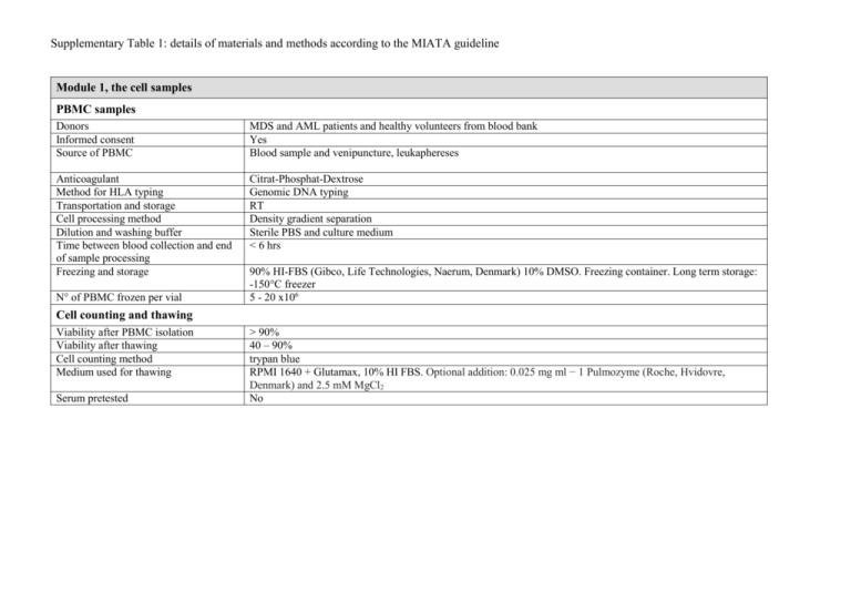
Supplementary Table 1: details of materials and methods according to the MIATA guideline Module 1, the cell samples PBMC samples Donors Informed consent Source of PBMC MDS and AML patients and healthy volunteers from blood bank Yes Blood sample and venipuncture, leukaphereses Anticoagulant Method for HLA typing Transportation and storage Cell processing method Dilution and washing buffer Time between blood collection and end of sample processing Freezing and storage Citrat-Phosphat-Dextrose Genomic DNA typing RT Density gradient separation Sterile PBS and culture medium < 6 hrs N° of PBMC frozen per vial 90% HI-FBS (Gibco, Life Technologies, Naerum, Denmark) 10% DMSO. Freezing container. Long term storage: -150°C freezer 5 - 20 x106 Cell counting and thawing Viability after PBMC isolation Viability after thawing Cell counting method Medium used for thawing Serum pretested > 90% 40 – 90% trypan blue RPMI 1640 + Glutamax, 10% HI FBS. Optional addition: 0.025 mg ml − 1 Pulmozyme (Roche, Hvidovre, Denmark) and 2.5 mM MgCl2 No Module 2, the assays CD8 T cells and CD34 tumor cells mix Culture medium Isolation Buffer X-Vivo 15 (Lonza), 10% HI-Human Serum (Sigma) MACS CD8 positive selection and CD34 negative selection kits (Miltenyi Biotec) PBS with 2% HI-FBS (Gibco) Peptide stimulation and combinatorial encoding MHC-multimer staining Culture medium Peptide concentration for stimulation Peptides provided by X-vivo 15 (Lonza) with 5% HI-Human Serum (Sigma), IL-2 (20 U/ml, Proleukin Novartis) and IL-7 (5 ng/ml, Peprotech) 10 μM Pepscan Ltd, NL. NK cell killing capacity assay Isolation Resting medium MACS untouched NK cell kit followed by CD56+ kit (Miltenyi Biotec) X-vivo 15 (Lonza) with 10% HI-Human Serum (Sigma), IL-2 (200 U/ml, Proleukin Novartis) and IL-15 (40 U/ml, Peprotech) Frequency of measured cell populations CD8 T cell and CD34 tumor cell mix 0.1-1.5 % CD107+aCD8+ cells (figure 1) MHC-multimer assay (figure 2) 0.001-0.20% multimer+CD8+ direct ex vivo, CTA-specific T cells 0.013-0.36% multimer+CD8+ after in vitro peptide pre-stimulation 0.015-3.36% multimer+CD8+ direct ex vivo, virus-specific T cells Cell counts and functionality assays 56-710 x 106 cells/L of CD8+ T cells (figure 3) 130-1478 x 106 cells/L of CD4+ T cells 10-2115 x 106 cells/L of CD56+ NK cells NK subpopulation and killing capacity assay analyses (figure 4) Tregs assay (figure 5) M-MDSCs assay (figure 5) 1.4-20% CD107a+CD8+ cells 0.1-3.0 % CD107a+CD4+ cells 0.1-3.0 % CD107a+CD56+ cells 20-573 6 x 10⁶ cells/L of CD56+CD16+ cells 5.5-119 x 106 cells/L of CD56+CD16+CD158b+ cells 0.043-6.5 x 10⁶ cells/L of CD56+CD16+CD158d+ cells 25-63% killing in effector:target ratio 10:1 5.2-31.6 x 10⁶ cells/L of CD4+CD25+CD127-FoxP3+CD49d0.038-195 x 106 cells/L of HLA-DR-lin-CD33+CD11b+CD14highCD15low Module 3, data acquisition Flow cytometer Software for acquisition Instrument settings and performance control Number of cells acquired Lasers and filters BD LSR II SORP Diva CS&T beads, daily performance check, compensation with beads All in the tube LSRII: UV laser (355nm, 60 mW): detector A: 710/50 and 680LP, detector B: 605/12 and 595LP, detector C: 580/30. Remaining detectors empty. Violet laser (405nm, 100 mW): detector A: 655/6 and 635 LP, detector B: 625/20 and 610 LP, detector C: 450/50. Blue laser (488 nm, 100 mW): detector A: 780/60 and 735 LP, detector B: 710/50 and 685LP, detector C: 670/30 and 635LP, detector D: 610/20 and 600LP, detector E: 585/15 and 570LP, detector F: 525/50 and 505LP, detector G: 488/10. Yeloow green laser (561 nm, 50 mW): detector A: 800/30 and 770LP, detector B: 695/40, detector C empty. Red laser (640 nm, 40 mV): detector A: 780/60 and 735 LP, detector B: 725/50 and 710 LP, detector C: 660/20. FACSCanto II: Violet laser (405 nm, 25 mW): detector A: 510/50 and 502LP, detector B: 450/50, detector C empty. Blue Laser (488 nm, 20 mW): detector A: 780/60 and 735LP, detector B: 670LP and 655LP, detector C: 610LP, detector D: 585/42 and 556LP, detector E: 530/30 and 502LP, detector F: 488/10. Red laser (633 nm, 17mW): detector A: 780/60, detector B: 685LP, detector C: 660/20. Filters were produced by BD Biosciences and were changed according to daily performance check. Module 4, data processing Software for analysis Gating strategy for measurements of viability and CD8+ cells Gating between experiments Any data excluded Positivity criteria Raw data provided on demand Diva Software v6.1.3 singlets FSC-A/H or FSC-A/W, lymphos FSC-A/SSC-A, living lymphos FSC-A/dead cell dye (gate). Example on gating strategy is shown in figure S1. One mastergate set in comparison analyses. No n.a. Yes Module 5, Lab conditions Guidance of lab operations Trained personal Accreditation of the lab Participation to proficiency panels Status of protocols Status of assays Exploratory research Yes No Yes, CIP Established lab protocols Qualified FBS = fetal bovine serum; HI = heat- inactivated; n.a. = not applicable; RT = room temperature; PBS: phosphate buffer; CTA: cancer-testis antigen; LP: longpass filter; FCS: forward scatter; SSC: side scatter; CIP: CIMT (The Association for Cancer Immunotherapy) Immunoguiding program
