Indications
advertisement
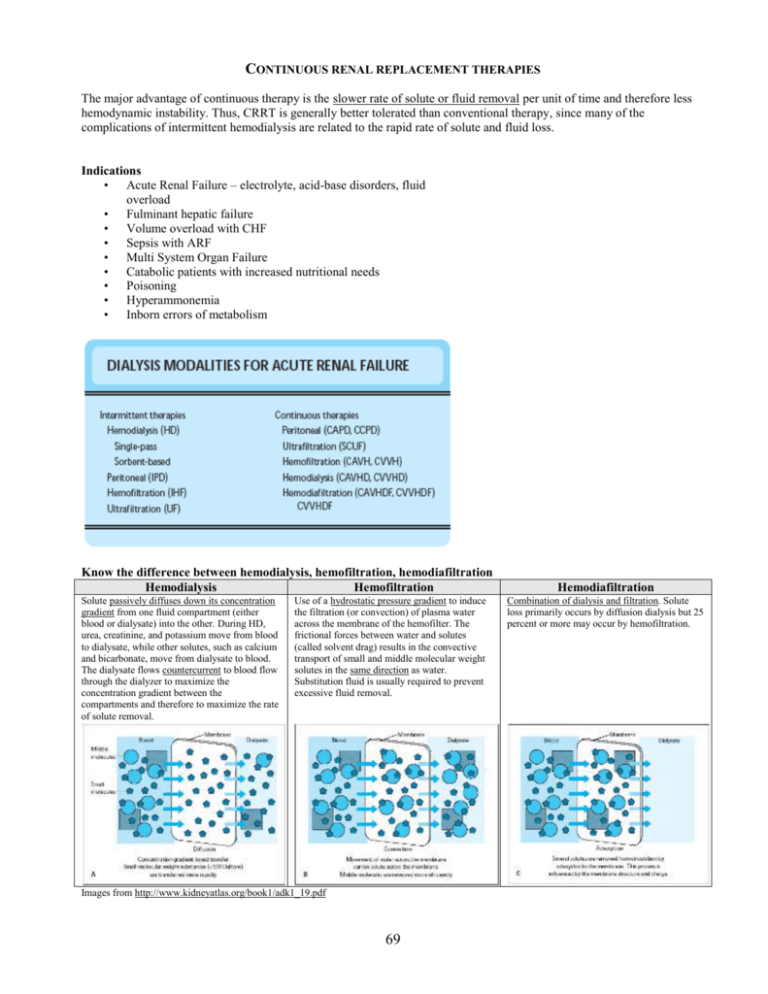
CONTINUOUS RENAL REPLACEMENT THERAPIES The major advantage of continuous therapy is the slower rate of solute or fluid removal per unit of time and therefore less hemodynamic instability. Thus, CRRT is generally better tolerated than conventional therapy, since many of the complications of intermittent hemodialysis are related to the rapid rate of solute and fluid loss. Indications • Acute Renal Failure – electrolyte, acid-base disorders, fluid overload • Fulminant hepatic failure • Volume overload with CHF • Sepsis with ARF • Multi System Organ Failure • Catabolic patients with increased nutritional needs • Poisoning • Hyperammonemia • Inborn errors of metabolism Know the difference between hemodialysis, hemofiltration, hemodiafiltration Hemodialysis Hemofiltration Solute passively diffuses down its concentration gradient from one fluid compartment (either blood or dialysate) into the other. During HD, urea, creatinine, and potassium move from blood to dialysate, while other solutes, such as calcium and bicarbonate, move from dialysate to blood. The dialysate flows countercurrent to blood flow through the dialyzer to maximize the concentration gradient between the compartments and therefore to maximize the rate of solute removal. Use of a hydrostatic pressure gradient to induce the filtration (or convection) of plasma water across the membrane of the hemofilter. The frictional forces between water and solutes (called solvent drag) results in the convective transport of small and middle molecular weight solutes in the same direction as water. Substitution fluid is usually required to prevent excessive fluid removal. Images from http://www.kidneyatlas.org/book1/adk1_19.pdf 69 Hemodiafiltration Combination of dialysis and filtration. Solute loss primarily occurs by diffusion dialysis but 25 percent or more may occur by hemofiltration. PERITONEAL DIALYSIS Ultrafiltrate Dialysate Introduction Peritoneal Dialysis works by infusing a dialysate into the peritoneal cavity and allowing it to dwell for a set period of time. Peritoneal dialysis solutions consist of water, osmotic agents, electrolytes and minerals and are sometimes fortified with different substances. While it is there, electrolytes and waste products (i.e. BUN, Cr) are allowed to diffuse down their concentration gradient for excretion. Excess fluid is removed (the ultrafiltrate) by an osmotic gradient set up by the high dextrose concentration of the dialysate. The dialysate is then drained and disposed of and new fluid is replaced. In pediatric patients this is done overnight for 8-10 hours while the patient is sleeping. Orders should include fluid composition, fill volume, fill time, dwell time, drain time, ultrafiltrate, and the number of cycles Daily notes should include Pre- and Post-dialysis weight Fill time, Dwell time, and Drain time Volumes, Number cycles Ultrafiltrate Problems for PD during the night include problems with filling and draining. Often positional or as simple as the tubing becoming kinked. First thing is to make sure that patient has been repositioned and all lines are free of obstruction. The RNs are trained to do this and will probably have already done so. Bladder distention and constipation can also interfere with fluid drainage. After that, call for advice. Peritonitis Peritonitis Signs/Symptoms 1. Cloudy effluent 2. Abdominal pain 3. An empiric diagnosis of peritonitis should be make if: Peritoneal effluent is cloudy and Effluent leukocyte > 100 or usually with neutrophils >50% One of the main complications of PD - contamination of the fluid during hook-up or disconnect. The agent can be bacterial or fungal, but most commonly it is skin flora and gram negative enterics. The earliest sign is cloudy fluid followed by abdominal pain and fever. The fluid should be sent for cell count and culture. Putting antibiotics directly into the PD fluid can treat it (Cefazolin, Vanc/Gent). o Gram-positive organisms may be covered by vancomycin or a cephalosporin o Gram-negative organisms may be covered by a 3rd-gen cephalosporin or an aminoglycoside. Serum drug levels for vancomycin and aminoglycosides should be monitored o Re-dose Vancomycin once serum levels fall to 15 µg/mL (roughly Q 4-5 days) 70 PERITONEAL DIALYSIS SOLUTIONS We use Reg Calcium and Low Mag Volume - Solutions are commonly available in 1 liter, 2 liter, 2.5 liter, 3 liter, 5 liter, and 6 liter bags. Dextrose concentrations — Three different dextrose concentrations are currently available: 1.5 %, 2.5 %, and 4.25 % dextrose solutions. The osmolality of these solutions is 346, 396, and 485 respectively. The adequate management of volume status in a peritoneal dialysis patient involves alternating the different concentrations of dextrose containing solutions to achieve dry weight and blood pressure control. Sodium — The sodium concentration is often 132 mmol/L (varies 130 to 137 mmol/L.) Calcium — We use reg calcium dialysate. Monitor risks of hypercalcemia from concurrent administration of calcium-containing phosphate binders and vitamin D analogues. However, hypocalcemia may develop in patients with poor compliance with calcium containing phosphorus binders. Magnesium — We often use low magnesium dialysate (0.5 mEq/L), but can range 0.5 mEq/L to 1.5 mEq/L. Potassium — Potassium is usually not added in the commercial dialysate; However for patients who develop hypokalemia, 1 to 4 mEq/L of potassium can be added to the dialysate. Other possible additives Antibiotics - to treat peritonitis Heparin - to prevent fibrin formation in PD fluid, esp during peritonitis episodes, when there is ↑ fibrin production and deposition of debris (may obstruct catheter). Does not lead to systemic anticoagulation. Insulin (diabetics) - to help control hyperglycemia and offset glucose load from the dextrose containing solutions. Intraperitoneal insulin has been found to be as good as subcutaneous insulin in management of DM in PD patients. 71 HEMODIALYSIS Introduction Filter is used to clear the blood from the metabolites and the toxins which accumulate when the kidneys fail Dialyzer Main Components: Vascular access * Double-lumen, non-cuffed, non-tunneled catheter OR * Cuffed tunneled catheters Dialyzer- Composed of a polyurethane capsule or shell within which hollow fibers or parallel membrane plates are suspended in dialysate. The fibers or plates function as a semipermeable membrane across which blood and dialysate flow. Dialysis machine: Controls flow of blood, dialysate fluid and amount of water, sodium and other noxious substances which need to be removed during 3-4 hours of dialysis. Possible Complications Catheter thrombosis —To prevent thrombus formation, both lumens of catheter are instilled with heparin following hemodialysis. The amount injected should only fill the catheter lumen to minimize systemic heparinization. Infection —Often cause by skin flora, staphylococcus and streptococcus species Hypotension – May be from excessive ultrafiltration, air embolus, hemorrhage, anaphylactoid reaction Note: HD catheters are to be used for hemodialysis ONLY (except in the case of an emergency). If the patient is actively crumping and needs boluses and pressors via the HD catheter, this must be cleared w/the attending first. These catheters are high flow catheters that are needed long term. Using it for small volume infusions increases the likelihood that they will clot, and the risk of infection goes up with the number of times the catheter is accessed. 72 73 Notes on Hemodialysis: (Urea 60 D, Creatinine 113 Da, B2-microglobulin 11800 Da) 60% TBW 1/3 ECF 25% Intravascular 75% Extravascular 2.3 ICF • During HD urea removed immed/efficiently only from intravascular b/c circuit connected only to vascular access • Equilibrium b/w intra and extravascular parts of ECF instantaneous b/c urea easily diffuses across vascular wall • In contrast, ICF ECF limited by cellular transport chx urea Urea Dysequilibrium (persists up to 1 hour) Anticoagulation: Initial bolus 2000 IU/m2 before connection Then 400 IU / m2 / hr during HD ACT Goal 1.25-1.5 x no 180 – 200s (nl ~ 90-140s) Anticoagulation in patients with increased bleeding risk - Minimal Heparin (50% usual dose) but you increase clotting time by 40% - Heparin free – but requires ↑ BFR and intermittent flushing of dialyzer 100-200 ml NS q15-30 min - Citrate – but monitor Ca and Na levels; Dilaysate should be Ca-free Estimate V (600 mL/kg) C1/C0 = e –kt/v Kt/V ~ -ln (C1/C0) URR 1st treatment goal 30% (kt/v 0.7) C1/C0) 2nd treatment goal 30% (kt/v 0.1) 3rd treatment goal 30% (kt/v 1.2) Amikacin Amikacin Dialysis 7.5 mg/kg load If level < 10 give more Amikacin 5 hours 1 hour after Equilibrium If at Dialysis level was 15, and now < 10, give 5mg/kg Check post 2.5 hours Amikacin Trough < 10 ; Amikacin Peak 20-30 Vancomycin Initial 15 mg/kg; Then use 10 mg/kg for redoseing for levels < 15 Other Medications: • Venofer (iron sucrose injection) • Hectoral (Vitamin D pro-hormone) iStat K Pre-Dialysis 3-4 4-5 5-6 >6 Dialysis Bath 3K 2K 1K 0 Monitor Homocysteine, Carnitine, IgG Catheter sizes: F3, F4, F6, 140 H (Gambro) For Optimal blood purification, ratio Dilaysis Fluid: BFR should be at least 1.5-2 (higher little additional benefit; lower ↓ clearance); most machines have fixed dialysate flow rate 500 ml/min requiring 120 L of dialysate per standard 4 hour session 74 Plasmapheresis (54656) Tube Station 724 Blood – Plasma (55%) + RBC/plts (45%) Plasma – Alb, Fibrinogen, Globulins, Coagulation factors, water (90%), Nutrients (vitamin, cholesterol, lipids), hormones, urea, lytes, glu Citrate in Replacement Fluids TTP • TTP pts have unusually large vWF multimers in plasma b/c lack plasma protease ADAMTS-13 responsible for breakdown of these large vWF multimers • So cryopoor plasma (depleted of high-molecular weight vWF multimers) used Complications • Hypotension • Dehydration secondary to inadequate fluid replacement • Electrolyte imbalance • Citrate-induced hypocalcemia, Citrate-induced metabolic alkalosis • Infection • Hemorrhage • Anaphylaxis • Problems with vascular catheters • Angiotensin converting enzyme inhibitors Biocompatibility • contact system of plasma (e.g. activated C3a, C5a) activated by (-) charged surfaces of biomaterials • activation cleavage kininogen by kallikrein release bradykinin (normally inactivated immed by kininase I and ACE) • Bradykinin vasodilation, stimulation vascular permeability, induction inflamm, hyperemia, edema, pain Catheter Size 4 yo (16kg) – 9 French Double lumen; also need central line for Calcium infusions 75 76 77 78 79 80 CONTINUOUS VENO-VENOUS HEMOFILTRATION (CVVH) Catheter Choice: Arteriovenous or venovenous Arteriovenous (AV) Arterial catheter allows blood to flow into circuit by virtue of systemic blood pressure venous catheter returns blood Does not require an extracorporeal blood pump Venovenous (VV) Both catheters or one dual lumen catheter placed in veins An extracorporeal blood pump is required to circulate blood through circuit. CVVH Flow Sheets 81 NOTE ON ANTICOAGULATION ON CVVH Instead of heparin, we anticoagulate the system with Citrate, which binds Ca++ and prevents progression of coagulation. The citratecalcium complex is removed across the dialyzer and a Ca++ free dialysate can also be used to further reduce free Ca++ level in the blood. Taken from Up-To-Date Regional anticoagulation is achieved by the infusion of 5% calcium chloride into the venous return line at a rate of 0.5 mL/min. This rate is constantly adjusted according to frequent measurements of plasma calcium concentration to prevent hypocalcemia or hypercalcemia. The major problem with regional citrate anticoagulation are hypocalcemia or hypercalcemia, hypernatremia (due to the hypertonic sodium citrate solution), and metabolic alkalosis (due to bicarbonate generated during the metabolism of citrate) that may require hydrochloric acid infusion. If closely monitored, however, the complication rate is relatively low Citrate toxicity Patients with severe liver disease or lactic acidosis are at risk for developing citrate toxicity. The resultant excess citrate can lead to a potentially fatal reduction in the ionized calcium level. One clue that may point to elevated citrate levels in the blood is a high "calcium gap", i.e. high total calcium with a low ionized calcium level. Clotting is a calcium dependent mechanism, removal of calcium from the blood will inhibit clotting • Adding citrate to blood will bind the free calcium (ionized) calcium in the blood thus inhibiting clotting • Common example of this is blood banked blood • Measure patient and system iCa in 2 hours then at 6 hr increments • Pre-filter infusion of Citrate - Aim for system iCa of 0.3-0.4 mmol/l • Systemic calcium infusion - Aim for patient iCa of 1.1-1.3 mmol/l Complications of Citrate: “Citrate Lock: • Seen with rising total calcium with dropping patient ionized calcium • Essentially delivery of citrate exceeds hepatic metabolism and CRRT clearance • Rx of “citrate lock” - Decrease or stop citrate for 3-4 hrs then restart at 70% of prior rate Citrate Pearls • Frequent clotting is a vascular access problem. • High flow CVVHDF is more effective at clearing citrate from circulation….keep dialysate + replacement = 40 – 50 ml/min/1.73 m2 • Keep circuit [Ca++] levels around .30 for best results. • Lock catheter with tPA between every circuit change. 82
