Infection with Mikrocytos mackini
advertisement
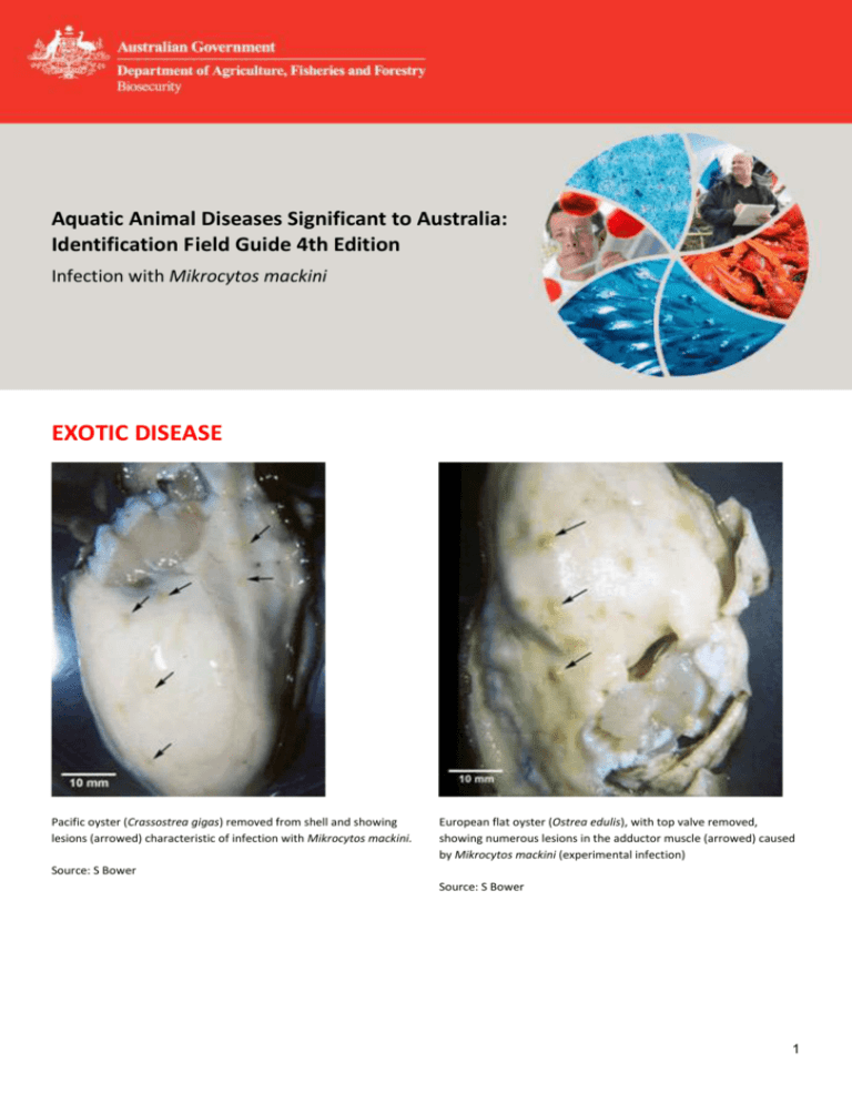
Aquatic Animal Diseases Significant to Australia: Identification Field Guide 4th Edition Infection with Mikrocytos mackini EXOTIC DISEASE Pacific oyster (Crassostrea gigas) removed from shell and showing lesions (arrowed) characteristic of infection with Mikrocytos mackini. European flat oyster (Ostrea edulis), with top valve removed, showing numerous lesions in the adductor muscle (arrowed) caused by Mikrocytos mackini (experimental infection) Source: S Bower Source: S Bower 1 Aquatic Animal Diseases Significant to Australia: Identification Field Guide, 4th edition Pacific oyster (Crassostrea gigas) removed from shell, showing lesions (arrowed) seen during later stages of infection with Mikrocytos mackini. Typically, M. mackini can no longer be found in oysters at this advanced stage of the disease. Source: S Bower Signs of disease Important: Animals with disease may show one or more of the signs below, but the pathogen may still be present in the absence of any signs. Diseases caused by any of the microcell species are similar, with few or no clinical or gross signs present with light infection. Identification of the Bonamia or Mikrocytos species requires histological laboratory examination and molecular diagnostic techniques. Disease signs at the farm, tank or pond level are: mortalities. Gross pathological signs are: focal yellow–green lesions up to 5 mm in diameter within the body wall or on surfaces of the gonad, labial palps, gills or mantle brown scars on the shell adjacent to lesions on the mantle surface gaping oysters (due to impaired adductor muscle contraction). Microscopic pathological signs are: focal intracellular infection, mainly of vesicular connective tissue cells, resulting in haemocyte infiltration and tissue necrosis intracellular microcell protozoa, 2–3 µm in diameter, in vesicular connective tissue cells immediately adjacent to lesions. Disease agent Mikrocytos mackini is an intracellular protozoan parasite that causes lethal infection of certain oysters. M. mackini is the only species described in the genus and is unrelated to Bonamia spp. 2 Aquatic Animal Diseases Significant to Australia: Identification Field Guide, 4th edition Host range Species known to be susceptible to infection with M. mackini are listed below. Common name Scientific name American oyster European flat oyster Olympia oystera Pacific oystera Crassostrea virginica Ostrea edulis Ostrea conchaphila Crassostrea gigas a Naturally susceptible (other species have been shown to be experimentally susceptible) Presence in Australia EXOTIC DISEASE—not present in Australia. Epidemiology Severe infections appear to be restricted to oysters over 2 years old. The disease is associated with low temperature and high salinity. Most mortalities occur during April–May (spring in the Northern Hemisphere). There is a 3–4 month pre-patent period when temperatures are less than 10 °C. The Pacific oyster appears to be more resistant to the disease than other species challenged experimentally under laboratory and field conditions. Differential diagnosis The list of similar diseases below refers only to the diseases covered by this field guide. Gross pathological signs may be representative of a number of diseases not included in this guide, which therefore should not be used to provide a definitive diagnosis, but rather as a tool to help identify the listed diseases that most closely account for the gross signs. Similar diseases Infection with Bonamia spp., B. ostreae and B. exitiosa There may be few or no visual cues to the presence of this disease other than poor condition, shell gaping and increased mortality. Any presumptive diagnosis requires further laboratory examination. Light microscopy can contribute diagnostic information, but further laboratory examination and molecular diagnostic techniques are required for a definitive diagnosis. Sample collection Due to the uncertainty in differentiating diseases using only gross pathological signs, and because some aquatic animal disease agents might pose a risk to humans, only trained personnel should collect samples. You should phone your state or territory hotline number and report your observations if you are not appropriately trained. If samples have to be collected, the state or territory agency taking your call will provide advice on the appropriate course of action. Local or district fisheries or veterinary authorities may also provide advice regarding sampling. Emergency disease hotline The national disease hotline number is 1800 675 888. This number will put you in contact with the appropriate state or territory agency. 3 Aquatic Animal Diseases Significant to Australia: Identification Field Guide, 4th edition Further reading Further information can be found on the following websites: disease pages of Fisheries and Oceans Canada: www.pac.dfo-mpo.gc.ca/science/species-especes/shellfishcoquillages/diseases-maladies/index-eng.htm Centre for Environment, Fisheries and Aquaculture Science (Cefas) International Database on Aquatic Animal Disease (IDAAD): www.cefas.defra.gov.uk/idaad/disocclist.aspx European Union Reference Laboratory for Molluscs Diseases website (at the French Research Institute for Exploration of the Sea—IFREMER): wwz.ifremer.fr/crlmollusc/Main-activities/Tutorials/Mikrocytos-mackini. These hyperlinks were correct and functioning at the time of publication. Further images (1) Haematoxylin and eosin stained section through a lesion caused by M. mackini on the mantle of a Pacific oyster (Crassostrea gigas). This intracellular protozoan (not visible at this magnification) usually occurs in the intact vesicular connective tissue cells immediately surrounding the periphery of the lesion (arrows). Source: S Bower (2) Many M. mackini (arrows) within vesicular connective tissue cells next to a lesion characterised by an accumulation of haemocytes and necrotic cells (haematoxylin and eosin stain) Source: S Bower 4 Aquatic Animal Diseases Significant to Australia: Identification Field Guide, 4th edition (3) Oil immersion magnification of M. mackini (arrows) within the cytoplasm of vesicular connective tissue cells of a Pacific oyster (Crassostrea gigas) (haematoxylin and eosin stain) (4) As for Figure 3 but from a different specimen. Because of the small size of this parasite, it is very difficult to visualise and photograph in histological preparations. (haematoxylin and eosin stain) Source: S Bower Source: S Bower (5) M. mackini (A) within fibres of the adductor muscle of a Pacific oyster (Crassostrea gigas). One M. mackini is located close to the nucleus (B) of a muscle cell. (haematoxylin and eosin stain) (6) Clusters of M. mackini (A), isolated and partially purified from a heavily infected Pacific oyster (Crassostrea gigas), among host cell debris (B) (Hemacolor® stain) Source: S Bower Source: S Bower 5 Aquatic Animal Diseases Significant to Australia: Identification Field Guide, 4th edition (7) Electron micrograph of a Pacific oyster vesicular connective tissue cell containing M. mackini (arrows) from a Pacific oyster (Crassostrea gigas) (uranyl acetate and lead citrate stain) 8) M. mackini (arrows), each containing a nucleus with a pronounced nucleolus and lacking mitochondria (uranyl acetate and lead citrate stain) Source: S Bower Source: S Bower 6 Aquatic Animal Diseases Significant to Australia: Identification Field Guide, 4th edition (9) Proposed developmental cycle of M. mackini, indicating host cell type and host organelle affiliation for the three recognised morphological forms: quiescent cell (QC), vesicular cell (VC) and endosomal cell (EC) Source: S Bower © Commonwealth of Australia 2012 This work is copyright. It may be reproduced in whole or in part subject to the inclusion of an acknowledgement of the source and no commercial usage or sale. +02 2 6272 3933 AAH@daff.gov.au daff.gov.au
