4.3 Appendicular Skeleton
advertisement
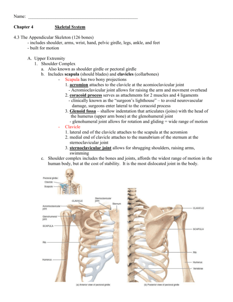
Name: ________________________________________________ Chapter 4 Skeletal System 4.3 The Appendicular Skeleton (126 bones) - includes shoulder, arms, wrist, hand, pelvic girdle, legs, ankle, and feet - built for motion A. Upper Extremity 1. Shoulder Complex a. Also known as shoulder girdle or pectoral girdle b. Includes scapula (should blades) and clavicles (collarbones) - Scapula has two bony projections 1. acromion attaches to the clavicle at the acomioclavicular joint - Acromioclavicular joint allows for raising the arm and movment overhead 2. coracoid process serves as attachments for 2 muscles and 4 ligaments - clinically known as the “surgeon’s lighthouse” – to avoid neurovascular damage, surgeons enter lateral to the coracoid process 3. Glenoid fossa – shallow indentation that articulates (joins) with the head of the humerus (upper arm bone) at the glenohumeral joint - glenohumeral joint allows for rotation and gliding = wide range of motion - Clavicle 1. lateral end of the clavicle attaches to the scapula at the acromion 2. medial end of clavicle attaches to the manubrium of the sternum at the sternoclavicular joint 3. sternoclavicular joint allows for shrugging shoulders, raising arms, swimming c. Shoulder complex includes the bones and joints, affords the widest range of motion in the human body, but at the cost of stability. It is the most dislocated joint in the body. Name: ________________________________________________ 2. Arm a. Single bone in the upper arm is the Humerus - second largest bone in the body (femur is 1st) - head of the humerus attaches to the scapula at glenohumeral joint b. forearm consists of Radius and Ulna - Radius articulates with wrist thumbside – it rotates around the ulna - Ulna is the larger and stronger of the two bones 1. Bony projection on the proximal end of the ulna is called the Olecranon process and articulates with the humerus at the humeroulnar joint (elbow) 2. Articulaes to the wrist on the side of the little finger - Radius and Ulna are connected their length by the interossius membrane and jointed at both proximal and distal ends by radioulnar joints - Distal end of the Radius and Ulna connect to the wrist at the Styloid process Name: ________________________________________________ 3. Wrist and Hand (27 bones in each) a.Wrist includes 8 carpal bones arranged in two rows - row 1 (forearm end, pinky to thumb) – Pisiform, Triquetrum, Lunate, Scaphoid - row 2 (finger end, pinky to thumb) – Harnate, Capitate, Trapezoid, Trapezium b. Hand includes 5 metacarpal bones numbers 1-5 beginning at the thumb c. Fingers include 14 phlalanges bones - four fingers have 3 phalanges each = proximal, medial, and distal phalanx - thumb has 2 = proximal distal phalanx Name: ________________________________________________ B. Lower Extremity – designed for weight bearing and walking, running, jumping, etc.. 1. Pelvic Girdle – female wider than male, main skeleton ID feature a. Bony encasement of reproductive organs, bladder and part of large intestine b. Includes four regions: 1. two large coxal bones (left and right) – hip bones formed by the fusion of the following parts: - ilium = most of the coxal bone, connects to sacrum at the sacroiliac joint, upper edge (iliac crest) can be felt by touch (palpation) - ischium = inferior portion of the coxal bone, supports the weight of the body when sitting. Holds the acetabulum (bony socket for the head of the femur) - pubis = anterior portion of coxal bone, both pubis bones fuse at the pubis symphysis with a disc of hyaline cartilage in between 2.Sacrum (5 fused spinal vertebrae) 3.Coccyx (4 fused spinal vertebrae - terminal) Name: ________________________________________________ 2. Leg a. Upper leg – one bone = femur 1. Longest and strongest bone in the body 2. Head of femur fits into the acetabulum of the hip a. ball and socket fit allows for an extremely stable joint b. weakest point on femur is the neck - neck injuries in older individuals often leads to reduced mobility and additional complications accelerating physical deterioration and death b. Lower leg – two bones = tibia and fibula 1. Tibia is the thick, strong, shin bone = weight bearing 2. Fibula is the thinner of the two bones a. it serves no special motion capability but holds various muscle attachments b. not articulated at the knee but on the distal end = lateral malleolus (protruding ankle bone) not really the ankle however 3. Tibia and Fibula are connected along their length by interosseous membrane and are joined (articulated) at both ends of the Fibula Kneecap = patella – triangular bone that protects the front of the knee Name: ________________________________________________ Lower leg Name: ________________________________________________ 3. Ankle and Foot a. Ankle (hindfoot) = 7 tarsal bones 1. Talus - large weight bearing 2. Calcaneus (heel) - large weight bearing 3. Navicular, lateral cuneiform, intermediate cuneiform, medial cuneiform, cuboid b. Midfoot – 5 metatarsals numbered 1 – 5, #1 being the large toe - Configuration of these bones froms two important arches = longitudinal arch (lengthwise) and the transvers arch (perpendicular to longitudinal arch) - These arches act as springs when you walk c. Toes – three phlanges each, only 2 on big toe (14 phlanges) – increase surface area of the foot giving greater body stability Name: ________________________________________________ Bone Accounting Location Skull: Facial Names Amount _____ Cranium Spine _____ Ribcage _____ Axial skeleton Total ______________ Shoulder Girdle _____ Upper extremity (x2) Upper arm _____ Lower arm _____ Wrist and hand _____ Pelvic Girdle _____ Lower extremity (x2) Upper leg _____ Lower leg _____ Ankle and foot _____ Appendicular skeleton Total ______________ Human Skeleton Total ______________

